Categories
Letters Articles

What comes next after #interncrisis?

Will last year be a turning point or just another chapter in the saga of medical workforce planning in Australia? The story, so ably publicised by the high profile grassroots #interncrisis campaign, [1] AMSA and the AMA, covered the lapse of planning by jurisdictional health authorities to provide graduates from Australian medical schools with internships. [2]

The reality was significantly more complex and will pose broader questions about the entire medical education continuum. As with many arguments in health, the complexity arises from the divide in funding, and therefore in control, of different levels of medical training.

Medical graduate numbers have increased by more than 200% in the last decade. [3] This increase was fuelled by both increases in Commonwealth supported places, and the unregulated expansion in international student numbers by many universities to cross-subsidise programs. Between 2003 and 2011, vocational medical training places have more than doubled in order to accommodate this growth. [3] However, capacity at all levels has supposedly been reached, with state and territory governments, who fund post-graduate training, now unwilling to match ongoing graduate increases. This has resulted in not only the well-publicised intern crisis but also a lack of post-internship prevocational positions in Queensland and the ever-increasing competition for specialty training places nationally. The disconnect between national and state medical workforce goals has never been clearer.

The crisis has far deeper themes than the simplistic media message typified by statements such as ‘unemployed doctors driving taxis’. As shown with the recent deployment of 60 regional internships, funded by the Commonwealth Department of Health and Ageing, bonding mechanisms are being used to attain better workforce geographic distribution. The ultimate prize is solving the chronic maldistribution of the medical workforce, both in terms of location and specialty. The goal is to create a sustainable medical workforce capable of delivering quality health care to the broader Australian community.

The events occurring at the end of 2012 contrast the conflicting goals of the Commonwealth with those of the states and territories. The Commonwealth Government considered the bigger picture of medical workforce reform and sought to retain the graduates of Australian medical schools, and yet was thwarted by the brinksmanship of the states and territories. The creation of additional medical internships was welcomed but ultimately, given the late hour, saw little uptake, as graduating students activated other plans. This was an opportunity missed but not one that should be forgotten as a new cohort nears graduation. The potential to partner with the private sector to open new pathways for training at both the prevocational and vocational levels is obvious, but, as always, will require funding.

It is clear that states and territories are planning and investing only for their own immediate need, and forgoing the potential of meeting Australia’s medical workforce requirements for short-term budgetary reasons. These decisions will have real world consequences including limiting accessibility to core services, leading to poorer health outcomes and unnecessary hospital admissions, particularly for those already disadvantaged, especially by distance. [4]

There is abundant evidence to support the employment of all Australian medical graduates. Health Workforce 2025 modelled the future health workforce required for Australia and proposed a number of possible futures. [5] In the baseline scenario, based upon current graduate trends, Australia will be short of more than 2000 doctors by 2025. The geographic maldistribution of the medical workforce and poor alignment of training pathways to the health requirements of the community is anticipated to further amplify perceived shortages.

We agree that Australia’s medical training system must change. Coordinating internship allocation at a national level, addressing the bottlenecks of entry into specialty training, and ensuring that community need shapes the medical workforce is just the beginning. Students and doctors-in-training also have an integral responsibility for creating a balanced, responsive medical workforce in their choices of specialty and location of training and practice.

Some action is already underway, with jurisdictions agreeing to the creation of the National Medical Training Advisory Network, but this will not be a panacea. Standing Council on Health meetings need to move beyond perpetuating the blame game, and create a timetable for reform while opening a discourse to consult meaningfully with the profession.

Innovative solutions to training issues must be influenced and informed by the experiences of students and doctors-in-training if they are to have the intended impact on the future medical workforce. Moreover, as the issue descends on states, ensuring that members of parliament are aware of the issues and their implications for the future of health service delivery will be a key opportunity for students to support long-term reform.

Conflict of interest

None declared.

Correspondence

M Bonning: michaelbonning@gmail.com

References

[1] Medical Student Action on Training. Interncrisis. 2013 [accessed 30 March, 2013]. Available from: http://interncrisis.org

[2] Australian Health Ministers’ Advisory Council. Communique 18 June 2012. Perth: AHMAC, 2012.

[3] Australian Government Department of Health and Ageing.  Medical Training Review Panel Fifteenth Report.  Canberra: DoHA, 2012.

[4] Australian Institute of Health and Welfare. Australia’s Health: 2010. Canberra. p.248

[5] Health Workforce Australia. Health Workforce 2025. Volume 3. 2012.

Categories
Review Articles Articles

Oncolytic Virotherapy: The avant-garde approach to oncological treatment via infectious agents

Over the past twenty years, advances in translational medicine have resulted in new and exciting treatments in the area of oncology. New modalities have arisen out of the need to address existing limitations in conventional treatments such as chemotherapy and radiotherapy. What started out as an outrageous idea in the 20th century to use potentially dangerous infectious agents such as viruses to kill cancer cells has gradually evolved into a maturing field, which has the promising potential to incorporate conventional and immunological aspects of treatment within a microbial-based system. Finally, in 2006, the introduction of the world’s first approved oncolytic virus by China heralded a milestone in the clinical application of this approach. This article will examine use of oncolytic viruses in cancer treatment with emphasis on its current status and strategies, possible immune mechanism and future considerations.

Introduction

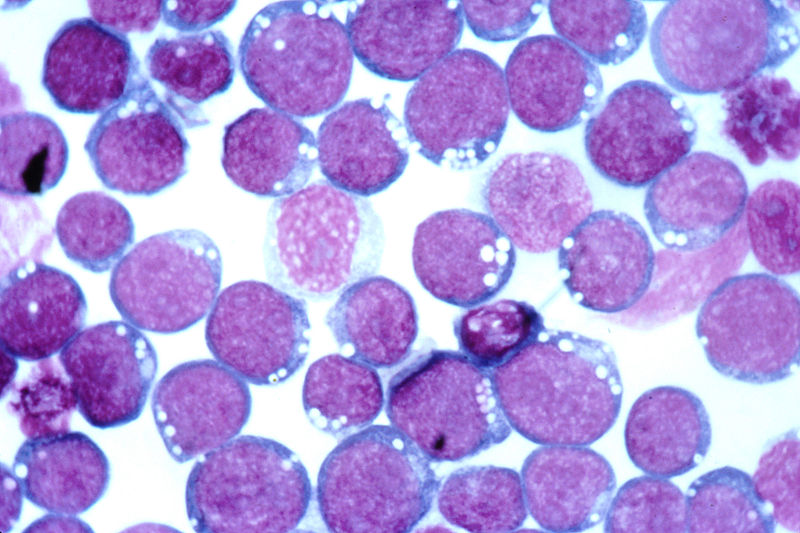
Oncolytic viruses are self-replicating viruses which can target and lyse cancer cells specifically. [1] Since the early 1900s, it was recognised that natural viral infections in cancer patients are sometimes associated with tumour regression. Indeed, case reports noted instances where influenza or measles infections in leukemia patients resulted in remissions. [2] Interest in utilising these ‘cancer-killing’ viruses peaked in the 1950-60s but the rise of chemotherapy and radiotherapy meant that progress in this field stagnated until the 1990s, when genetic engineering and better understanding of viruses and tumours revived the development of oncolytic viruses. [3] A breakthrough in the clinical translation of oncolytic viruses finally came in 2006 with the world’s first approved oncolytic virus- H101 (a genetically modified adenovirus) for head and neck cancers. [4]

Why oncolytic virotherapy?

Conventional treatments such as chemotherapy and radiotherapy have been the cornerstone of oncological management for many years. While we have achieved a considerable amount of success in many cancers, there are often criticisms against conventional treatments in terms of their limitations (e.g. transient effects against metastasis) and flaws (e.g. poor toxicity profile). [5] In recent years, gene therapy and immunotherapy have emerged as alternatives but results have been mixed. In 2002, the development of leukaemia in x-linked severe combined immunodeficiency (X-SCID) patients due to insertional mutagenesishas severely affected public confidence in gene therapy. [6] While immunotherapy remains promising, the current emphasis on specific targeting neglects the ability of tumour cells to mutate and change antigen profiles, resulting in variable clinical outcomes. [7]

In view of these insufficiencies, there has been renewed interest in oncolytic virotherapy, an interesting cross-disciplinary approach to treatment based virology, genetic engineering and immunology. The initial thinking behind this approach was simple- certain viruses exhibit tropism for cancer cells, which either express specific receptors for viral entry or lack anti-viral mechanisms that are normally intact in normal cells. [8] Once viral entry is achieved, replication of viruses continues until cell lysis occurs; allowing their progeny to infect other cancer cells.  If viral spread is homogenous, the oncolytic effect can be amplified many times and this effectively destroys the whole tumour. [8] As therapeutic genes encoding pro-apoptotic and immune effectors can be incorporated into the viral genome, an effective anti-tumour response may be initiated and magnified with each replication. [9] The ingenuity of this idea is that it exploits the infectious nature of viruses and uses it as a carrier and amplifier of other therapeutic agents. The latter may be crucial in exploiting synergistic anti-tumour effects between distinct treatment modalities.

Current status of oncolytic virotherapy

A variety of natural occurring and genetically modified viruses have been tested in clinical trials (Table 1).

Natural occurring oncolytic viruses are chosen for their low pathogenicity and inherent specificity for tumour cells. [10] Conversely, genetically modified viruses are those that are modified to promote tumour specificity, for example through use of tumour-specific promoters and gene deletions, or reduce pathogenicity by serial passage through cell culture. [8] Based on clinical data, it has been shown that virotherapy has a favourable toxicity and safety profile as compared to conventional treatment; the most common side effects being fever,flu-like symptoms and safety issues mainly concerning viral shedding and mutation-induced pathogenesis. [1,11] For the latter, dosing limitations and use of pro-drug activating suicide genes have addressed many of these issues. [1,6]

A straightforward dose-response relationship is not often observed as viral replication occurs in a heterogeneous tumour microenvironment and depends on factors such as availability of cell surface receptors and anti-viral responses. [12] Efficacy varies between different viruses but is reasonable at this early stage of development. The clinical trial for H101 reported complete remissions and partial responses in three and eleven out of forty-six patients respectively while another modified adenovirus- ONYX-15 was also used in head and neck cancer trials and achieved tumour growth stabilisation in eight out of twenty-two patients and tumour necrosis in five out of twenty-two patients.  [11,13]

It appears that limitations in efficacy were due to certain barriers. Firstly, viruses are not adept at surviving in the circulation. They are subjected to neutralising antibodies, complement and sequestration by the reticuloendothelial system. [8] In some cases, previous viral exposure may result in pre-existing anti-viral antibodies. For example, almost all individuals have antibodies to measles while reovirus infections are prevalent in about half the population. [14,15] Potent anti-viral responses such as type 1 interferons (IFNs) may also inhibit viral replication within the tumour. [8] Secondly, viruses have to endure acidotic and hypoxic conditions,transverse necrotic tumour regions and areas of poor vasculature in order to survive and infect tumour cells. [1] Thereafter, the availability of cell receptors may become a limiting factor in viral entry. [8] These obstacles are expected as viruses are foreign but this does not mean they are unsuitable therapeutic agents. On the contrary, viruses have the advantage of alerting the immune system to attack their infected target(s).

Enhancing oncolytic virotherapy via protective strategies

Protective strategies are aimed at improving delivery of viruses and avoiding viral clearance. The systemic delivery of viruses can be improved by preventing uptake of viruses by liver Kupffer cells (specialised macrophage cells). In mouse studies, viral delivery can be enhanced by clodronate-containing liposomes. [1] Clodronate is a selective macrophage-depleting agent that can temporarily inhibit viral uptake by Kuppfer cells, thereby allowing more virus particles to reach the tumour site. [1] Recent interest is focused on cell-carrier based delivery of oncolytic viruses, which aims to protect viruses from systemic and intra-tumoural barriers by packaging within a cell carrier that supports viral replication and targets tumour cells, its microenvironment or the tissue/organ in which the tumour resides. [16]

Cell-carriers targeting tumour cells include tumour-infiltrating lymphocytes (TILs) and cytokine-induced killer (CIK) cells. [16] TILs are T cells which accumulate in tumours and possess T cell receptors (TCRs) that recognise tumour-associated antigens (TAAs) in the context of major histocompatibility complex (MHC). [17] Since TILs are inherently cytotoxic to T cells, using such a carrier synergistically enhances the anti-tumour effects of oncolytic viruses. Highly-specific TAAs are rare and use of less-specific TAAs may lead to non-specific targeting of normal cells. [18] Production of TILs against TAAs is also an expensive and tedious process, which argues against its widespread clinical application. [16] Conversely, although cytotoxic lymphocytes like CIKs have a lower tumour-specificity, these cells are non-MHC dependent and can proliferate ex vivo without antigen stimulation. [18] Thorne et al. injected vaccinia virus-containing CIKs into nude mice and found that the VV/CIK combination was able to accurately target tumour cells and also improved the survival rate of mice as compared to VV administration alone. [19] To improve specificity, Yoon et al. engineered Her-2/neu expressing CIKs which can target ovarian cancer cells in nude mice with high affinity. Results suggest that this approach was more effective in killing cancer cells than administering Herceptin alone. [20] Nonetheless, mechanisms underlying the tumour-specificity of CIKs remain unclear and should be studied further.

In comparison, cell carriers targeting the tumour microenvironment have well-studied. Examples include mesenchymal stem cells (MSCs) and tumour-associated macrophages (TAMs). [18] MSCs are often attracted by inflammatory chemokines expressed in the microenvironment while TAMs tend to accumulate in hypoxic areas and regions of chronic inflammation in the tumour. [16] Mader et al. reported that in mouse studies, intra-peritoneal injection of a measles virus-MSC combination can prolong the survival period of mice with ovarian cancer. [21] Similarly, a clinical study found that intravenous injection of autologous MSCs carrying the modified adenovirus-ICOVIR 5 into four children with metastatic neuroblastoma was found to induce a complete clinical response in one child who also achieved complete remission within 3 years. [22] However, as MSCs are potentially tumourigenic, there is a trade-off between exploiting its propensity for tumour accumulation and the risk of enhancing tumour growth. The main criticism against targeting the tumour microenvironment relates to the inability of these cell carriers to deliver viruses directly into tumour cells. However, modifying the microenvironment through the engineering of viruses containing genes encoding pro-apoptotic and pro-inflammatory cytokines may circumvent this limitation by disrupting tumour-promoting interactions between the microenvironment and cancer cells. [16] Furthermore, the administration of proteases such as relaxin to degrade the extracellular matrix or fusogenic membrane glycoproteins to promote cell-to-cell fusion before oncolytic therapy may facilitate intratumoural spread of viruses. [23,34]

Lastly, the targeting of tumour-associated tissues and organs is also achieved by carriers such as dendritic cells (DCs) and peripheral blood lymphocytes (PBLs). [18] These carriers are attractive because they circulate through lymphoid organs such as the lymph nodes and spleen, which are sites of micrometastases and T-cell priming. [16] DC or PBL mediated delivery of VSV and reoviruses have been shown to purge metastases in lymphoid organs. Qiao et al. found that a VSV/PBL combination partially purged B16 metastases in mice 2-3 days after administration. [25] The oncolysis of metastatic cells by VSV also primed anti-tumour T cell responses effectively and probably contributed to fast purging. [16] Targeting of tissues/organs is the least specific but this negates the requirement for highly-specific tumour markers. In addition, the circulatory paths of these cell carriers are well-characterised, allowing better prediction of their tumour trafficking patterns. [18]

The mechanisms of viral loading, amplification and transfer are equally important in enhancing cell-carrier based strategies. Willmon et al. suggested that the loading of viruses depends on the multiplicity of infection (MOI), which is the ratio of infectious agent to infection target (i.e. cell carrier). [16] In high MOI loading, a higher viral loading density may be achieved but many viral particles will be stuck to the cell’s external surface and become susceptible to neutralising antibodies. [26] Conversely, in low MOI loading, most viral particles will be internalised although the viral loading density may be lower. [26] This approach may help avoid neutralisation and is suitable for individuals who have pre-existing antibodies against the oncolytic virus (e.g. measles and reovirus).

The carrier’s ability to support viral replication determines the amount of virus delivered. As viral replication can be affected by intact IFN responses in normal cells and also requires synchronous timing with carrier bursting, tumour cells have been implicated as possible carriers. [27] A successful example has been shown in the use of VSV-infected carcinoma cells to target lung metastases in mice but safety issues concerning the tumourigenity of tumour cell-based carriers remain. [28]

The transfer of virus from cell carrier to tumour cells is crucial as exposed viral particles are susceptible to neutralisation. In some viruses such human immunodeficiency virus (HIV), viral spread is mediated by a virological synapse (a specialised form of immunological synapse) between TCRs on T cells and MHC on adjacent cells. [29] By identifying viruses which utilises a virological synapse, oncolytic viruses can transfer safely between cells.

Besides cell-carrier based strategies, immunosuppression has been considered as a means of inhibiting anti-viral immunity. In rat glioma models, cyclophosphamide and cyclosporin A (CPA) have been shown to enhance HSV-mediated oncolysis by inhibiting tumour-mediated phagocyte infiltration. [30] However, recent studies suggest that such agents can be immunostimulatory and there is increasing recognition that anti-viral immunity also contribute to effective anti-tumour responses; implying that immune mechanisms of oncolytic virotherapy may need to be examined further. [31]

Immune mechanisms of oncolytic virotherapy

The direct oncolytic effects of oncolytic virotherapy are well appreciated. Successful infection and efficient spread of oncolytic viruses determine the extent of tumour lysis; leading to emphasis on developing viruses that replicated robustly and extensively. [31] However, the lack of a straightforward dose-response relationship suggests that other oncolytic mechanisms are present. The immune system may play paradoxical roles in enhancing or impeding anti-tumour responses mediated by oncolytic viruses. [32]

Innate immune responses have been shown to inhibit viral replication in rat glioma models as indicated by rapid decrease in HSV/VV titers with concomitant increase in natural killer (NK) cell infiltration following oncolytic virotherapy. [30,33] However, viral-mediated recruitment of NK cells is advantageous as NK cells are cytotoxic and associated with tumour regression. NK cells and DCs are also involved in reciprocal interactions. [31] In vitro experiments involving Mel888 melanoma cell lines showed that reovirus-infected DCs induced IFN-β production, which in turn activated NK cells. [34] Activation of NK cells resulted in cytotoxic effects against Mel888 cells and reciprocal maturation of DCs. [34] As DCs are involved in antigen presentation to T cells, DC maturation may also promote adaptive anti-tumour responses. However, DC functions are virus-dependent as studies showed that wild-type measles and adenoviruses are inhibitory and neutral respectively. [31] It appears that the timing of viral clearance is crucial and prolonging this therapeutic window by immunosuppression may be beneficial. This is because some immunosuppressive agents may suppress anti-viral responses while stimulating anti-tumour responses. For example, similar to HSV, rat glioma studies indicate that CPA may promote VV replication while inducing a cytokine storm, which enhances activity of tumour-associated cytotoxic lymphocytes. [35]

Adaptive anti-tumour responses may be shaped by two models of immune activation: the infectious non-self (INS) and ‘danger’ models. [31] The former refers to the provision of pathogen-associated molecular patterns (PAMPs) such as viral nucleic acids to pattern-recognition receptors (e.g. toll-like receptors) on antigen-presenting cells (APCs) while the latter refers to the release of endogenous ‘danger’ signals such TAAs to APCs. [31] In the INS model, the presence of viral PAMPs induces activation and proliferation of antibodies and T cells upon antigen presentation. Infection of tumour cells is therefore not a pre-requisite for anti-tumour responses, which may instead be due to bystander effects of anti-viral responses. This was illustrated by Breitbach et al. in a murine colorectal cancer model whereby administration of HSV and VV infected only a small number of cancer cells but triggered massive destruction of non-infected cancer cells. [36] Conversely, the ‘danger’ model is more in line with oncolysis of tumour cells. Greiner et al. showed that an attenuated VV was capable of lysing human melanoma cells with subsequent development of an anti-TAA response. [37] These two models are not mutually exclusive, suggesting that the actual anti-tumour effect may be mediated by both, with their relative contributions dependent on the immunogenicity of the oncolytic virus or the tumour. [31] It is therefore apparent from an immunological perspective that effective oncolyic virotherapy may capitalise on the use of highly immunogenic viruses in a bystander effect or alternatively, promoting efficient anti-TAA responses via engineering of TAA-expressing viral vectors in poorly immunogenic viruses.  [31,38]

The mechanisms involved in oncolytic virotherapy are summarised in Figure 1.

Future work

The current development of oncolytic virotherapy is based on rational designing but this approach may not always lead to the most selective and potent viruses. Buazon and Hermiston suggested that directed evolution might help researchers identify viruses with these desired characteristics. This involves growing diverse viruses in conditions that enhance diversity and passaging them through conditions mimicking the tumour microenvironment. [39] Application of directed evolution to colon cancer cell lines resulted in the adenovirus ColoAd1, which was 2-3 logs more potent than the advanced ONYX-15 and also had a therapeutic window 3-4 logs greater than the standard Ad5. [4,40] Furthermore, ColoAd1 is more sensitive to the anti-viral cidofovir (CDV) than either of its parents (Ad11p and Ad3) and this was due to directed evolution. [41] Thus, such an approach has a promising safety profile.

Integrating oncolyic virotherapy with existing treatments must be considered. Current studies have indicated promising results with radiotherapy and chemotherapy. A combination of modified HSV and radiotherapy have shown additive cell-killing effects in colorectal cancer assays and increased tumour regression in human glioma xenograft models. [42,43] It was suggested that radiotherapy could have improved viral replication and spread through irradiation-induced cellular changes. Similarly, pre-clinical studies have indicated that combination of ONXY-15 with cisplatin and 5-flurouracil in oesophageal cancers had a 39% increase in response rate as compared to chemotherapy alone (79% versus 40%). [44] Although both radiotherapy and chemotherapy are immunosuppressive to some extent, synergistic effects can be achieved by selecting the right virus and radiation/drug dosage. Combination of immunotherapy and virotherapy is at an early stage but success has been seen with VSV therapy combined with IL-2 and regulatory T cell (Treg) depletion in terms of enhanced NK cell activity and increased viral delivery. [45]

In the short term, the conflict between intra-tumoural and intravenous/intra-peritoneal administration of oncolytic viruses needs to be resolved. Mastrangelo et al showed that intra-tumoural injection of a GM-CSF-modified VV was able to induce regression in distant non-injected metastatic sites in melanoma patients but systemic effects were absent in intra-tumoural injections of ONYX-15 or reovirus. [46] Intravenous administration of the latter viruses was efficacious but resulted in thrombocytopenia and transaminitis respectively. [47] Therefore, when deciding the route of administration, a clear clinical endpoint must be established (targeting primary tumour or metastatic sites) and this will guide the type of virus used and effects (beneficial and detrimental) observed, and eventually how the patient is managed.

To conclude, oncolytic virotherapy has its antecedent in early observations and experiments detailing viral-mediated tumour regressions. Despite being neglected for decades, its resurgence reflects a current trend towards exploring new oncological treatments and a genuine hope that it can deliver better clinical outcomes.  Encouraging early results and the increasing availability of solutions to its problems suggest that it is well-poised to be the avant-garde of next-generation cancer therapeutics.

Conflict of interest

None declared.

Correspondence

K Ho: koho2292@uni.sydney.edu.au

References

[1] Prestwich RJ, Errington F, Harrington KJ, Pandha HS, Selby P, Melcher A. Oncolytic Viruses:  Do they have a role in anti-cancer therapy? Clin Med Oncol 2008; 2:83-96.

[2] Pelner L, Fowler GA, Nauts HC. Effects of concurrent infections and their toxins on the course of leukemia. Acta Med Scand Suppl 1958; 338: 1–47.

[3] Kelly E, Russell SJ. History of Oncolytic Viruses: Genesis to Genetic Engineering. Mol Ther 2007; 15(4):651-9.

[4] Garber K. China approves world’s first oncolyic virus therapy for cancer treatment. J Natl Cancer Inst 2006; 98:298-300.

[5] Gavhane YN, Shete AS, Bhagat AK, Shinde VR, Bhong KK, Khairnar GA, et al. Solid Tumours: Facts, Challenges and Solutions. IJPSR 2011; 2(1):1-12.

[6] McCormick F. Future prospects for oncolytic therapy. Oncogene 2005; 24:7817-9.

[7] Waldmann TA. Immunotherapy: Past, Present and Future.  Nat Med 2003; 9(3):269-77.

[8] Wong HH, Lemonine NR, Wang Y. Oncolytic viruses: Overcoming the Obstacles. Viruses 2010; 2(1):78-106.

[9] Chen L, Chen D, Gong M, Na M, Li L, Wu H, et al. Concomitant use of Ad5/35 chimeric oncolytic adenovirus with TRAIL gene and taxol produces synergistic cytotoxicity in gastric cancer cells. Cancer Lett 2009; 284:141-8.

[10] Cervantes-Garcia D, Ortiz-Lopez R, Mayek-Perez N, Rojas-Martinez A. Oncolytic virotherapy. Ann Hepatol 2008; 7(1):34-45.

[11] Lu W, Zheng S, Li XF, Huang JJ, Zheng X, Li Z. Intra-tumour injection of H101, a recombinant adenovirus, in combination with chemotherapy in patients with advanced cancers: a pilot phase II clinical trial. World J Gastroenterol 2004; 10(24):3634-8.

[12] Aghi M, Martuza RL. Oncolytic viral therapies-the clinical experience. Oncogene 2005; 24:7802-16.

[13] Ganly I, Kirn D, Eckhardt G, Rodriguez GI, Soutar DS, Otto R, et al. A phase I study of Onyx-015, an E1B attenuated adenovirus, administered intratumourally to patients with recurrent head and neck cancer. Clin Cancer Res 2000; 6(3):798-806.

[14] Tyler KL. Mammalian reoviruses. In Lippencott Fields Virology, Williams and Wilkens: Philidelphia; 2001.

[15] Iankov ID, Blechacz B, Liu C, Schmeckpeper JD, Tarara JE, Federspiel MJ, et al. Infected cell carriers: a new strategy for systemic delivery of oncolytic measles in cancer virotherapy. Mol Ther 2007; 15:114-22.

[16] Wilmon C, Harrington K, Kottke T, Prestwich R, Melcher A,Vile R. Cell carriers for oncolytic viruses: Fed Ex for cancer therapy. Mol Ther 2009; 17(10):1667-76.

[17] Goff SL, Smith FO, Klapper JA, Sherry R, Wunderlich JR, Steinberg SM, et al. Tumour infiltrating lymphocyte therapy for metastatic melanoma: analysis of tumours resected for TIL. J Immunother 2010; 33(8):840-7.

[18] Su X, Zhang L. Advances in cell carriers for oncolytic viruses in cancer therapy. Tumour 2011; 31(1):85-8.

[19] Thorne SH, Negrin RS, Contag CH. Synergistic antitumour effects of immune cell-viral biotherapy. Science 2006; 331(5768):1780-84.

[20] Yoon SH, Lee JM, Woo SJ, Park MJ, Park SJ, Kim HS, et al. Transfer of Her-2/neu specificity into cytokine-induced killer (CIK) cells with RNA encoding chimeric immune receptor (CIR). J Clin Immunol 2009; 29(6):806-14.

[21] Mader EK, Maeyama Y, Lin Y, Butler GW, Russell HM, Galanis E, et al. Mesenchymal stem cell carriers protect oncolytic measles viruses from antibody neutralization in an orthotopic ovarian cancer therapy model. Clin Cancer Res 2009; 15(23):7246-55.

[22] Garcia-Castro J, Alemany R, Cascallo M, Martinez-Quintanilla J, Arriero-Mdel M, Lassaletta A, et al. Treatment of metastatic neuroblastoma with systemic oncolyic virotherapy delivered by autologous mesenchymal stem cells: an exploratory study. Cancer Gene Ther 2010; 17(7):476-83.

[23] Kim JH, Lee YS, Kim H, Huang JH, Yoon AR, Yun CO. Relaxin expression from tumour-targeting adenoviruses and its intra-tumoural spread, apoptosis induction, and efficacy. J Natl Cancer 2006; 98:1482-93.

[24] Bateman A, Bullough F, Murphy S, Emiliusen L, Lavillette D, Cosset FL, et al. Fusogenic membrane glycoproteins as a novel class of genes for the local and immune-mediated control of tumour growth. Cancer Res 2000; 60:1492-7.

[25] Qiao J, Kottke T, Willmon C, Galivo F, Wongthida P, Diaz RM, et al. Purging metastases in lymphoid organs using a combination of antigen-nonspecific adoptive T cell therapy,oncolytic virotherapy and immunotherapy. Nat Med 2008; 14(1):37-44.

[26] Kottke T, Diaz RM, Kaluza K, Pulido J, Galivo F, Wongthida P, et al. Use of biological therapy to enhance both virotherapy and adoptive T-cell therapy for cancer. Mol Ther 2008; 16:1910-8.

[27] Ottolino-Perry K, Diallo JS, Lichty BD, Bell JC, McCart JA. Intelligent design: combination therapy with oncolytic viruses. Mol Ther 2010; 18(2):251-63.

[28] Garcia-Castro J, Martinez-Palacio J, Lillo R, Garcia-Sanchez F, Alemany R, Madero L, et al. Tumour cells as cellular vehicles to deliver gene therapies to metastatic tumours. Cancer Gen Ther 2005; 12(4):341-9.

[29] Groot F, Welsch S, Sattentau QJ. Efficient HIV-1 transmission from macrophages to T cells across transient virological synapses. Blood 2008; 111:4660-3.

[30] Fulci G, Breymann L, Gianni D, Kurozomi K, Rhee SS, Yu J, et al. Cyclophosphamide enhances glioma virotherapy by inhibiting innate immune responses. Proc Natl Acad Sci 2006; 103:12873-8.

[31] Prestwich RJ, Errington F, Diaz RM, Pandha HS, Harrington KJ, Melcher AA, et al. The Case of Oncolytic Viruses Versus the Immune System: Waiting on the Judgment of Solomon. Hum Gene Ther 2009; 20(10):1119-32.

[32] Prestwich RJ, Harrington KJ, Pandha HS, Vile RG, Melcher AA, Errington F. Oncolytic viruses: a novel form of immunotherapy. Expert Rev Anticancer Ther 2008; 8(10):1581-8.

[33] Lun XQ, Jang JH, Tang N, Deng H, Head R, Bell JC, et al. Efficacy of systemically administered oncolytic vaccinia virotherapy for malignant gliomas is enhanced by combination therapy with rapamycin or cyclophosphamide. Clin Cancer Res 2009; 15:2777-88.

[34] Prestwich RJ, Errington F, Ilett EJ, Morgan RS, Scott KJ, Kottke T, et al. Tumour infection by oncolytic reovirus primes adaptive anti-tumour immunity. Clin Cancer Res 2008; 14:7358-66.

[35] Brentjens RJ, Riviere I, Hollyman D, Taylor C, Nikhamin Y, Stefanski J, et al. Unexpected Toxicity of Cyclophosphamide Followed by Adoptively Transferred CD19-Targeted T Cells in a Patient with Bulky CLL. Molecular Therapy. 2009; 17(Suppl 1):S157.

[36] Breitbach CJ, Paterson JM, Lemay CG, Falls TJ, Mcguire A, Parato KA, et al. Targeted inflammation during oncolytic virus therapy severely compromises tumour blood flow. Mol Ther 2007; 15:1686-93.

[37] Greiner S, Humrich JY, Thuman P, Sauter B, Schuler G, Jenne L. The highly attenuated vaccinia virus strain modified virus Ankara induces apoptosis in melanoma cells and allows bystander dendritic cells to generate a potent anti-tumoural immunity. Clin Exp Immunol 2006; 146:344-53.

[38] Diaz RM, Galivo F, Kottke T, Wongthida P, Qiao J, Thompson J, et al. Oncolytic immunovirotherapy for melanoma using vesicular stomatitis virus. Cancer Res 2007; 67:2840-8.

[39] Bauzon M, Hermiston TW. Oncolytic Viruses: The Power of Directed Evolution. Adv Viro 2012; doi:10.1155/2012/586389.

[40] Kuhn I, Harden P, Bauzon M, Chartier C, Nye J, Thorne S, et al. Directed evolution generates a novel oncolytic virus for the treatment of cancer treatment. PLoS ONE 2008; 3(6): e2409. doi:10.1371/journal.pone.0002409.

[41] Bauzon M, Jin F, Kretschmer P, Hermiston T. In vitro analysis of cidofovir and genetically engineered TK expression as potential approaches for the intervention of ColoAd1-based treatment of cancer. Gene Ther 2009; 16(9):1169-74.

[42] Stanziale SF, Petrowsky H, Joe JK, Roberts GD, Zager JS, Gusani NJ, et al. Ionizing radiation potentiates the antitumour efficacy of oncolytic herpes simplex virus G207 by upregulating ribonucleotide reductase. Surgery 2002; 132:353-9.

[43] Advani SJ, Sibley GS, Song PY, Hallahan DE, Kataoka Y, Roizman B. Enhancement of replication of genetically engineered herples simplex viruses by ionizing radiation: a new paradigm for destruction of therapeutically intractable tumours. Gene Ther 1998; 5:160-5.

[44] Heise C, Sampson-Johannes A, Williams A, McCormick F, Von Hoff DD, Kim DH. ONYX-15, an E1B gene-attenuated adenovirus, causes tumour-specific cytolysis and antitumoural efficacy that can be augmented by standard chemotherapeutic agents. Nat Med 1997; 3:639-45.

[45] Kottke T, Galivo F, Wongthida P, Diaz RM, Thompson J, Jevremovic D, et al. Treg depletion-enhanced IL-2 treatment facilitates therapy of established tumours using systemically delivered oncolytic virus. Mol Ther 2008; 16:1217-26.

[46] Mastrangelo MJ, Maguire HC Jr, Eisenlohr LC, Laughlin CE, Monken CE, Mccue PA, et al. Intratumoural recombinant GM-CSF-encoding virus as gene therapy in patients with cutaneous melanoma. Cancer Gene Ther 1999; 6:409-22.

[47] Liu TC, Kirn D. Systemic efficacy with oncolytic virus therapeutics: clinical proof-of-concept and future directions. Cancer Res 2007; 67(2):429-32.

Categories
Letters Articles

National Leadership Development Seminar: developing the health care leaders of the future

The vast field of medicine transcends the mere finding of cures for ailments, seeking approaches to prolonging life, and undertaking research in the pursuit of wellbeing, important as these duties are. However, medicine’s empathetic pledge to the ill requires us to exercise leadership, amongst other qualities, as an important tool to advance the interests of both the individual and the population at large. Practical leadership and advocacy is the cornerstone of the increasingly complex environment in which 21st century healthcare is provided. Patients, institutions and communities often perceive doctors as agents of change i.e. leaders. However, some physicians may have been marginalised by the healthcare system because they either do not receive good leadership and management training, or they occupy positions that require leadership and managerial skills, which they initially do not possess. This apparent lack of appropriate leadership and management development may preclude doctors from participating in essential roles to shape the delivery of health services. [1] The problem can be traced back to medical school, where relatively little, or perhaps non-existent emphasis is given to nourish medical students’ attitudes towards leadership. Current medical curricula offer students little leadership education of the kind considered necessary to develop competences essential in becoming actively involved in the planning, implementation and provision of patient care. [2]

The Australian Medical Students’ Association (AMSA), being the peak advocating body for key affairs that concern medical students across the country, has identified this issue. It has responded by establishing the National Leadership Development Seminar (NLDS), an initiative aimed to assist motivated students interested in leading the medical profession. Each year, since its inception in 2005, NLDS attracts hundreds of applications from bright students who are keen to enhance their leadership skills. The seminar allows for approximately 90 applicants to participate annually. The NLDS program is carefully constructed to equip attendees with knowledge, skills and attitudes regarding leadership, advocacy and management, with a focus on current national health issues. The three-day seminar, which is held in Canberra, integrates guest speaker presentations, small group activities and interactive workshops to teach students how to link necessary leadership competencies with actual service opportunities.

The NLDS focus on leadership is closely aligned with Health LEADS Australia, a health professional leadership framework draft that has recently been published by Health Workforce Australia. The framework describes some of the most important leadership attributes that health workers who are involved in building a flourishing and sustainable health system should embrace and promote. This leadership framework is divided into five arms, including emotional intelligence and self-performance reflection, acknowledging the abilities of others whilst helping them to develop, and concentrating on achieving goals and pursuing innovative change. [3]

Although long term evaluation data is required to assess the effectiveness of NLDS (especially in meeting the key objective competences as outlined by Health LEADS Australia and Domain 4 of the Australian Medical Council’s Graduate Outcome Statements [4]), this program offers an innovative model of a leadership-based course. This can have a positive impact on leadership skills development among medical school students and can be incorporated into the medical school curriculum. We understand that NLDS has some limitations in terms of its primordial structure compared to other more established leadership programs in the realm of business and economics. These limitations include the program’s exclusivity to only a minor number of students per year, non-exhaustive coverage of all aspects of what it takes to become a successful leader in the clinical arena, and lack of networking past the event’s conclusion. Despite these drawbacks, NLDS is a unique national attempt to illustrate the importance of leadership in medical education. We invite medical schools to look at NLDS as a template whilst designing an innovative, socially accountable curriculum to engage students in the practices of advocacy, management and leadership.

Conflict of interest

None declared.

Correspondence

L Ngu: lngu45@student.monash.edu

References

[1] Abbas MR, Quince TA, Wood DF, Benson JA. Attitudes of medical students to medical leadership and management: a systematic review to inform curriculum development. BMC Med Educ. 2011;11:93.

[2] O’Connell MT, Pascoe JM. Undergraduate medical education for the 21st century: leadership and teamwork. Fam Med. 2004 Jan; 36 Suppl:S51-S56.

[3] Health LEADS Australia: Consultation for an Australian Health Leadership Framework: Health Workforce Australia; 2012. [cited July 2013]. Available from: http://hwaleadershipframework.net.au/files/HWA-HealthLeadershipFramework-forConsultation.pdf

[4] Accreditation Standards for Primary Medical Education Providers and their Program of Study and Graduate Outcome Statements: Australian Medical Council; 2012. [cited July 2013]. Available from: http://www.amc.org.au/images/Accreditation/FINAL-Standards-and-Graduate-Outcome-Statements-20-December-2012.pdf

Categories
Letters Articles

International medical electives: time for a rethink?

International medical electives (IMEs) are rapidly growing in popularity. A recent study by Law and colleagues [1] conducted across Australia reported that 53% of graduate entry program students and 35% of high school entry students undertook IMEs, of which just over half were in developing countries. In some medical schools the majority of students head overseas for their electives. [2] This phenomenon is not restricted to Australia; in the United Kingdon (UK) and United States (US) roughly 40% of students reported having spent some time in developing countries. [3,4] Many universities across Australia now have global health interest groups, and an increasing proportion of graduating medical students report having some experience in overseas health. [4] Traditionally, these electives are unstructured and arranged on an ad hoc basis between local partners and medical students.

There are undeniable benefits to practicing medicine in an unfamiliar, foreign setting. Students often describe IMEs as one of the highlights of their time at medical school, and it can be an opportunity for unparalleled personal and professional development. On a personal level, students report increased confidence, broadened perspectives, increased cultural competence, and improved communication skills. [5] Professionally, students benefit from being exposed to uncommon conditions and the opportunity for more hands-on experience. [6,7] IMEs also have the potential to influence future practice, with students more likely to enter public service, serve underprivileged populations, and participate in volunteering. [8,9]

However, the results of the aforementioned studies have to be interpreted with caution. Unlike other aspects of the medical course, IMEs tend to be student driven and lack a structured curriculum. Therefore many of the outcome measures are highly subjective and were assessed with unvalidated questionnaires. Given the observational nature of these studies, it is difficult to establish a causative relationship between IMEs and outcome measures. There is also the potential for selection bias (for example, where IME participants were chosen based on their commitment to global health) and publication bias in this area. [8,10] Given the subjectivity of the current literature, it remains unclear if there are indeed any long-term benefits for medical students.

Of note, much of the research on IMEs has revolved around medical students from OECD (Organisation for Economic Cooperation and Development) nations instead of host institutions or patients. Given the short term, transient nature of many IMEs, it seems unlikely that there will be any long term benefits to the local institution. There is potential for limited, temporary benefits such as increased supply of resources, incorporation of new teaching ideas, and positive support from local communities. [5] However, even this can turn out to be a double edged sword, as local institutions develop reliance on visiting medical students. Furthermore, there are cases where the donated equipment end up draining more of the hospitals resources in the long run, or are unable to be maintained. This is not to say that there are not examples of IMEs that have had a positive impact. [11] However, these programs tend to be structured, continuous partnerships between hosts and visiting students with a clear long-term goal.  Unfortunately, the vast majority of IMEs lack such a structure.

A significant proportion of IMEs involve students from developed countries heading to less developed countries. These include pre-clinical students with little to no practical medical training. As students they require proper supervision and this puts added strain on already scarce resources in developing countries. In addition to this problem, many students perceive electives as a holiday, tending to be ill-prepared both culturally and medically for the experience. [3] In worst case scenarios, the student may be placed in a position where he expected to take on the role of a qualified physician and is given responsibility for their own patients. [12] There are several reports in the literature of junior medical students being asked to participate in potentially risky procedures such as lumbar puncture and tubal ligation. [13] Students often try to rationalise this by adopting a utilitarian viewpoint, arguing that no one would look after these patients if they did not step up to the plate. The moral boundaries in these situations are vague and to date there are few established guidelines. However medical students must bear in mind that practicing beyond one’s competency is a serious breach of medical ethics. Students risk doing more harm than good, particularly when they may not be fully aware of the complexities associated with unfamiliar medical conditions and treatments.

To further aggravate this problem, patients in developing countries tend to be vulnerable and greatly disadvantaged. The risk of students developing their skills at the expense of vulnerable patients is a very real one that is probably under-reported in the literature. [14] Anecdotally, we often hear of medical students speak proudly about having been able to perform surgeries or risky procedures on their own with little supervision. There is often a lack of critical reflection surrounding this phenomenon, and clear ethical guidelines should be developed for students.

The motivation behind IMEs is slowly evolving. Traditionally, altruistic reasons were often quoted as the driving factor in medical students pursuing IMEs where students had a genuine interest in serving resource poor areas. [15,16] However, gaining a competitive advantage with the increasing demand for experiences in developing countries has become an important motivating factor. Global health programs look good on a CV and with training programs becoming more competitive, the proportion of students participating in IMEs for this reason will increase.

The threat to medical students’ well-being during electives is often an aspect that is overlooked. Medical students are often drawn by the sense of adventure, opportunity for travel, and the chance for a unique experience different to that back home. At the turn of the century, there was strong concern due to the lack of preparation by visiting British medical students to areas with a high prevalence of HIV. [17] There are often reports of a range of infectious diseases, ranging from schistosomiasis, thyphoid fever, malaria, and dysentery. [2] Literature now demonstrates that adverse events go beyond the risk of HIV and other infectious diseases. Deaths and serious injury have occurred due to risks associated with overseas travel (such as road traffic accidents), suicide, crime and political issues. [18] Aside from physical harm, psychological trauma has also been reported. [18]

Numerous studies encourage pre-departure training as a way to increase awareness of  ethical issues, encourage critical self-reflection, and practical preparation. [13,19] In spite of the physical dangers and ethical dilemmas that are sometimes posed by IMEs, studies have shown that basic practical and ethical preparations for students travelling abroad was low. [20] Only three quarters of Australian medical schools offer pre-departure training, however only half of these are mandatory. [1] The average duration of pre-departure training was 4.7 hours. Only half of Australian medical schools offered post-elective debriefing, out of which roughly half was mandatory. [1] The average duration of post-elective debriefing was 1.2 hours. [1] Medical schools have a duty of care towards medical students and it seems surprising that there is a significant lack of preparation for what is often a unique and unusual experience.

With the increasing ease and affordability of international travel, IMEs will continue to have a growing appeal to medical students. However there is a dangerous lack of critical thought and reflection in terms of the ethical aspect of IMEs, as well as the possible threat to student well-being. Given the strong consensus in the literature for more structured global health education, medical schools should consider developing training programs aimed at enabling students to conduct considered, structured and sustainable IMEs.

Acknowledgement

Saion Chatterjee for his assistance and feedback in editing the draft manuscript.

Conflict of interest

None declared.

Correspondence

Y Foong: Yichao.Foong@utas.edu.au

References

[1] Law IR, Worley PS, Langham FJ. International medical electives undertaken by Australian medical students: current trends and future directions. The Medical journal of Australia. 2013;198(6):324-6. Epub 2013/04/03.

[2] Goldsmid JM, Sharples N, Bettiol SS. A Preliminary Study on Travel Health Issues of Medical Students Undertaking Electives. Journal of Travel Medicine. 2003;10(3):160-2.

[3] Dowell J, Merrylees N. Electives: isn’t it time for a change? Medical Education. 2009;43(2):121-6.

[4] Association of American Medical Colleges. Medical School Graduation Questionnaire Report: 2000. Washington, DC: October 2000.

[5] Mutchnick IS, Moyer CA, Stern DT. Expanding the Boundaries of Medical Education: Evidence for Cross-Cultural Exchanges. Academic Medicine. 2003;78(10):S1-S5.

[6] Thompson MJ, Huntington MK, Hunt DD, Pinsky LE, Brodie JJ. Educational effects of international health electives on US and Canadian medical students and residents: a literature review. Academic Medicine. 2003;78(3):342-7.

[7] Drain PK, Holmes KK, Skeff KM, Hall TL, Gardner P. Global Health Training and International Clinical Rotations During Residency: Current Status, Needs, and Opportunities. Academic Medicine. 2009;84(3):320-5 10.1097/ACM.0b013e3181970a37.

[8] Ramsey AH, Haq C, Gjerde CL, Rothenberg D. Career influence of an international health experience during medical school. Fam Med. 2004;36(6):412-6. Epub 2004/06/08.

[9] Matar WY, Trottier DC, Balaa F, Fairful-Smith R, Moroz P. Surgical residency training and international volunteerism: a national survey of residents from 2 surgical specialties. Canadian journal of surgery Journal canadien de chirurgie. 2012;55(4):S191-9. Epub 2012/08/03.

[10] Imperato PJ. A third world international health elective for U.S. medical students: the 25-year experience of the State University of New York, Downstate Medical Center. Journal of community health. 2004;29(5):337-73. Epub 2004/10/09.

[11] Vora N, Chang M, Pandya H, Hasham A, Lazarus C. A student-initiated and student-facilitated international health elective for preclinical medical students. Medical education online. 2010;15. Epub 2010/02/27.

[12] Banatvala N, Doyal L. Knowing when to say “no” on the student elective: Students going on electives abroad need clinical guidelines. BMJ: British Medical Journal. 1998;316(7142):1404.

[13] Petrosoniak A, McCarthy A, Varpio L. International health electives: thematic results of student and professional interviews. Medical Education. 2010;44(7):683-9.

[14] Radstone SJ. Practising on the poor? Healthcare workers’ beliefs about the role of medical students during their elective. Journal of medical ethics. 2005;31(2):109-10. Epub 2005/02/01.

[15] Powell AC, Casey K, Liewehr DJ, Hayanga A, James TA, Cherr GS. Results of a national survey of surgical resident interest in international experience, electives, and volunteerism. Journal of the American College of Surgeons. 2009;208(2):304-12.

[16] Huish R. The Ethical Conundrum of International Health Electives in Medical Education. Journal of Global Citizenship & Equity Education. 2012;2(1).

[17] Wilkinson D, Symon B. Medical students, their electives, and HIV. BMJ (Clinical research ed). 1999;318(7177):139-40. Epub 1999/01/15.

[18] Tyagi S, Corbett S, Welfare M. Safety on elective: a survey on safety advice and adverse events during electives. Clinical medicine (London, England). 2006;6(2):154-6. Epub 2006/05/13.

[19] Dharamsi S, Osei-Twum J-A, Whiteman M. Socially responsible approaches to international electives and global health outreach. Medical Education. 2011;45(5):530-1.

[20] Bozorgmehr K, Schubert K, Menzel-Severing J, Tinnemann P. Global Health Education: a cross-sectional study among German medical students to identify needs, deficits and potential benefits (Part 1 of 2: Mobility patterns & educational needs and demands). BMC Medical Education. 2010;10(1):66.


Categories
Letters Articles

Australian Medical Student Journal Volume 4 Issue 2: Editor’s Welcome

Welcome to Volume 4, Issue 2 of the Australian Medical Student Journal.

Coming into our eighth issue, we are proud to announce that the journal continues to be a showcase of the outstanding quality of medical research done by students across Australia. We have continued to focus on the issues relevant to local medical students whilst maintaining a stringent peer review system. However, we feel that this is an opportune time to expand our horizons, and we’re pleased to announce that from the next issue onwards we will be trialing online publication of suitable international submissions. These are exciting times for the AMSJ and we look forward to what the future brings for the journal

The number of submissions have continued to grow with each issue, with a number of high quality submissions. Highlights include a unique 11 year audit of melioidosis in the Torres Strait Islands and a timely letter on the high profile #interncrisis campaign. Given the rising importance of melioidosis as a cause of infective disease in Northern Territories and Far North Queensland, the article provides us with vital statistics regarding the situation in the Torres Strait Islands. Of note, the authors conclude that the incidence of melioidosis is one of the highest in Australia and internationally.  Other notable submissions include an original research article on social phobia in children, reviews on the role of viruses in carcinogenesis and the use of viruses as oncological treatment, a report on an internship at the World Health Organisation, and a case report on dynamic compression of the motor ulnar nerve branch in the hand caused by a mobile segment of the hamulus.

We have been fortunate to receive articles from respected Australians in this issue of the AMSJ, namely Dr Helen Caldicott, Professor John Mattick and Professor David Wilkinson. Dr Helen Caldicott is a prominent Australian physician and a leading anti-nuclear activist, who presents her opinions on the impact of the recent Fukushima nuclear crisis on global health. Professor John Mattick is the executive director of the Garvan Institute and a internationally recognised leader in the field of genetics, and his article provides us with timely and well-placed advice on the rise of genomic medicine. Last but not least, Professor David Wilkinson has a wealth of experience in medical education, and is currently the Deputy Vice Chancellor at Macquarie University. We believe that his article on medical student assessment will be of great interest and relevance to Australian medical students.

As a medical student journal, we are reliant on the voluntary work of our student staff. This issue is a culmination of many months of hard work by staff members all across Australia, who have managed to put together a high quality, peer-reviewed journal whilst maintaining full time medical studies. A sincere thank you to all of our staff. I’d also like to take this opportunity to thank the previous Editor-in-Chief, Dr Michael Thompson, for his invaluable guidance and support, whilst wishing the incoming Editor-in-Chief, Saion Chatterjee, the best of luck with the following issue. We’re also indebted to our peer reviewers, most of whom are full-time professionals who took time out of their busy schedules to review our articles. We’re pleased to be able to acknowledge them in this issue, and look forward to working with them in future issues. Last but not least, we’d also like to thank our authors for their outstanding contributions, which provide the basis for the continued success of our journal. We hope you enjoy this issue of the AMSJ, and look forward to your feedback and future contributions.

Categories
Case Reports Articles

Mobile segment of the hamulus causing dynamic compression of the motor ulnar nerve branch in the hand

This paper is the first to document the mechanism of how a mobile segment of the hook of hamate can dynamically compress the motor branch of the ulnar nerve. Presented is the case of a professional golfer who experienced pain on the ulnar aspect of his right hand that he attributed to weakness and inability to control his hand. Imaging revealed the rare condition of os hamulus proprius causing a dynamic compression of the ulnar nerve when in power grip.  Provided is a review of wrist anatomy with particular focus on the peculiar case of the bipartite hamulus.

Introduction

Anatomy of the wrist

The wrist comprises a proximal and distal carpal row. The distal carpal row consists of the trapezium, trapezoid, capitate and hamate and acts as a base for the metacarpals. The proximal carpal row consists of the scaphoid, lunate, triquetrum and pisiform bone. These function as an intercalated segment, balancing the hand on the radius and ulna. [13]

Hamate anatomy and function

The hamate articulates with the triquetrum proximally and the bases of the 4th and 5th finger metacarpals distally. The hook of the hamate is an important structure in the hand.  Protruding from the volar surface of the hamate, it anchors the distal transverse carpal ligament, acting as a pulley for the ulnar flexor tendons and protecting the motor branch of the ulnar nerve. This branch of the ulnar nerve courses dorsally and distally around the hook of the hamate to supply nearly all the intrinsic muscles of the hand. [14]


Guyons Canal and the Ulnar Nerve

Felix Guyon described a potential space [15], which is a fibro-osseous tunnel, protecting the ulnar nerve and artery and veins as they enter the hand. The boundaries of Guyon’s canal are the pisiform bone, the tip of the hook of the hamate, the piso-hamate ligament and the transverse carpal ligament.

Os Hamulus proprium

The os hamulus ossifies from a primary ossification center in the body of the hamate; however, occasionally a secondary ossification center in the hook of the hamate is also present. [1] Rarely, the secondary ossification center in the hook of the hamate does not unite with the primary ossification center in the body of the hamate. [2] When the tip of the hook of the hamate does not fuse with the body of the hamate the result is a separate ossicle known as the os hamulus proprium or a bipartite hamulus. Whilst an os hamulus proprium or bipartite hamulus is often congenital a similar appearance can sometimes be the result of a non-union of a fracture of the hook of the hamate. [3]

Ossification of the hamate is not complete until the early teenage years. [4] Bone growth and maturation usually takes place via a single ossification center. However, a secondary ossification center independent from associated underling bone occasionally develops giving rise to an accessory ossicle. [9] This lack of fusion has been observed involving the hamulus and the hamate and is known as either os hamulus proprium or bipartite hamulus. Such cases are often congenital in nature; however, depending on the patient’s history, trauma or degenerative etiology should be considered. [10]

A study [5] conducted in 2005 on 3,218 hand radiographs revealed that variations are more prevalent than previously thought. 96 participants were found to have variations of the hook of hamate of which 42 patients had a bipartite hook, 50 had a hypoplastic hook and 4 had an aplastic hook. Furthermore, 93 of these cases presented with carpal tunnel syndrome symptoms.

In 1981, Greene et al. [6] identified a single case of bipartite hamulus with ulnar tunnel syndrome. However, since then there have been no other accounts of the os hamulus proprius, associated with dynamic ulnar neuropathy.

Case Study

History

The patient was a 37 year old professional right handed golfer with an unremarkable medical record.

He presented with an eight-week history of pain in the ulnar side of the right hand with loss of fine motor control requiring the use of his contralateral left hand to perform activities of daily living. The patient reported no other neurological symptoms at the time.

Physical examination revealed wasting of the intrinsic muscles of the right hand, most pronounced in the first dorsal interosseous muscles with weak intrinsic movements when comparison to the left side. Following initial examination a series of investigation and imaging was conducted:

It is not uncommon for golfers to fracture the hook of hamate based on the type of grip and dynamics of the golf swing. Furthermore, they can develop stress fractures of the hook of the hamate, which subsequently do not unite. [11,12]

Whilst this may have been the mechanism for the development of injury, an alternative explanation implicates a congenital anomaly where the primary ossification center the hamate fails to unite with the hook of the hamate giving rise to a bipartite bone (os hamulus proprius). [3]

Findings

This patient had a well-established long-standing asymptomatic non-union of the hamate or an os hamuli proprius, which subsequently became symptomatic following a motor vehicle accident in January 2005 resulting in an acute eight-week history of fine motor control deficit in the right hand.

Surgical intervention

A mobile segment of the hook of the hamate was identified.  Pressure over the mobile segment of the hook of the hamate compressed the motor branch of the median nerve as it traversed around the ulnar and distal hook of the hook of the hamate. The motor branch of the median nerve was swollen proximal to the point where the mobile segment of the hook of the hamate dynamically impacted on the nerve. This had the appearance of a ‘neuroma in continuity’ commonly seen from failure of regenerating nerve growth cone to reach peripheral targets.

The ulnar nerve was released in Guyon’s Canal. The motor branch of the ulnar nerve was identified and dissected as it coursed around the hook of the hamate. The hook of the hamate was very mobile and unstable. Manipulation of the mobile hook of the hamate demonstrated how it impacted and compressed the motor branch of the median nerve distal to the swollen segment of the motor branch of the median nerve. This was surgically excised.

The patient noticed a marked improvement of symptoms within two days post-operatively commenting on a return of ‘power and movement’. Following rehabilitation through daily grip strength exercises; this was further demonstrated on clinical examination at eighteen days confirming a return of intrinsic muscle power in the right hand.

The following five images describe the surgical repair of Os Hamulus Proprius as performed in this case.

Discussion

The hook of the hamate is an important structure providing mechanical stability on the ulnar aspect and protecting the motor branch of the ulnar nerve as it traverses deep into the hand from Guyon’s canal. It is also an important structure for insertion of the flexor retinaculum and as a result the muscles on the ulnar side of the hand. [16]

It is very likely that this abnormality of the hook of the hamate was present prior to his injury. The most likely explanation is that it is a secondary ossification center of the hook of the hamate (os hamulus proprius) which went on to unite. However, it is not possible to completely rule out that this represents a long standing non-union of the hook of the hamate and at some stage in the past he may have sustained a stress fracture which resulted in a non-union. [1,3,6,8,11,17]

Clinical examination plays a crucial role in isolating cases of os hamulus proprius. Patients will often present with clinical signs suggesting ulnar neuropathy such as intrinsic muscle weakness and altered sensation of the hand. In differentiating a case of bipartite hamulus, there will also be marked local tenderness over the hook of hamate with symptomatic pain due to dynamic compression such as when performing a power grip. Further hand and upper limb evaluation can compliment the diagnosis by quantifying and comparing loss of strength in the hand. [17]

The patient had marked motor (intrinsic hand muscles) weakness and some minor impairment of sensation in the ulnar distribution, which is consistent with the electrophysiological abnormalities in the hand. Surgery to remove the mobile segment of hamulus resulted in major improvement – particularly in terms of the level of his symptoms and restoration of normal power to the intrinsic muscles of the hand. Excision of the mobile os hamulus proprius has restored control and sensation of his left hand and enabled him to resume his career as a professional golfer.

Ulnar nerve compression in the hand could be due to a multitude of factors, including a tumour, a ganglion cyst, a fracture of either the pisiform or the hamate, compression in Guyon’s Canal, and an aneurysm of the ulnar artery. [18] To discriminate between a congenital bipartite hamulus or a non union of the hook of the hamate five criteria [17] have been described:

  • Bilaterally similar bipartite hamulus
  • Absence of history or signs of previous trauma
  • Equal size and uniform signal intensity of each part on imaging
  • Absence of progressive degenerative changes between the two components of the hamate or elsewhere in the wrist
  • Smooth well corticated and rounded margins of the hamate and mobile separate hook

Treatment

There are a limited number of options to treat a mobile hamulus segment causing ulnar nerve compression. [8] Initial splinting of the hand can be trialed to prevent dynamic compression of the nerve in the hope that pain and weakness resolve. [5] Furthermore, avoidance of sports relying on grip strength may provide symptomatic relief. If these interventions do not result in the resolution of symptoms, then there is the option of surgically excising the accessory ossification center on the tip of the hook of the hamate with subsequent decompression and release of the ulnar nerve such as presented in this case.

Consent declaration

Informed consent was obtained from the patient for publication of this case report and accompanying figures. IMAGE ONE is taken from http://upload.wikimedia.org/wikipedia/commons/3/31/Gray422.png. This image is in the public domain because its copyright has expired. This applies worldwide.

Acknowledgments

This paper was written under the supervision of Jeff Ecker from the Western Orthopaedic Clinic in Perth, WA.

Conflict of interest

None declared.

Correspondence

S Moniz: monizsheldon@gmail.com

References

[1] Andress M, Peckar V. Fracture of the hook of the hamate. British Journal of Radiology. 1970;43(506):141-143.

[2] Blum AG, Zabel J-P, Kohlmann R, Batch T, Barbara K, Zhu X, et al. Pathologic Conditions of the Hypothenar Eminence: Evaluation with Multidetector CT and MR Imaging1. Radiographics. 2006;26(4):1021-1044.

[3] Bianchi S, Abdelwahab I, Federici E. Unilateral os hamuli proprium simulating a fracture of the hook of the hamate: a case report. Bulletin of the Hospital for Joint Diseases Orthopaedic Institute. 1990;50(2):205.

[4] Grave K, Brown T. Skeletal ossification and the adolescent growth spurt. American journal of orthodontics. 1976;69(6):611-619.

[5] Chow JC, Weiss MA, Gu Y. Anatomic variations of the hook of hamate and the relationship to carpal tunnel syndrome. The Journal of hand surgery. 2005;30(6):1242-1247.

[6] Greene M, Hadied A. Bipartite hamulus with ulnar tunnel syndrome–case report and literature review. The Journal of hand surgery. 1981;6(6):605.

[7] O’Driscoll SW, Horii E, Carmichael SW, Morrey BF. The cubital tunnel and ulnar neuropathy. Journal of Bone & Joint Surgery, British Volume. 1991;73(4):613-617.

[8] Pierre-Jerome C, Roug I. MRI of bilateral bipartite hamulus: a case report. Surgical and Radiologic Anatomy. 1998;20(4):299-302.

[9] Garzón-Alvarado D, García-Aznar J, Doblaré M. Appearance and location of secondary ossification centres may be explained by a reaction–diffusion mechanism. Computers in biology and medicine. 2009;39(6):554-561.

[10] Freyschmidt J, Brossmann J. Koehler/Zimmer’s Borderlands of Normal and Early Pathological Findings in Skeletal Radiography. TIS; 2003.

[11] Koskinen SK, Mattila KT, Alanen AM, Aro HT. Stress fracture of the ulnar diaphysis in a recreational golfer. Clinical Journal of Sport Medicine. 1997;7(1):63.

[12] Torisu T. Fracture of the hook of the hamate by a golfswing. Clinical orthopaedics and related research. 1972;83:91-94.

[13] Viegas SF, Patterson RM, Hokanson JA, Davis J. Wrist anatomy: incidence, distribution, and correlation of anatomic variations, tears, and arthrosis. The Journal of hand surgery. 1993;18(3):463-475.

[14] Berger R, Garcia-Elias M. General anatomy of the wrist. In: Biomechanics of the wrist joint: Springer; 1991.

[15] SHEA JD, McCLAIN EJ. Ulnar-nerve compression syndromes at and below the wrist. The Journal of Bone & Joint Surgery. 1969;51(6):1095-1103.

[16] LaStayo P, Michlovitz S, Lee M. Wrist and hand. Physical Therapies in Sport and Exercise. 2007:338.

[17] Evans MW, Gilbert ML, Norton S. Case report of right hamate hook fracture in a patient with previous fracture history of left hamate hook: is it hamate bipartite? Chiropractic & osteopathy. 2006;14(1):1-7.

[18] Zeiss J, Jakab E, Khimji T, Imbriglia J. The ulnar tunnel at the wrist (Guyon’s canal): normal MR anatomy and variants. AJR. American journal of roentgenology. 1992;158(5):1081-1085.

Categories
Case Reports Articles

Adult Onset Still’s Disease – a diagnostic dilemma

Introduction

ASOD is characterised by fever, an evanescent skin rash, polyarthralgia, hepatosplenomegaly, leucocytosis, liver enzyme elevation and a high serum ferritin level. [1,2,3] It is a difficult diagnosis to make, as there is no pathognomonic test for the disease and it is a great mimicker of other conditions, such as autoimmune disorders and haematological malignancies.

Despite being a separate clinical entity to JIA and rheumatoid arthritis, there is evidence to suggest that AOSD as well as JIA are triggered by viral infections. [2,3,4] The following case demonstrates a young man who was diagnosed with AOSD following an infection with Epstein Barr Virus. This is impetus for a discussion of the interplay between AOSD and a viral aetiology, and the innate and adaptive immune responses in guiding effective therapy.

Case Presentation

In 2012, a previously healthy 20 year old male presented with a sore throat, malaise, tender cervical lymphadenopathy and fever, consistent with infectious mononucleosis. He was transferred to a secondary referral hospital where paired EBV serology was positive for an active infection despite a negative monospot test. The patient’s travel history and past medical history were unremarkable apart from regular alcohol binge drinking.

After being discharged, he began to experience intermittent fevers and night sweats. In addition to this, he had ongoing malaise and was forced to stop work as a mechanic. Weight loss of 10kg occurred during a two month period, along with a persisting microcytic anaemia, with a haemoglobin level of approximately 8.0 g/dL.

His polyarticular pain was distributed mainly to his ankles, knees, shoulders and wrists, and associated with morning stiffness and visible swelling. The pain was partially responsive to regular ibuprofen. He also complained of intermittent pleuritic chest pain. Over a course of two months, his weight stabilised and night sweats improved, but his anaemia and polyarthralgias persisted.

Approximately two months after his initial diagnosis of infectious mononucleosis, the patient represented to hospital with severe polyarthralgias and was unable to walk. During this admission, he was afebrile but had some mild tender cervical lymphadenopathy with no hepatosplenomegaly. A pleural rub was auscultated. He had a salmon-coloured non-blanching rash on the medial aspect of both legs that felt like a ‘sunburn’; this was biopsied. Although the diagnosis of ASOD had previously been considered, the patient was investigated for other causes for these symptoms. The results of these investigations are presented in Table 1. His investigations included a bone marrow and trephine biopsy, which revealed a markedly hypercellular bone marrow. The skin biopsy of the rash on his legs showed a leucocytoclastic vasculitis with perivascular neutrophilic invasion, but negative staining for complement. This finding is non-specific to the condition and can occur due to drug reaction, immune-complex deposition or be idiopathic. [5] As test results did not indicate another likely cause for his symptoms, the patient was commenced on treatment for ASOD and was referred to a rheumatologist.

Case Discussion

This case illustrates the unique clinical and laboratory picture of AOSD, with its intermitting and remitting fevers, polyarthralgias, myalgias, lymphadenopathy, transient macular rash and pleuritis. It is likely that the patient had a degree of pleuritis, as suggested clinically with a pleural rub and on CT imaging. Serositis manifesting as pleuritis, pleural effusions or pericarditis can be encountered in ASOD, but is rare. [3,6] The rash is fleeting and may only last for hours or days, and skin biopsies generally reveal a non-specific perivascular inflammation. [1] Our patient’s thrombocytosis and markedly elevated serum ferritin are reactive changes. The serum ferritin level has been suggested as a predictive marker for AOSD as it is invariably elevated and often higher than levels found in other autoimmune or inflammatory diseases, with a five-fold increase in serum ferritin being 41% specific and 80% sensitive as a diagnostic test. [9] The markedly high ferritin level in AOSD has been attributed to hyper-production by the reticuloendothelial system or hepatocyte damage, and is unrelated to iron metabolism. [8] The patient’s blood results illustrated a microcytic anaemia, although the iron studies point towards an inflammatory reaction.

The leukocyte count appears to correlate well with the activity of illness. The underlying mechanism of this is probably bone marrow granulocyte hyperplasia, as demonstrated on bone marrow biopsy in our patient. It is not uncommon to see marked reductions in red cell counts, weight loss and hypoalbuminaemia in active disease. [8]

In our patient, causes of fever of unknown origin with or without rash were considered, such as endocarditis, haematological malignancies and systemic vasculitides. The single cytopaenia, normal LDH and bone marrow biopsy excludes leukaemia, lymphoma and myelodysplasia. It is unlikely he had a protracted course of EBV due to the nature of his symptoms and degree of anaemia, in addition to the negative EBV IgM serology. Given the recent heavy rainfall, migrating polyarthritic conditions such as Ross River and Barmah Forest viruses were considered in the differentials.

The diagnosis of ASOD is made after taking into account the patient’s medical history and risk factors for other infectious agents, environment and relevant infectious diseases epidemiology. Although being a diagnosis of exclusion, there are two commonly used clinical criteria in practice, that being Yamaguchi (Table 2), which has been shown to be most sensitive (93.5%) followed by Cush’s (80.6% sensitivity). [7,8]

In regards to the aetiology of AOSD, there have been numerous case reports of AOSD following viral infection, [4,10] with one citing an older female patient diagnosed with AOSD after EBV infection. [2] Other implicated viruses include rubella, mumps, cytomegalovirus, parainfluenza, human herpes virus 6, echovirus, parvovirus B19, and bacterial infections like mycoplasma pneumoniae, chlamydia pneumonia, yersinia enterocolitica and borrelia. [2,8] Although relevant to our patient, the link between infections and AOSD has not been robustly established from an aetiological perspective, [10] and probably only forms part of the multifaceted pathogenesis, that being a dysregulated immune system combined with susceptible HLA loci. However, no consistent associations between AOSD and particular HLA loci have been elucidated, although HLA-B17, HLA-B18, HLA-B35 and HLA-DR2 have been implicated. [6]

It does appear that pathogenesis of the condition overlies autonomous activity of both innate and adaptive immune systems. Patients with AOSD often show hypercomplementaemia, and serum levels of IL-1β, IL-6, IL-18, TNFα, IFN Ɣ and macrophage-colony stimulating factor (M-CSF) have been found to be considerably higher than compared with controls. [6,7,11] These cytokines also appear to share a role in increasing the production of ferritin. [1,12] IL-18 is predominantly secreted by macrophages and has been implicated in hepatotoxicity [13] and joint disease, [7] and may be the cause of liver enzyme derangement characteristic of AOSD. Serum IL-18 levels also appear to correlate significantly with serum ferritin levels. [8] Furthermore, IL-18 may be seen as the part of the bridge between activation of the innate and adaptive immune systems in AOSD, as it facilitates the Th1 response and induces other cytokines like IL-1 β, TNFα and IFNƔ. [6] Pro-inflammatory cytokines such as IL-6, TNFα and IFN Ɣ also increase the expression of Toll-like receptors (TLR), and high circulating levels of cytokines leads to a higher sensitivity of TLR to anti-microbial or viral peptides, thus creating a self-perpetuating cycle of inflammatory response and augmentation. [14]

On the adaptive immunity side of the pathogenesis, the role of T cells in pathogenesis has been well documented. [11,14] Dysregulated production of a particular subset of T helper cells, called Th17 cells, that secrete IL-17 have been implicated in the development of autoimmune diseases. [15] Significantly higher levels of Th17 cells and serum IL-17 levels were found in both AOSD and SLE patients, and there was a parallel decrease with clinical remission. [10] IL-17 stimulates monocytes to produce IL-6 and IL-1β, which are also principle cytokines involved in the differentiation of CD4+ T cells into Th17 cells. [6] These therefore augment and maintain the inflammatory cascade. [16]

Non-steroidal anti-inflammatory drugs (NSAIDS) had previously been the first line medication for ASOD, despite only being effective monotherapy in less than 15% of patients. [10] The benefits of corticosteroids are higher when patients have more pronounced joint disease, with a response rate of two thirds of the patient population. [10]

Highlighting the implicated cytokines, namely IL-1β, IL-6 and TNFα, [17,18] will guide the use of targeted therapies such as the disease modifying anti-rheumatic drugs (DMARDS). There have been favourable results with corticosteroids, and more than two thirds of patients require corticosteroids after NSAIDs are attempted as symptom relief. [6] The use of DMARDS are indicated where the condition is refractory to corticosteroids without signs of remission, or in combination as corticosteroid-sparing agents. This includes methotrexate, which has indirect actions on TNFα and IL-6. Although there is a lack of robust evidence regarding TNF in the pathogenesis of ASOD compared to rheumatoid arthritis, the use of etanercept and infliximab have shown significant improvement in disease in several case series. [10] Of particular note, there is increasing evidence to suggest that anakinra, an IL-1 receptor antagonist, is well tolerated, and several case series have yielded positive results in ameliorating the disease at a haematological, biochemical and cytokine level. An excess of IL-1β inducing factor has been demonstrated in JIA, a condition that also shares similar pathogenesis to that of AOSD. [6,19]

The clinical course of AOSD is heterogeneous, with patients falling into one of three clinical patterns. The first group which affects about 60% of patients [8] is a monocyclic systemic group with only one episode of systemic manifestations, with complete remission within one year of the onset of symptoms. The second group is polycyclic systemic, whom experience more than one episode which is followed by partial or total remission. The third group is a chronic articular group, with persistent polyarthritis lasting longer than 6 months. [6] In the chronic group, the average duration of disease is 10 years, the symptoms appear to be less permanent than other rheumatological diseases and the disease shows less propensity to interfere with social functioning or time off from work despite disability and the need for long-term medication. [20]

Patient outcome

The patient improved satisfactorily with regular ibuprofen and prednisolone 20 mg daily and was discharged after day 7 with a tapering steroid dose.

He was able to resume work, but continued to experience mild intermittent polyarthralgias with no other significant systemic symptoms. Six months post-admission, deterioration in arthritic symptoms prompted the addition of methotrexate.

Key points

  • Adult Onset Stills Disease is a rare systemic inflammatory disorder that mainly affects people aged 16-35 years old.
  • It is a difficult diagnosis to make, and one that must be questioned continually, as it is a mimicker of other disorders, including other causes of fever of unknown origin, infectious diseases and malignancy.
  • It is characterised by both clinical and laboratory manifestations like fever, evanescent rash, polyarthritis and polymyalgias, microcytic anaemia, leucocytosis, thrombocytosis and marked hyperferritinaemia.
  • Treatment is based on clinical course and is similar to that of rheumatoid arthritis. A more targeted biological disease modifying therapy should be chosen with consideration of likely pathogenic pro-inflammatory cytokines.

Consent declaration

Consent from the patient was gained for the writing and distribution of this article for education purposes.

Acknowledgements

Thank you to Dr. Hedley Griffiths, Consultant Rheumatologist.

Conflict of interest

None declared.

Correspondence

S Ooi: soo@deakin.edu.au

References

[1] Mehrpoor G, Owlia M. Adult – onset Still’s disease: A review. Indian J Med Sci. 2009;63(5):207-21.

[2] Schifter T, Lewinski U. Adult onset Still’s disease associated with Epstein-Barr virus infection in a 66-year-old woman. Scand J Rheum. 1998;27(6):458-60.

[3] Mert A, Ozaras R, Tabak F, Bilir M, Ozturk R, Ozdogan H, et al. Fever of unknown origin: a review of 20 patients with adult-onset Still’s disease. Clin Rheum. 2003;22(2):89-93.

[4] Wouters J, van der Veen J, van de Putte L, de Rooij D. Adult onset Still’s disease and viral infections. Ann Rheum Dis. 1988;47(9):764-7.

[5] Koutkia P, Mylonakis E, Rounds S, Erickson A. Leucocytoclastic vasculitis: an update for the clinician. Scand J Rheum. 2001;30(6):315-22.

[6] Kontzias A, Efthimiou P. Adult-onset Still’s disease: pathogenesis, clinical manifestations and therapeutic advances. Drugs. 2008;68(3):319-37.

[7] Bagnari V, Colina M, Ciancio G, Govoni M, Trotta F. Adult-onset Still’s disease. Rheumatol Int. 2010;30(7):855-62.

[8] Kádár J, Petrovicz E. Adult-onset Still’s disease. Best Pract Res Cl Rh. 2004;18(5):663-76.

[9] Lian F, Wang Y, Yang X, Xu H, Liang L. Clinical features and hyperferritinemia diagnostic cutoff points for AOSD based on ROC curve: a Chinese experience. Rheumatol Int. 2012;32(1):189-92.

[10] Mavragani C, Spyridakis E, Koutsilieris M. Adult-Onset Still’s Disease: From Pathophysiology to Targeted Therapies. Int J Inflamm. 2012;2012:1-10.

[11] Efthimiou P, Georgy S. Pathogenesis and management of adult-onset Still’s disease. Semin Arthritis Rheu. 2006;36(3):144-52.

[12] Mehrpoor G, Owlia MB, Soleimani H, Ayatollahi J. Adult-onset Still’s disease: a report of 28 cases and review of the literature. Mod Rheumatol. 2008;18(5):480-5.

[13] Kasper D, Braunwald E, Fauci A, Hauser S, Longo D, Jameson J, Loscalzo J. Harrison’s principles of internal medicine (17th ed.). New York: McGraw-Hill Medical Publishing Division; 2008.

[14] Kramer M, Joosten L, Figdor C, van den Berg W, Radstake T, Adema GJ. Closing in on Toll-like receptors and NOD-LRR proteins in inflammatory disorders. Future Rheumatol. 2006;1(4):465-79.

[15] Lichtman M, Kipps T, Seligsohn U, Kaushansky K, Prchal J. Williams Hematology. 8th ed. USA: McGraw-Hill Companies; 2010.

[16] Chen D, Chen Y, Lan J, Lin C, Chen H, Hsieh C. Potential role of Th17 cells in the pathogenesis of adult-onset Still’s disease. Rheumatology. 2010;49(12):2305-12.

[17] Nordstrom D, Knight A, Luukkainen R, van Vollenhoven R, Rantalaiho V, Kajalainen A, et al. Beneficial Effect of Interleukin 1 Inhibition with Anakinra in Adult-onset Still’s Disease. An Open, Randomized, Multicenter Study. J Rheumatol. 2012 October 1, 2012;39(10):2008-11.

[18] Efthimiou P, Kontzias A, Ward C, Ogden N. Adult-onset Still’s disease: can recent advances in our understanding of its pathogenesis lead to targeted therapy? Nat Clin Prac Rheumatol. 2007;3(6):328-35.

[19] Allantaz F, Stichweh D, Pascual V. Interleukin-1 as a therapeutic target in systemic-onset juvenile idiopathic arthritis. Future Rheumatol. 2007;2(3):305-12.

[20] Sampalis J, Esdaile J, Medsger Jr T, Partridge A, Yeadon C, Senécal J, et al. A controlled study of the long-term prognosis of adult still’s disease. Am J Med. 1995;98(4):384-8.


Categories
Case Reports Articles

Acute viral bronchiolitis in the setting of extensive family history of asthma

This case report describes a previously healthy eleven-month old ex-preterm female with a severe presentation of acute viral bronchiolitis with an extensive family history of asthma. The link between viral bronchiolitis and asthma has always been controversial despite extensive research. Several studies have linked respiratory syncytial virus (RSV) bronchiolitis to the development of persistent wheezing or asthma later in childhood, even suggesting that a dose-response relationship may exist between the two entities. Some studies have also demonstrated that severe lower respiratory infections in the first year of life are important contributors to asthma, particular in those sensitized during infancy. On the other hand, it has also been studied as to whether an individual at risk of asthma has any impact on the severity of bronchiolitis. Despite numerous studies, results have largely been inconclusive, and the question of whether it is RSV that directly results in asthma, or if the susceptibility to RSV is conferred due to predisposing pulmonary pathology, still remains unknown.

Case

Sally was an eleven-month old ex-preterm (35 weeks) female who presented to the Emergency Department (ED) with symptoms of fever, coryzal symptoms and a wheeze, subsequently diagnosed as viral bronchiolitis.

History

Sally had become acutely febrile two nights prior to presentation, developing coryzal symptoms the following morning. Sally was initially treated for an upper respiratory tract infection (URTI) and acute otitis media with amoxicillin by her general practitioner, but began to worsen over the subsequent 24 hours, with laborious and wheezy breathing, coupled with a persistent fever of 38.4oC. Despite two doses of salbutamol, her breathing continued to deteriorate, leading to her presentation at the ED. There were no apnoeic or cyanotic episodes, rigors or any associated inspiratory stridor. During this period, Sally was anorexic, with subsequently fewer nappy changes, and was reported by her parents to be far less active than usual. She was previously well, with no known sick contacts, and her vaccinations were up-to-date.

Sally had a similar episode of bronchiolitis in at eight months of age but was treated then as an outpatient. There was an extensive family history of asthma (Figure 1), and both the patient’s siblings had bronchiolitis as infants. At eleven months, Sally was meeting all the developmental parameters for her age. There were no known drug allergies and other than salbutamol PRN, the patient was not on any other medication. There was no remarkable social history.

Examination

Sally appeared lethargic and was in respiratory distress, with tachypnoea, an audible wheeze and classical signs of increased breathing effort (nasal flaring, tracheal tug and subcostal recession). She had a respiratory rate of 78 and was saturating at 100% on 8L supplementary oxygen, which decreased to 90% on room air, and was febrile at 38.5oC. She was tachycardic at a heart rate of 170. There were no clinical signs of dehydration. On auscultation, air entry was reduced bilaterally, with an expiratory wheeze and diffuse crackles present. There was no evidence of stridor, increased vocal resonance, or dullness to percussion. An ear, nose and throat examination revealed an erythematous pharynx but was otherwise unremarkable. All other examination findings were unremarkable.

Workup and Progress

The presenting symptoms suggested a diagnosis of acute viral bronchiolitis. However, with the extensive family history of asthma, Sally’s presentation could be her first virus- triggered asthma attack. While the clinical presentation suggested otherwise, there were concerns over the possibility of pneumonia, which had to be ruled out in the workup. In consideration of the severity of her initial presentation and the likely further deterioration until its peak at day 2-3, basic investigations were ordered. These included a full blood count, urea, electrolytes and creatinine parameters, a chest X-ray and a nasopharyngeal aspirate. There were no abnormal findings.

Sally was commenced on immediate supportive therapy. An IV line was inserted and she was commenced on 75% maintenance fluids as per guidelines to avoid Syndrome of Inappropriate Antidiuretic Syndrome. [1] She was also commenced initially on 2L oxygen as per guidelines for respiratory distress, [2] but failed to saturate appropriately until given 8L of humidified oxygen via nasal prongs, where she maintained 100% O2 saturation. While the efficacy of short-acting beta agonists (SABAs) in the acute management of bronchiolitis has been inconclusive despite extensive research, current guidelines recommended a trial of bronchodilators in infants >6 months. [3] Sally was administered six puffs of Salbutamol MDI (100mcg/ puff) via spacer [1] but with no results, hence the regime was discontinued.

The Royal Children’s Hospital (RCH) provides further guidance regarding management based on clinical signs and symptoms. Admission to the intensive care unit (ICU) was indicated for Sally to allow continuous cardiorespiratory monitoring and supportive management. Observations were performed hourly and with only supportive management, her oxygen requirements were weaned down to 2L over 24 hours.

Discussion

Bronchiolitis during infancy and asthma in childhood – is there a causal link? Should infants at high risk of asthma receive Palivizumab immunization?

Several studies have linked respiratory syncytial virus (RSV) bronchiolitis to the development of persistent wheezing or asthma later in childhood. In a long-term prospective cohort study, there was a relative risk of 2.8 of developing wheezing at 5.5 years in children who had had bronchiolitis. [4] Sigurs et al. (2005) also reported a ten-fold excess of asthma in a similar study. [5]

It has also been suggested that a dose-response relationship exists between bronchiolitis and asthma. In a study involving 90,341 children, Carroll et al. (2009) demonstrated that the odds ratios (OR) for asthma as a child were 1.86 (95% CI, 1.7-2.0), 2.41 (95% CI, 2.2-2.6) and 2.8 (95% CI, 2.6-3.0) in the outpatient, ED, and hospitalization groups, respectively, compared to children without bronchiolitis. [6] Henderson et al. (2005) also noted an OR of 2.5 (95% CI 1.4-4.3) of developing asthma with hospitalization for RSV bronchiolitis, [7] as did two prospective studies, which showed a 30-40% likelihood of subsequent asthma. [8]

RSV bronchiolitis as a direct cause of asthma

In an extensive seven-year REBEL prospective cohort study, Bacharier et al. (2012) reported that increased Chemokine (C-C motif) Ligand 5 (CCL5) expression in nasal epithelial cells during RSV infection carried an OR of 3.8 (95% CI, 1.2-2.4) for developing asthma. [9] This is in concordance with studies demonstrating increased CCL5 levels in subjects with asthma, [10] as well as in vitro studies demonstrating increased expression and transcription by RSV. [11] Unfortunately, the study failed to measure CCL5 levels prior to infection and thus the causal relationship has not been established. Hence, whether it was RSV that directly resulted in asthma, or if the susceptibility to RSV was conferred due to predisposing pulmonary pathology, [12] still remains unknown.

A five-year cohort study on children at high risk of atopy by Kusel et al. (2007) demonstrated that severe lower respiratory infections in the first year of life are important contributors to asthma, particular in those sensitized during infancy. [13] These findings suggest that protecting high-risk individuals from infection during infancy may be considered for long-term asthma prevention.

Effect of family history of asthma or atopy on severity of bronchiolitis

It has also been studied whether an individual at risk of asthma has any impact on the severity of bronchiolitis. This was particularly relevant in the Sally’s case, with her significant family history of asthma. Results in this field have been conflicting, with most studies not eliciting any significant association. However, Gurwitz et al. (1981) demonstrated that hospitalized cases were associated with a higher incidence of first-degree relatives with bronchial hyper-responsiveness. [14] A study by Trefny et al. (2000) also demonstrated similar results. [15]

Should high-risk atopic individuals receive Palivizumab immunization during RSV season for prevention of asthma?

Passive immunization with Palivizumab is currently recommended only for high-risk infants to prevent serious complications arising from RSV infections. [16] However, a recent double-blinded RCT in the Netherlands has begun examining its preventive effect on recurrent wheeze in healthy preterm children 33-35 weeks gestational age (MAKI trial), based on a non-randomized trial suggesting a prevention of wheeze in 50% of preterm children. [17] Such a study would complement this case study’s patient profile, and would be especially relevant in the context of her rich family history of asthma which puts her at high risk, and the abovementioned association but inconclusive causation between bronchiolitis and asthma.

From an economic standpoint, studies assessed the cost-effectiveness of Palivizumab, albeit in the context of high-risk premature infants (32-35 weeks). Unfortunately, the predisposition of these infants to a higher disease burden and costlier hospitalizations constitutes a higher cost per QALY [18]      compared to this case study’s patient, but even then there is still considerable controversy over its cost-effectiveness, especially across various healthcare systems.

Conclusion

In summary, this was a case of severe viral bronchiolitis warranting ICU admission for supportive management, on a background of an extensive family history of asthma. While studies have shown a clear association of bronchiolitis with asthma, causation has not been conclusively established, with family history of atopy possibly interacting in the development of asthma. Current research is lacking in the area of Palivizumab prophylaxis in the interest of asthma prevention in healthy children, but the evidence would suggest that it is likely to be cost-ineffective.

Conflict of interest

None declared.

Correspondence

G Yong: glenn.yong.kw@gmail.com

References

[1] Royal Children’s Hospital. Bronchiolitis- Ongoing Management. http://www.rch.org.au/rchcpg/hospital_clinical_guideline_index/Bronchiolitis_Ongoing_Management/#Fluid_requirements (accessed 11 March 2013)

[2] Royal Children’s Hospital. Oxygen Delivery. http://www.rch.org.au/rchcpg/hospital_clinical_guideline_index/Oxygen_delivery/ (accessed 11 March 2013)

[3] [Guideline] Diagnosis and management of bronchiolitis. Pediatrics. 2006 Oct;118(4):1774-93.

[4] Murray M, Webb MS, O’Callaghan C, Swarbrick AS, Milner AD. Respiratory status and allergy after bronchiolitis. Arch Dis Child. 1992 April;67(4): 482-7.

[5] Sigurs N, Bjarnason R, Sigurbergsson F, Kjellman B. Respiratory syncytial virus bronchiolitis in infancy is an important risk factor for asthma and allergy at age 7. Am J Respir Crit Care Med. 2000;161:1501–7.

[6] Carrol KN, Wu P, Gebretsadik T, Griffin MR, Dupont WD, Mitchel EF, Hartert TV. The severity- dependent relationship of infant bronchiolitis on the risk and morbidity of early childhood asthma. Journal of Allergy & Clinical Immunology 2009;123:1055-61.

[7] Henderson J, Hilliard TN, Sherriff A, Stalker D, Al Shammari N, Thomas HM. Hospitalization for RSV bronchiolitis before 12 months of age and subsequent asthma, atopy and wheeze: a longitudinal birth cohort study. Pediatric Allergy Immunology. 2005;16:386–92.

[8] Sign AM, Moore PE, Gern JE, Lemanske RF, Hartert TV. Bronchiolitis to asthma A review and call for studies of gene-virus interactions in asthma causation. Am J. Respir. Crit Care Med. January15 2007;175(2):108-19.

[9] Bacharier LB, Cohen R, Schweiger T, Yin-Declue H, Christie C, Zheng J et al. Determinants of asthma after severe respiratory syncytial virus bronchiolitis. Journal of Allergy & Clinical Immunology 2012;130:91-100.

[10] Humbert M, Ying S, Corrigan C, Menz G, Barkans J, Pfister R et al. Bronchial mucosal expression of the genes encoding chemokines RANTES and MCP-3 in symptomatic atopic and nonatopic asthmatics: relationship to the eosinophilactive cytokines interleukin (IL)-5, granulocyte macrophage-colony-stimulating factor, and IL-3. Am J Respir Cell Mol Biol 1997;16:1-8.

[11] Koga S, Novick AC, Toma H, Fairchild RL. CD81T cells produce RANTES during acute rejection of murine allogeneic skin grafts. Transplantation 1999;67:854-64.

[12] Adamko DJ, Friesen M. Why does respiratory syncytial virus appear to cause asthma? Journal of Allergy & Clinical Immunology 2012 Jul;130(1):101-2.

[13] Kusel MM,  de Klerk NH, Kebadze T Vohma V, Holt PG, Johnston SL et al. Early-life respiratory viral infections, atopic sensitization, and risk of subsequent development of persistent asthma. Journal of Allergy & Clinical Immunology 2007;119:1105-10.

[14] Gurwitz D, Mindorff C, Levison H. Increased incidence of bronchial reactivity in children with a history of bronchiolitis. J Pediatr 1981;98:551–5.

[15] Trefny P, Stricker T, Baerlocher C, Sennhauser FH. Family history of atopy and clinical course of RSV infection in ambulatory and hospitalized infants. Pediatric Pulmonology 2000;30:302–6.

[16] Wang D, Byliss S, Meads C. Palivizumab for immunoprophylaxis of respiratory syncytial virus (RSV) bronchiolitis in high-risk infants and young children: a systematic review and additional economic modelling of subgroup analyses. Health Technol Assess 2011 Jan;15(5):1-124.

[17] Broughton S, Bhat R, Roberts A, Zuckerman M, Rafferty G, Greenough A. Diminished lung function, RSV infection, and respiratory morbidity in prematurely born infants. Arch Dis Child Jan 2006;91(1):26-30.

[18] Smart KA, Paes BA, Lanctot KL. Changing costs and the impact of RSV prophylaxis. Journal of Medical Economics. 2010;13(4):705-8.


Categories
Case Reports Articles

A case of solid pericardial metastases causing constrictive pericarditis in a patient with non small cell lung cancer

Introduction: Cardiac metastases are rarely diagnosed in patients with cancer despite being a common finding at autopsy. We report on a case of pericardial metastases in a patient with non small cell lung cancer (NSCLC) in the setting of coexisting direct tumour invasion into the superior vena cava (SVC) and right atrium. Case: We present a case of a 61-year old gentleman with metastatic adenocarcinoma of the lung. He presented with 2 – 3 days of progressive dyspnoea associated with pre-syncope. His physical examination was significant for bilateral elevated jugular venous pressure (JVP), bilateral arm oedema and hypotension. These clinical signs were explained by solid pericardial metastases causing constrictive pericarditis in the absence of significant pericardial effusion, and direct invasion of tumour into the SVC and right atrium producing SVC obstruction. His admission was complicated by an episode of supraventricular tachycardia (SVT), presumably caused by compromise of the electrical conduction system within the right atrium. Discussion: Cardiac metastasis is a common occurrence in advanced neoplastic disease, but is often clinically silent. The mechanism of cardiac metastases is believed to be primarily through lymphatic channels and metastasis is most commonly located in the pericardium. Symptoms of cardiac metastasis can be understood with respect to their anatomical position and are best diagnosed using transoesophageal echocardiogram (TOE). The most concerning complication of metastasis to the heart is pericardial effusion leading to life-threatening cardiac tamponade. This is an oncological emergency and is treated with pericardiocentesis and follow up preventative measures. Ultimately, cardiac metastasis signals advanced disease and poor prognosis.

Introduction

Cardiac metastases occur in 20-30% of patients with non small cell lung cancer (NSCLC) [1] but are clinically silent in the majority of cases. [2] We report on a case of constrictive pericarditis caused by solid pericardial metastases concurrent with direct invasion of tumour into the superior vena cava (SVC) and right atrium. The clinical picture was complicated by SVC obstruction and right atrial compromise causing supraventricular tachycardia (SVT).

The case

The patient was a 61-year old store worker with a previous 60 pack-year smoking history.  He was admitted with progressive dyspnoea over 2-3 days, associated with pre-syncope, on a background of metastatic NSCLC (T4N0M1b).

Past Medical History

The patient initially presented with a six month history of pain when abducting his right arm, which was associated with dyspnoea and a productive cough. A chest computed tomography (CT) identified a large right hilar lesion causing right middle lobe bronchus occlusion and collapse of the right lung. The tumour had invaded the mediastinum, and was attenuating the SVC and compromising the right atrium. Metastases to the liver, the right sternoclavicular joint and the 4th, 9th and 10th ribs were also identified.

Pathology from the bronchoscopy biopsy demonstrated that the tumour was p63 negative and Thyroid Transcription Factor 1 (TTF-1) positive via immunohistochemical staining, consistent with adenocarcinoma of the lung. Epidermal Growth Factor Receptor (EGFR) screening was negative, hence the tumour was not sensitive to treatment to EGFR tyrosine kinase inhibitors. His management plan, following multidisciplinary team discussion, was radiotherapy to his chest and clavicle for pain management, and palliative chemotherapy (carboplatin/paclitaxel).

Three months post initial diagnosis, restaging CT of the chest, abdomen, pelvis and lumbar spine was organised after completion of radiotherapy and two cycles of chemotherapy. CT scans of the chest, abdomen and pelvis showed disease progression with new right adrenal metastases and new pericardial metastases (Figures 1 and 2). CT of his lumbar spine revealed disease in L1-2 and S1. In light of his latest CT results, radiotherapy to the lumbar spine and pericardium was planned.

Medications and Allergies

At time of admission the patient’s medications included oxycodone/naloxone, omeprazole and dexamethasone. He was allergic to penicillin.

Inpatient Admission

The patient was admitted after worsening dyspnoea was noticed during radiotherapy to his lumbar spine. Initial physical examination revealed mild bilateral pitting oedema at the ankle and no other signs. His initial investigations included full blood examination (FBE), urea, electrolytes and creatinine (UEC), liver function tests (LFTs), and calcium, magnesium and phosphate (CMP). All were normal except for a decrease in haemoglobin (Hb: 83). A provisional diagnosis of anaemia secondary to chemotherapy or neoplastic disease was made based on his haemoglobin, and he was given two units of packed red blood cells.

The following morning, the patient’s dyspnoea had not improved with transfusion.  On examination, his jugular venous pressure (JVP) was elevated bilaterally at four centimetres, his arms were swollen bilaterally and his blood pressure (BP) was 95/55. He was not tachycardic. An urgent electrocardiogram (ECG) and transthoracic echocardiogram (TOE) was performed to exclude cardiac tamponade. ECG was normal and a pericardial effusion, though noted on TOE, was trivial and deemed insufficient to cause cardiac tamponade. However, the echocardiogram demonstrated pericardial metastases overlying the left and right ventricular apex as well as the lateral left ventricular wall and inferior right ventricular wall. Significant echocardiography findings consistent with constrictive pericarditis included abnormal septal motion with marked septal bounce, annulus reversus on tissue Doppler, and left diastolic dysfunction with shortened deceleration time.

On day three of his admission, the patient had an episode of supraventricular tachycardia. His tachycardia was asymptomatic and he was treated conservatively with fluid hydration. Fluid resuscitation was unsuccessful and he remained tachycardic and hypotensive. Amiodarone 200mg was delivered and his heart rate and BP normalised gradually over eight hours. Electrolyte replacement was also initiated after UEC and CMP results revealed mildly decreased potassium and magnesium.

Outcome

The patient remained as an inpatient for a further two weeks, amiodarone was gradually reduced from 200mg three times daily to 200mg daily and he had no further episodes of SVT. He completed radiotherapy to his lumbar spine and pericardium. Chemotherapy was ceased due to disease progression and functional decline.

Before discharge, the patient enquired about his prognosis which was carefully explained to him and communicated to his family. He was discharged to a palliative care unit, where he died six days later.

Discussion

Cardiac metastases are rare clinical ante-mortem diagnoses, as they are silent in more than 90% of patients. [2] Most cases of cardiac metastases are diagnosed post-mortem and, as a result, most epidemiological data regarding cardiac metastases are from autopsy results. The reported incidence according to the literature up to 15% [3] in oncology patients.  An increased incidence has been reported due to modern diagnostic tools and improved survival of cancer patients, secondary to improved treatment and change in the natural cancer history. [2]

In theory, any primary malignancy has the potential to spread to the heart. The rates of metastasis in different tumour types were reported by Bussani et al. in 2007 [4] in a review of post-mortem studies performed at the University of Trieste, Italy, where over 80% of in-hospital deaths are examined by autopsy. They reviewed data from 1994 to 2003 and their reported rates of cardiac metastasis in different tumour types is summarised in the table below (Table 1). Currently, the only tumours which have not been demonstrated to metastasise to the heart are central nervous system tumours. [5]

Tumours can spread to the heart via one of four routes: 1) direct invasion, 2) haematogenous spread, 3) lymphatic spread and 4) intracavitary diffusion. [4] Tumours which originate near the heart, such as bronchial and oesophageal tumours, can directly invade the heart. Lymphatic channels facilitate pericardial metastases, whereas haematogenous routes seed myocardial metastases. [5] Endocardial metastases arise from a combination of haematogenous and intracavitary diffusion through other layers of the heart. [4]

The most common site of cardiac metastases is the pericardium, followed by the myocardium and endocardium. [4-7] With a preference for lymphatic spread, lung and breast carcinomas commonly spread to the pericardium, whereas lymphomas, leukaemias, sarcomas and malignant melanomas spread haematogenously and seed in the myocardium. [5] Only in isolated cases has there been tumour spread to the valves. [4,7]

Clinical evidence of cardiac metastases is variable ante-mortem. However, common presenting symptoms of cardiac involvement include dyspnoea, cough, palpitations, syncope and chest pain. [2] Presentations of cardiac metastases may be obscured by symptoms of advancing primary malignancy, but they can also present as life-threatening emergencies, such as cardiac tamponade, myocardial rupture, ventricular arrhythmia and, rarely, acute myocardial infarction. [2] In some cases, the rise of symptoms from cardiac involvement may be the only indication of an underlying malignancy. [8,9]

In our case, cardiac metastases presented with dyspnoea and elevated JVP. Relevant differentials for dyspnoea associated with elevated JVP included intracardiac SVC obstruction, cardiac tamponade, constrictive pericarditis, radiation pericarditis and restrictive cardiomyopathy. CT chest demonstrated SVC obstruction, and echocardiography findings were suggestive of constrictive pericarditis caused by solid pericardial metastases. As such, the patient’s dyspnoea and elevated JVP were likely to have been caused by a combination of tumour compression of the SVC reducing venous return to the right atrium and impaired diastolic filling due to an inelastic pericardium in constrictive pericarditis.

Presentations of cardiac metastases can be explained by the anatomical position of the metastases. Pericardial lesions cause pericarditis, which lead to serosanguineous or haemorrhagic pericardial effusions and, in most cases, cardiac tamponade. [5] Replacement of the myocardium and endocardium with tumour can cause systolic or diastolic heart failure, particularly if the ventricles are involved. [4] Myocardial infarctions occur when a neoplasm-induced embolus occludes the coronary circulation, or when coronary arteries are directly compressed or invaded by tumour or pericardial effusion. [4] Arrhythmias are common in the setting of any neoplastic involvement of the heart. [10]

The investigation of choice in detecting cardiac metastases is transoesophageal echocardiography. [5] Pericardial involvement is strongly indicated by a thickened pericardium, or in some cases, as a cauliflower-like projection into the pericardial fluid space. [11] Pericardial effusions can be detected with high sensitivity, and pericardiocentesis can be immediately performed under ultrasound guidance, quickly verifying the diagnosis of metastatic disease. Other imaging modalities such as MRI and CT can determine the size and extension of the tumour more precisely, and provide information on the characteristics of the lesion. [5,12] As such, myocardial metastases are better demarcated by CT and MRI over ultrasonography.

ECG findings in cardiac metastases are non-specific, although more than two thirds of patients with cardiac metastases show some degree of abnormality. [2] Similarly, chest radiography has limited use, but may reveal an increased cardiac silhouette from pericardial effusion or pericardial tumour. Chest radiography may demonstrate a primary lung tumour or pleural effusion resulting from heart failure. [5] There have been rare cases of osteogenic sarcoma metastases to the heart which contained bone and were visualised on the chest radiography.  [13]

Biopsy of cardiac metastases is rarely indicated, as less invasive imaging techniques are usually adequate to suggest tumour type and determine if surgery is feasible. However, biopsies of the heart can be done using fluoroscopy- or ultrasound-guided techniques, or through open surgery. [5] Coronary angiogram studies have value in surgical planning. However, the above techniques are rarely utilised in secondary cardiac tumours, and are more significant in the evaluation of likely primary tumours. [5,14]

Surgical treatment of cardiac tumours is uncommon and reserved for those with good long-term prognosis. Radiotherapy is commonly used to relieve local symptoms, provide local control and obtain haemodynamic stability. [2] Chemotherapy is also employed if the tumour is chemo-sensitive, as in the case of lymphomas, leukaemias and germ cell tumours. The life expectancy of pericardial metastases without treatment is reported to be 1.75 weeks. [15] With treatment, namely radiotherapy and periocentesis when necessary, life expectancy was extended to 22.5 weeks. [15]

Immediate treatment is required in patients presenting with pericardial effusions leading to life-threatening cardiac tamponade. Drainage of the pericardial fluid by pericardiocentesis is required, but effusions return in up to 60% of cases. [16] Thus, treatment of the initial effusion is combined with prevention of recurrence, which can be achieved with prolonged catheter drainage, obliteration of the pericardial space or creation of a permanent pericardial window which drains into the pleural or peritoneal cavity. The utilisation of sclerosing agents and instillation of chemotherapeutic agents in the pericardium have also been shown to prevent effusions. [2]

Conclusion

This case demonstrates salient features of cardiac involvement in metastatic lung cancer, including primary invasion into the SVC and right atrium as well as metastatic involvement of the pericardium. While most cardiac metastases are silent, or obscured by advanced disease, this case has highlighted clinical complications of cardiac involvement, including SVC obstruction, SVT and constrictive pericarditis.  Suspicion of cardiac metastases should always be high in oncology, as it allows prompt treatment and optimal comfort of the patient.

Consent declaration

Informed consent was obtained from the patient for publication of this case report and accompanying figures.

Acknowledgements

The authors would like to thank Dr. Vishal Boolell and Dr. Peter Briggs for their advice and supervision.

Conflict of interest

None declared.

Correspondence

J Ng: jwng9@student.monash.edu

References

[1] Tamura A, Matsubara O, Yoshimura N, Kasuga T, Akagawa S, Aoki N. Cardiac metastasis of lung cancer. A study of metastatic pathways and clinical manifestations. Cancer 1992;70(2):437-42.

[2] Al-Mamgani A, Baartman L, Baaijens M, de Pree I, Incrocci L, Levendag PC. Cardiac metastases. Int J Clin Oncol 2008;13(4):369-72.

[3] Mukai T, Shinka T, Tominaga K, Shimosata Y. The incidence of secondary tumours of the heart and pericardium: a 10-year study. Jpn J Clin Oncol 1988;18(3):195–201.

[4] Bussani R, De-Giorgio F, Abbate A, Silvestri F. Cardiac metastases. J Clin Pathol 2007;60(1):27-34.

[5] Reynen K, Kockeritz U, Strasser RH. Metastases to the heart. Ann Oncol 2004;15(3): 375-81.

[6] Klatt EC, Heitz DR. Cardiac Metastases. Cancer 1990;65(6):1456-9.

[7] Malaret GE, Aliaga P. Metastatic disease to the heart. Cancer 1968;22(2):457-66.

[8] Imazio M, Demichelis B, Parrini I, Favro E, Beqaraj F, Cecchi E, Pomari F, Demarie D, Ghisio A, Belli R, Bobbio M, Trinchero R. Relation of acute pericardial disease to malignancy. Am J Cardiol. 2005;95(11):1393-4.

[9] Sosinska-Mielcarek K, Sosvinska-Mielcarek K, Senkus-Konefka E, Jassem J, Kulczycka J, Jendrzejewski J, Jaskiewicz K. Cardiac involvement at presentation of non-small-cell lung cancer.J Clin Oncol. 2008;26(6):1010-1.

[10] Nakamura A, Suchi T, Mizuno Y. The effect of malignant neoplasms on the heart: a study on the electrographic abnormalities and the anatomical findings in cases with and without cardiac involvement. Jpn Circ J. 1975;39(5):531-42.

[11] Chandraratna PA, Aronow WS. Detection of pericardial metastases by cross-sectional echocardiography. Circulation 1981;63(1):197–199.

[12] Chiles C, Woodard PK, Gutierrez FR, Link KM. Metastatic involvement of the heart and pericardium: CT and MR imaging. Radiographics  2001;21(2):439-49.

[13] Seibert KA, Rettenmier CW, Waller BF, Battle WE, Levine AS, Roberts WC. Osteogenic sarcoma metastatic to the heart. Am J Med 1982; 73(1):136–141.

[14] Vander Salm TJ. Unusual primary tumours of the heart. Semin Thorac Cardiovasc Surg 2000;12(2):89-100.

[15] Quaraishi MA, Constanzi JJ, Honkanson J. The natural history of lung cancer with pericardial metastases. Cancer 1983;51(4):740-2.

[16] Tsang TS, Seward JB, Barnes ME, Bailey KR, Sinak LJ, Urban LH, Hayes SN. Outcomes of primary and secondary treatment of pericardial effusion in patients with malignancy. Mayo Clin Proc 2000;75(3):248-53.

Categories
Original Research Articles Articles

Social phobia in children – risk and resilience factors

Introduction: Anxiety disorders account for one third of psychiatric complaints that young people present to their general practitioners with. Social phobia (SP) is one of the most prevalent of these disorders, in children and adolescents. Methods: Sixty nine patients with carefully defined SP and a control group of 129 typically developing (TD) children were recruited through the Academic Child Psychiatry Unit, Royal Children’s Hospital. All completed the McMasters Family Assessment Device, Hopkins Symptom Checklist, and the Spanier Dyadic Adjustment Scale. Results: There were no clinically meaningful differences in family functioning between the SP group and TD group. Parents of children with social phobia reported higher rates of anxious (η2 = 0.10), obsessive compulsive (η2 = 0.12) and depressive (η2 = 0.13) symptoms, compared to parents of the control group. Furthermore, the relationships of parents with children who have SP appeared to be unhappier (η2 = 0.15) and they reported working together less (η2 = 0.14) than their counterparts. Discussion: Although family functioning per se is not associated with an increased risk of SP in children, the presence of dysfunction tends to lead to protracted SP. Moreover, the stress of having a family member with a mental illness can impact on the parental relationship, causing problems. This may or may not be related to parents of young people with SP displaying symptoms of anxiety, obsessive-compulsiveness and depression. This supports the need to consider both the parents and children when constructing a management plan, which can be initiated and executed by general practitioners.

Introduction

Mental health problems are prevalent amongst young people, with almost one in four experiencing some impairing difficulties in their adolescence. [1] While the first port of call for Australian children tends to be their general practitioners (GP), it is estimated that out of the 25% who seek help, there are at least twice as many who actually have mental health issues, since most young people present with somatic complaints. [1,2] Anxiety disorders are one of the commonest psychiatric problems and over a third of young people who present to their GPs have symptoms of anxiety and depression. [1-3] Social phobia (SP) is a social situation-dependent condition, characterised by persistent and exaggerated fear of embarrassment or humiliation in front of others. [2] It is among the most prevalent of anxiety disorders in children and adolescents, and can be significantly distressing and debilitating to its sufferers, causing social and academic impairment resulting in isolation, school avoidance and refusal. [2,3] Some studies have even shown a more serious side to this condition: anxiety disorders, including SP, are associated with increased risk of suicide attempts and deliberate self-harm. [4,5]

Existing literature found that family functioning is not associated with SP, but the persistence of SP in young people is greater when family functioning is dysfunctional. [6-8] Furthermore, parents of children with SP are likely to suffer from SP themselves, [6-8] there is an emerging association between SP and Bipolar Disorder (BPAD), while comorbid alcohol abuse in patients with BPAD and co-morbid SP seems to be recognised. [9-11] Interestingly, to date, there are no replicated findings about the impact of parental relationship factors on SP specifically.

Aim

The aim of this study is to investigate the potential risk and resilience factors in children with SP in the domains of family functioning, parental psychopathology and parental relationship.  The McMasters Family Assessment Device (FAD), Hopkins Symptom Checklist (HSCL) and Spanier Dyadic Adjustment Scale (DAS) were used to explore these three respective domains.

Hypotheses

The hypotheses that the research addresses are 1) that family functioning between the SP and TD groups would not differ; 2) that parents of children with SP would show features of SP and other anxiety disorders and 3) that parental relationship factors would not have a clear association with SP compared to TD young people.

Methods

This research represents a cross-sectional study and was conducted at the Academic Child Psychiatry Unit (ACPU), Royal Children’s Hospital (RCH) in Melbourne. The ACPU is a clinical research unit that provides comprehensive, standardised assessments and treatment for children and adolescents with internalising and externalising disorders. Prior to the assessments, informed consent was obtained from the parents and children, and a consent form was signed. The data used in the analysis were obtained from standardised questionnaires and structured clinical interviews completed by the parents and young people.

An ethics approval was not required for this paper as both the data analysis and the questionnaires used in this research did not involve the use of identifying information. In addition, the questionnaires utilised for the data are part of the full standard assessment that all patients referred to the ACPU are required to undertake as part of their management. Furthermore, this research project is not part of a Doctoral or Master’s degree.

Family Functioning

In order to assess family functioning, the McMasters Family Assessment Device (FAD) was used. Devised in the 1980s by Epstein and colleagues, the FAD described seven aspects of family functioning through a 52-item questionnaire: problem solving, communication, roles, affective responsiveness, affective involvement, behaviour control and general functioning. [12] The selection of responses for each item ranged from 1 to 4, where 1 = strongly agree, 2 = agree, 3 = disagree, 4 = strongly disagree. [13] The positively oriented items were then recoded and the total score could range from 12 to 48, where higher scores represent better functioning. [13]

Parental Psychopathology

To measure parental psychopathology, the 58-item Hopkins Symptom Checklist (HSCL), a self-report symptom inventory, was utilised. It was scored on parental distress from 1 to 4, where 1 = not at all and 4 = extremely, and it was reported from the five symptom dimensions of somatization, obsessive-compulsive, interpersonal sensitivity, depression and anxiety. [14] The outcome of the survey was in the form of raw data, i.e. mean factor scores and standard deviations, calculated using average-unit weight methods, which made it better geared towards use in clinical research. [14]

Parental Relationship

The Spanier Dyadic Adjustment Scale (DAS) is a 32-item, widely used measure of relationship quality between couples. [15] For the purpose of this study, the abbreviated seven-item version of this instrument, which has shown good internal consistency and is deemed psychometrically sound, was used. [16] The DAS-7 consists of six-point Likert-type scales with end-points of “always agree” to “always disagree” or “all the time” to “never”. [16] The last item on the questionnaire rates relationship satisfaction on a seven-point scale, with end-points of “extremely unhappy” to “perfectly happy”. [16]

Statistical Analysis

Age, social adversity status (SAS) and full-scale IQ (FSIQ) were analysed using univariate analysis of variance, while gender was controlled using the chi square test.  The HSCL, FAD and DAS variables were analysed using univariate analysis of covariance, controlling for SAS and FSIQ.  Partial eta squared was used to ascertain effect sizes for variables that differed between the groups.  The value at which a sample is considered to be clinically significant or large, was set at η2 ≥ 0.10.

Results

The 69 children diagnosed with SP and 129 TD children were identified using the Anxiety Disorders Interview Schedule for Children (A-DISC), which is a semi-structured interview conducted by clinically-trained interviewers. [17] The A-DISC comes in a parent (A-DISC-P) and child (A-DISC-C) form and is designed specifically to diagnose anxiety and other related disorders in individuals from 6-16 years of age. [17] Based on the parent and child account of the most distressing or interfering symptoms, the children are given a principle diagnosis and any other diagnoses fitting the criteria, as determined by the A-DISC. [17]   Patients with Full Scale IQ less than 70, and children living away from their parents were excluded. Patients with any coexisting DSM-IV-TR Axis I diagnosis were also excluded.

The SP and TD groups did not differ in their age or gender:  mean age of the children with SP was 11.01 while the mean age of the TDP was 10.50 years. Out of the experiment group, 40 of them were males and 29 females. Similarly there were more boys in the control group at 70, compared to girls, of which there were 53.

Family functioning

There were no clinically meaningful family functioning differences between the families of children with SP and the TD young people. This was based on scores of η2 = 0.06 for general functioning, η2 = 0.02 for problem solving, η2 = 0.03 for communication, η2 = 0.03 for roles and η2 = 0.03 for behaviour control, all of which are not clinically significant.

This suggests that families from both groups were able to effectively solve problems together and communicate, from a clinical standpoint. Furthermore, the results implied that the established roles and execution of those roles within families of either group were not dissimilar. Also, the way in which the expression and maintenance of behavioural regulation is achieved in the two groups was not different from a clinical perspective.

Parental psychopathology

The data revealed some interesting results in this regard, supporting previous literature that traits of anxiety are significantly present in parents of socially phobic children (F = 16.62, p < 0.0005, η2 = 0.10). Furthermore, it was found that parents of the control group displayed symptoms of an obsessive-compulsive (F = 20.08, p < 0.0005, η2 = 0.12) and depressive (F = 22.01, p < 0.0005, η2 = 0.13) nature. In addition, the effect size of the total HSCL score between the groups was η2 = 0.14.

This demonstrated that parents with children with SP also tended to have manifestations of anxiety, e.g. restlessness, nervousness, tension or even somatic signs like trembling. Moreover, these individuals tended to experience the presence of unwanted thoughts, impulses or actions more often than their counterparts. Interestingly, parents of SP children also appeared to suffer from more dysphoria, anhedonia, avolition and hopelessness than parents of the control group. Overall, the data showed that parents of socially phobic children seemed to have more symptomology of mental health problems than parents with TD children.

Parental relationship

Contrary to the hypothesis on parental relationship, the effect size of the total DAS scores of the two groups proved to be η2 = 0.11. Additionally, there were clinically significant problems with the happiness in the relationship (F = 20.41, p < 0.0005, η2 = 0.14) and ability of spouses with SP children to work together (F = 22.62, p < 0.0005, η2 = 0.15) compared to the control group.

This non-hypothesised result suggested that in families with children with SP, the relationships between the parents tended to be more strained, and they did not often collaborate on projects together.

Discussion

In general, the results of the data analysis were largely similar to the hypotheses put forth at the beginning of this paper. As supported by Knappe and colleagues in both their 2009 publications, family functioning was not associated with a risk of having offspring with SP. Earlier studies by Lieb et al. nearly a decade before also agreed that there was no connection between a child with SP and family functioning.

They did, however, discover that other parental factors, which were outside the scope of the measures used in this project, were associated with greater persistence of SP in children with the diagnosis. For instance, in cases where parents also had SP, negative parental rearing styles like parental overprotection coexisted (DSM-IV threshold SP: Beta = 0.23, T = 2.06, p = 0.043; at least sub threshold SP: Beta = 0.22, T = 2.07, p = 0.042). [8] In situations where there was an absence of disorders in parents, parental rejection (Beta = -0.42, T = -2.18, p = 0.032) also caused the persistence of SP in their offspring. [8] Furthermore, it was noted that, when families were dysfunctional in their functioning, SP tended to be more persistent in the children. [8]

According to the data produced in this study, parents of children with SP tend to have traits of anxiety and obsessive compulsive disorders themselves. Interestingly, the results also showed that a clinically significant portion of these also suffered from depressive symptoms.

It is well known in the literature that parents with SP themselves are at greater risk of having offspring with SP. [6-8] One study supported the findings of this paper, showing the risk of children developing SP is greater when their parents have SP (OR =3.3, 95% CI: 1.4-8.0), other anxiety disorders (OR =2.9, 95% CI: 1.4-6.1), depression (OR =2.6, 95% CI: 1.2-5.4), and even alcohol use disorders (OR =2.8, 95% CI: 1.3-6.1). [7]

This was not the first time alcohol abuse has been associated with SP. Studies by Perugi et al. found that patients with SP and co-morbid Bipolar Affective Disorder Type II (BPAD II) tended to develop alcohol abuse problems. [10] In that situation, however, they argued that the co-existence of BPAD and SP led to protracted anxiety in social situations, which may have explained their increased susceptibility to using alcohol as a social lubricant. [10-11]

Future research should seek to uncover whether the symptoms experienced by the parents are a direct result of raising children with SP, or whether their own psychopathology has contributed to their children’s condition.  Longitudinal study designs are needed.

It was hypothesised that the parental dyad would not be affected as a result of having a child with SP, due to the fact that SP, like many anxiety disorders, are internalising conditions. However, in this study these individuals ranked lower in relationship satisfaction and working on joint projects together. One explanatory theory could be that behavioural difficulties in children with SP, such as school refusal and poor academic performance, indirectly cause discord in the relationship of their parents. Conversely, a troubled parental relationship could potentially exacerbate or even contribute to symptoms of SP that their child.

Although no prior studies have been conducted exploring the use of the DAS as a parental relationship measure, the findings are not unreasonable. A study in 1997 by Friedman et al., which examined adaptive functioning in the families of patients with psychiatric disorders, agreed with this. Their research found that, regardless of diagnosis, having a family member in an acute phase of a psychiatric illness was a significant stressor and put them at risk of poor family functioning. [18]

It may be reasonable to conclude then, that having an offspring with SP puts stress on the family as a whole, and can therefore lead to difficulties within the parental relationship. For instance, the demands of caring for a child with SP in addition to other responsibilities may result in less time spent together as a couple, and hence less time spent working together on projects. Given enough time, this may lead to relationship dissatisfaction. Ideally, future research will recreate or produce more modern data looking into this area, allowing for better interpretation.

Relevance to general practice

As alluded to earlier in this paper, there is a darker side to suffering from SP: namely the risk of suicide and self-harm. Even if one disregards this aspect of the condition, it is undeniable that an individual’s development will be impaired if they are unable to fully participate with their peers socially and academically when growing up. In addition, this research supports the fact that the parents and family unit should not be forgotten when it comes to managing SP in young people. [1]

One of the more effective treatments for SP is Cognitive Behaviour Therapy (CBT). [3,19] In addition to treating SP in children, CBT is also useful in managing adult depression and anxiety disorders. [19] Also within the scope of CBT is dealing with issues related to marital distress. [19] CBT is a type of talking-therapy where a person’s emotions, thoughts and behaviours as linked to particular circumstances e.g. social situations, and negative thought patterns are challenged. [19]

While traditionally seen as a time consuming form of psychotherapy in the GP context, a recent article by Harden encourages GPs to reconsider. [19] She argues that CBT is among the least consuming of psychological therapies, due to its highly structured nature. [19] Furthermore, where CBT was once the domain of psychologists and psychiatrists, Harden outlines several resources for GPs to undergo training in basic CBT techniques, which will enable them to utilise this skill. [19]

Post-training, GPs should be well-equipped to handle the milder forms of SP and family dynamics, and still retain their ability to refer complex cases to specialists. [19] Moreover, they can serve as a bridge for more complex patients who are waiting for specialist appointments. [19] These GPs can gain satisfaction from enabling their patients to develop problem solving techniques, take more responsibility and make better choices. [19] As an added bonus, GPs trained in psychotherapy now receive greater rebates from the government as an incentive to participate in mental health care. [19]

Exposure therapy is another form psychotherapy which effectively manages SP, which GPs are able to execute. [2,20] This behavioural intervention, which incorporates activity scheduling, graded task assignment, distraction and relaxation, can be easily learned by both GPs and patients to a level of competence comparable to treatments conducted by mental health specialists. [2]

Another way to manage SP is using drug therapy, e.g. sertraline with or without psychotherapy. [20] Blomhoff and colleagues found that sertraline was one of more ‘GP-friendly’ psychiatric drugs, owing to its effectiveness and tolerability. They recommended a blend of sertraline and exposure therapy to manage SP in general practice, the latter more so in patients unsuitable for drug treatment or who do not respond to sertraline alone. [20]

Conclusion

In summary, although family functioning per se is not associated with an increased risk of SP in children, the presence of dysfunction can lead to protracted SP. Moreover, the stress of having a family member with a mental illness can impact on the parental relationship, causing problems. This may or may not be related to the parents of young people with SP displaying greater symptoms of anxiety, obsessive-compulsiveness and depression. These interplaying factors make it necessary to consider both the parents and child, when constructing a management plan.

The field of primary care is well-equipped to aid with the management of patients with SP and their families through the use of psychotherapies e.g. CBT and exposure therapy, as well as medications e.g. sertraline. This will be extremely beneficial due to the debilitating and sometimes serious nature of this problem.

Future research should be geared towards producing more modern data and exploring the areas of parental relationship and parental psychopathology in the context of SP, in more detail.

Acknowledgements

To Associate Professor Alasdair Vance – Head of the Academic Child Psychiatry Unit at The Royal Children’s Hospital. Your guidance, encouragement and support were integral to the completion of this project.

Conflict of interest

None declared.

Correspondence

S de Menezes: saramenezessldm@gmail.com

References

[1] Sanci L, Vance A, Haller D, Patton G, Chanen A. Common mental health problems in adolescents. In: Blashki G, Judd F, Piterman L, editors. General Practice Psychiatry

[2] Haug TT, Hellstrøm K, Blomhoff S, Humble M, Madsbu H-P, Wold JE. The treatment of social phobia in general practice. Is exposure therapy feasible? Family Practice. 2000 Apr;17(2):114-8

[3] Baer S, Garland EJ. Pilot study of Community-Based Cognitive Behavioral Group Therapy for adolescents with social phobia. Journal of the American Academy of Child and Adolescent Psychiatry. 2005 Mar;44(3):258-64.

[4] Nepon J, Belik S-L, Bolton J, Sareen J. The Relationship between anxiety disorders and suicide attempts: Findings from the epidemiological survey on alcohol and related conditions. Depression and Anxiety. 2010 Sep;27(9):791-8

[5] Chartrand H, Sareen J, Toews M, Bolton JM. Suicide attempts versus nonsuicidal self-injury among individuals with anxiety disorders in a nationally representative sample. Depression and Anxiety. 2012 Mar;29(3):172-9

[6] Lieb R, Wittchen H-U, Hofler M, Fuetsch M, Stein MB, Merikangas KR. Parental psychopathology, parenting styles and the risk of social phobia in offspring: A Prospective Longitudinal Community Study. Archives of General Psychiatry. 2000 Sep;57(9):859-66.

[7] Knappe S, Lieb R, Beesdo K, Fehm L, Low NCP, Gloster AT, et al. The role of parental psychopathology and family environment for social phobia in the first three decades of life. Depression and Anxiety.2009;26(4):363-70 .

[8] Knappe S, Beesdo K, Fehm L, Ho¨fler M, Lieb R, Wittchen H-U. Do parental psychopathology and unfavorable family environment predict the persistence of social phobia? Journal of Anxiety Disorders.2009 Oct;23(7):986-94.

[9] Pini S, Dell’Osso L, Amador XF, Mastrocinque C, Saettoni M, Cassano GB. Awareness of illness in patients with bipolar I disorder with or without comorbid anxiety disorders. Australian and New Zealand Journal of Psychiatry. 2003 Jun;37(3):355-61.

[10] Perugi G, Frare F, Madaro D, Maremmani I, Akiskal HS. Alcohol abuse in social phobic patients: is there a bipolar connection? Journal of Affective Disorders. 2002 Feb;68(1):33-9.

[11] Perugi G, Frare F, Toni C, Mata Bn, Akiskal HS. Bipolar II and unipolar comorbidity in 153 outpatients with social phobia. Comprehensive Psychiatry. 2001 Sep-Oct;42(5):375-81

[12] Epstein NB, Baldwin LM, Bishop DS. The McMaster Family Assessment Device. Journal of Marital and Family Therapy. 1983 Apr; 9(2) 171–180

[13] Georgiades K, Boyle MH, Jenkins JM, Sanford M. A multilevel analysis of whole family functioning using the McMaster Family Assessment Device. Journal of Family Psychology. 2008 Jun;22(3):344-54

[14] Derogatis LR, Lipman RS, Rickels K, Uhlenhuth EH, Covi L. The Hopkins Symptom Checklist (HSCL): A self-report symptom inventory. Behavioural Science. 1974 Jan;19(1):1-15

[15] Graham JM, Liu YJ, Jeziorski JL. The Dyadic Adjustment Scale: A reliability generalization meta-analysis. Journal of Marriage and Family. 2006; 68:701-717.

[16] Hunsley J, Best M, Lefebvre M, Vito D. The seven-tem short form of the Dyadic Adjustment Scale: Further evidence for construct validity. American Journal of Family Therapy. 2001;29(4):325-335.

[17] Lyneham H, Abbott MJ, Rapee RM. Interrater reliability of the Anxiety Disorders Interview Schedule for DSM-IV: Child and Parent version. Journal of the American Academy of Child and Adolescent Psychiatry. 2007; Jun;46(6):731-6.

[18] Friedman MS, McDermut WH, Solomon DA, Ryan CE, Keitner GI, Miller IW. Family functioning and mental illness: A comparison of psychiatric and nonclinical families. Family Process Journal. 1997Dec;36(4):357-67.

[19] Harden M. Cognitive Behaviour Therapy: Incorporating therapy into general practice. Australian Family Physician 2012 Sept;41(9):668-671

[20] Blomhoff S, Haug TT, Hellstrøm K, Holme I, Humble M, Madsbu HP, et al. Randomised controlled general practice trial of sertraline, exposure therapy and combined treatment in generalised social phobia. The British Journal of Psychiatry. 2001 Jul;179:23-30