Categories
Review Articles

Bispectral analysis for intra-operative monitoring of the neurologically impaired: a literature review

Introduction: The bispectral index (BIS) is a technology which uses a modified electroencephalogram (EEG) to predict the likelihood that an anaesthetised patient has awareness of their surroundings. This method of monitoring was developed by analysing the EEGs of approximately 1000 patients with normal neurological function. It therefore has questionable applicability to those with neurological disability which may cause abnormal EEG patterns. Aim: To review the literature and establish whether the BIS monitor can be used to measure depth of anaesthesia in patients with neurologic disability. Method: Databases including Ovid MEDLINE, the Cochrane Central Register of Controlled Trials, EMBASE and PubMed were searched to identify studies investigating the use of the BIS in patients with neurological disability causing atypical EEG patterns. Results: Four case reports and four observational studies were found describing patients with Alzheimer’s disease, vascular dementia, intellectual disability, epilepsy and congenitally low EEG, who were monitored with the BIS when undergoing anaesthesia. In general, these studies showed patients with neurologic disabilities score lower on the BIS even when fully aware than their non-disabled peers; however, relative changes in BIS score appear to reflect reasonably accurately changes in conscious state and likelihood of awareness. Conclusion: The BIS score fails to provide an absolute measure of level of consciousness in patients with neurological impairment and should not be relied upon as the sole measure of awareness. It can, however, provide a relative measure of change in consciousness.

“The anaesthetist and surgeon could have before them on tape or screen a continuous record of the electric activity of… [the] brain.” F. Gibbs, 1937 [1]

v6_i1_a5

Introduction

Originally, monitoring depth of anaesthesia involved the use of clinical signs considered proxies for consciousness, such as those described by Snow in 1847 and later by Guedel. [2,3] Subsequent calculations of the minimum alveolar concentration improved monitoring and reduced  the  incidence  of  awareness.  More  recently,  however,  it has been recognised that intra-operative awareness can occur independently of sympathetic responses or changes in end tidal concentration parameters. [4] Awareness under anaesthesia is defined as “consciousness under general anaesthesia with subsequent recall”, [5] which is commonly detected via patient self-reports or the use of a structured interview, such as a ‘Brice’ questionnaire. [6] The current incidence of awareness is estimated as occurring in 0.1-0.2% of surgical procedures. [7] Though uncommon, episodes of intra-operative awareness can have significant negative psychological consequences. [8] These consequences have the potential to be greater in patients with neurological disease as they may lack insight into their medical condition and the need for surgery.

The EEG was first suggested as a way to overcome the shortcomings of clinical measures of awareness in 1937. [1] Since then, there have been numerous attempts to achieve this, culminating with the production of the bispectral index (BIS) in 1996. The BIS uses a proprietary algorithm to transform the EEG into a single, dimensionless number between 0 and 100. 100 correlates to “awake”, 40 to 60 to “general anaesthesia” and 65-85 to “sedation”. The mathematics of bispectral analysis are beyond the purview of this paper but are detailed elsewhere. [9] A trial in patients at high-risk for awareness, but without neurological illness, found significant reductions in rates of intra-operative awareness, though similar successes have not been replicated elsewhere. [10,11]

Importantly, the algorithm underpinning the BIS was developed by analysing the normal electroencephalograms (EEG) of over 1000 healthy volunteers. Patients with neurologic disease, however, often have underlying structural or physiological abnormalities that manifest themselves as abnormal EEG findings. This has been demonstrated in a variety of psychiatric, degenerative and developmental disabilities. [12] Atypical EEG patterns not taken into consideration during the development of the algorithm can therefore influence BIS levels independently of the depth of anaesthesia. [13] Theoretically, this reduces the BIS’s ability to accurately measure depth of anaesthesia in patients with neurological disease. [14]

Aim

To review the literature and establish whether the BIS monitor can be used to measure depth of anaesthesia in patients with neurologic disability.

Search strategy

A search was undertaken of the medical literature. The following keywords and their alternative spellings were mapped to their medical subject headings: neurology, cognitive disability, intellectual disability, BIS, bispectral index and intra-operative monitoring. These keywords were combined with appropriate Boolean operators and used to search databases including Ovid MEDLINE, the Cochrane Central Register of Controlled Trials, EMBASE and PubMed.

Literature review

There were four case reports and four observational studies found. Conditions described in the literature were Alzheimer’s disease and vascular dementia (one observational study), intellectual disability (two observational studies), seizures (three case reports and one observational study), and congenital low amplitude EEG (one case report).

The results of a prospective observational study suggest the BIS may be of limited use in monitoring patients with Alzheimer’s disease or vascular dementia. [15] The study compared 36 patients with dementia with 36 age-matched controls. It found that patients with these conditions had an awake BIS on average of 89.1, 5.6 lower than age-matched controls with a baseline of 94.7, and below 90, considered the cut-off point indicating sedation. [16] These results indicate the BIS values corresponding to awareness validated in normal patients may not apply to those diagnosed with Alzheimer’s disease or vascular dementia. Participants in this study were not anaesthetised, so response in BIS to anaesthesia was unable to be assessed. Therefore, it could not be determined whether the BIS intervals which correspond to general anaesthesia and sedation in normal patients were applicable to Alzheimer’s patients or alternatively whether they would need to be anaesthetised to a lower BIS given their lower baseline level.

The BIS in intellectually-disabled patients has been investigated in two prospective, observational studies, though these provided conflicting results. The first compared 20 children with quadriplegic cerebral palsy and intellectual disability with 21 matched controls at a number of clinical endpoints. [17] The mean BIS of children with cerebral palsy was significantly lower at sedation (91.63 vs 96.79, p = 0.01), at an end-tidal sevoflurane concentration of 1% (48.55 vs 53.38, p = 0.03) and at emergence (90.73 vs 96.45, p = 0.032). The authors concluded validation of the BIS in children with intellectual disability may be tenuous. However, though the absolute BIS scores were different between these groups, the relative reduction in BIS score and pattern of change at increasing levels of anaesthesia was similar. The BIS may therefore not be a guide to the absolute depth of anaesthesia, but changes indicate increasing or decreasing awareness. It should be noted that this study was performed in children, for whom the BIS was not developed, as opposed to adults. The difference in EEGs between adults and children may therefore have confounded these results.

The  second  article described  a  prospective observational study  of 80 adolescent and adult patients with varying degrees of intellectual disability undergoing general anaesthesia for dental procedures. [18] The aetiology of intellectual disability varied between patients but was predominately due to autism, cerebral palsy or Down syndrome. The study found no statistically significant difference in BIS scores between patients with mild, moderate, severe or profound disability at eight different clinical endpoints (awake, induction of anaesthesia, intravenous catheter placement, tracheal intubation, start of surgery, end of surgery, awakening to commands, and tracheal extubation). The only statistically significant finding of the study was that patients with more severe intellectual disability took longer to emerge from anaesthesia. The BIS monitor, however, accurately predicted this and provided an additional clue to the anaesthetist of the time required until extubation. These results indicate that intellectual disability does not affect the BIS and support the authors’ hypothesis that the BIS score is “a measure of global neuronal function, not a measure of the aberrant neuronal connection” [18] and could therefore be applied to these patients.

Though these two studies provide conflicting results on whether intellectual  disability  affects  the  absolute  BIS  level,  both  provide good evidence that relative reductions in BIS scores correlate well with increasing depth of anaesthesia in these patients. The BIS may therefore have a role in monitoring changes in conscious states.

Despite the known ability of epilepsy to cause significant derangement of the EEG, only three case reports were found which dealt with this in relation to the BIS. The first describes a patient with pre-existing epilepsy undergoing surgery. [19] Despite no clinical change, the patient’s BIS score dropped sharply from 40 to 20 before recovering every five minutes. This occurred over a period of hours until the raw EEG was checked and found to show epileptiform activity. Anticonvulsants were given at which point the BIS stabilised. In another report, seizures were evoked using photic stimulation. [20] Despite the patient remaining conscious, the BIS level dropped to 63 during the seizure. In the third case report, a patient in status epilepticus had a measured BIS of 93 despite being unconscious. [21] This dropped to 23 with control of the seizures. These reports provide strong support for the assertion that “BIS values may not accurately reflect the actual level of consciousness when abnormal EEG activity is evoked in epileptic patients”. [20]

In these studies of epilepsy, when patients were not ictal, BIS scores provided measures of depth of anaesthesia that were as accurate as would be expected in non-epileptic patients. The seizures themselves were heralded by large, rapid changes in the BIS, as was their recovery. Epilepsy is not therefore a contraindication to monitoring with the BIS, but anaesthetists should be aware that abnormal BIS scores may be the result of seizures rather than changes in depth of anaesthesia. Furthermore, in instances of sudden changes in the BIS the raw EEG can be checked to determine if the change is due to seizure activity.

The final description of a neurological condition affecting the BIS found in the literature was a congenital, non-pathological low amplitude EEG. In one case report, a man with this condition, despite being fully conscious, had a recorded BIS of 40. [22] This is on the low edge of the level considered ideal for general anaesthesia. As many as 5-10% of the population may show this rhythm when attached to an EEG, which is genetically determined and not associated with any pathology. [23] The current BIS algorithm is incapable of distinguishing awareness from anaesthesia in these patients.

Conclusion

A search of the literature showed almost all neurological conditions which were studied cause abnormal BIS levels. Alzheimer’s disease, vascular dementia, intellectual disability, epilepsy and congenitally low amplitude EEG were studied and all disease states, except intellectual disability, in which the results were conflicting, were shown to affect the BIS. It is far from clear whether the BIS may have a role in intra- operative awareness in addition to standard clinical measures in patients  with  neurological  disease.  The  use  of  BIS  in  these  cases may therefore mislead the anaesthetist rather than help them. If the anaesthetist does choose to use the BIS to monitor these patients, the BIS should be measured at baseline as the relative reduction in BIS scores may be more important than the absolute value in these patients. Given the lack of published data on this subset of patients, further controlled trials or subgroup analysis of existing trials that compares the use of the BIS against anaesthetic outcomes in patients with neurological disease would be a worthy avenue of future research.

Acknowledgements

None.

Conflict of interest

None declared.

Correspondence

J Gipson: jsgip2@student.monash.edu.au

References

[1] Gibbs FA, Gibbs EL, and Lennox WG. Effect on the electro-encephalogram of certain drugs which influence nervous activity. Arch Intern Med 1937;60:154-66.

[2] Snow J. On the inhalation of the vapor of ether in surgical operations: containing a description of the various stages of etherization and a statement of the result of nearly eighty operations in which ether has been employed in St. George’s and University College Hospitals. London: Churchill J; 1847.

[3] Guedel A. Inhalational anesthesia, A fundamental guide. New York: Macmillan; 1937. [4] Domino KB, Posner KL, Caplan RA, Cheney FW.  Awareness during anesthesia: a closed claims analysis. Anesthesiology. 1999;90(4):1053-61.

[5] American Society of Anesthesiologists Task Force on Intraoperative, A., Practice advisory for intraoperative awareness and brain function monitoring: A report by the American Society  of  Anesthesiologists  Task  Force  on  Intraoperative  Awareness.  Anesthesiology. 2006;104(4):847-64.

[6] Pandit JJ, Andrade J, Bogod DG, Hitchman JM, Jonker WR, Lucas N et al. 5th National Audit  Project  (NAP5)  on  accidental  awareness  during  general  anaesthesia:  protocol, methods, and analysis of data. Br J Anaesth. 2014;113(4):540-8.

[7] Myles PS, Williams DL, Hendrata M, Anderson H, Weeks AM. Patient satisfaction after anaesthesia and surgery: results of a prospective survey of 10,811 patients. Br J Anaesth. 2000;84(1):6-10.

[8] Lennmarken C, Bildfors K, Eulund G, Samuelsson P, Sandin R. Victims of awareness. Acta Anaesthesiol Scand, 2002;46(3):229-31.

[9] Sigl JC, Chamoun NG. An introduction to bispectral analysis for the electroencephalogram. J Clin Monit. 1994;10(6):392-404.

[10] Myles PS, Leslie K, McNeil J, Forbes A, Chan MT. Bispectral index monitoring to prevent awareness during anaesthesia: the B-Aware randomised controlled trial. Lancet. 2004;363(9423):1757-63.

[11] Avidan MS, Zhang L, Burnside BA, Finkel KJ, Searleman AC, Selvidge JA et al. Anesthesia awareness and the bispectral index. N Engl J Med. 2008; 358(11):1097-108.

[12]  Hughes  JR,  John  ER.  Conventional  and  quantitative  electroencephalography  in psychiatry. J Neuropsychiatry Clin Neurosci. 1999;11(2):190-208.

[13] Dahaba AA. Different conditions that could result in the bispectral index indicating an incorrect hypnotic state. Anesth Analg. 2005;101(3):765-73.

[14] Bennett C, Voss LJ, Barnard JP, Sleigh JW. Practical use of the raw electroencephalogram waveform during general anesthesia: the art and science. Anesth Analg. 2009;109(2):539-50.

[15] Renna M, Handy J, Shah A. Low baseline Bispectral Index of the electroencephalogram in patients with dementia. Anesth Analg. 2003;96(5):1380-5

[16] Johansen JW Sebel PS, Development and clinical application of electroencephalographic bispectrum monitoring. Anesthesiology. 2000;93(5):1336-44.

[17] Choudhry DK, Brenn BR. Bispectral index monitoring: a comparison between normal children and children with quadriplegic cerebral palsy. Anesth Analg. 2002. 95(6):1582-5 [18] Ponnudurai RN, Clarke-Moore A, Ekulide I, Sant M, Choi K, Stone J et al. A prospective study of bispectral index scoring in mentally retarded patients receiving general anesthesia. J Clin Anesth. 2010;22(6):432-6.

[19] Chinzei M, Sawamura S, Hayashida M, Kitamura T, Tamai H, Hanaoka K. Change in bispectral index during epileptiform electrical activity under sevoflurane anesthesia in a patient with epilepsy. Anesth Analg. 2004;98(6):1734-6

[20] Ohshima N, Chinzei M, Mizuno K , Hayashida M, Kitamura T, Shibuya H et al. Transient decreases in Bispectral Index without associated changes in the level of consciousness during photic stimulation in an epileptic patient. Br J Anaesth. 2007;98(1): 100-4.

[21]  Tallach  RE,  Ball  DR,  Jefferson  P.  Monitoring  seizures  with  the  Bispectral  index. Anaesthesia. 2004;59(10): 1033-4.

[22] Schnider TW, Luginbuhl M, Petersen-Felix S, Mathis J. Unreasonably low bispectral index values in a volunteer with genetically determined low-voltage electroencephalographic signal. Anesthesiology. 1998;89(6):1607-8.

[23] Niedermeyer E. The normal EEG of the waking adult, in Electroencephalography : basic principles, clinical applications, and related fields. Philadelphia: Wolters Kluwer/Lippincott Williams & Wilkins Health; 2011. 1275p.

Categories
Guest Articles

Introducing JDocs, a competency framework for junior doctors

v6_i1_a4aIntroduction

The Royal Australasian College of Surgeons is pleased to announce the launch of JDocs, a competency framework supported by a suite of educational resources that have been designed to promote flexible and  self-directed  learning,  together with  assessment opportunities to record and log procedural experiences and capture evidence of personal achievements.  These resources will be available online later this year, and will continue to evolve and expand over time.   Some resources will be available for an annual subscription fee.

Why has College engaged in the prevocational space?

The College recognised the need and importance of re-engagement with prevocational junior doctors to provide guidance and education that would assist with their development towards a proceduralist career. Key to this was to ensure that the doctor entering any procedural speciality program would be well-prepared and clinically competent relevant to their postgraduate year. As a result, the College established JDocs, which is available to any doctor registered in Australia and New Zealand, from and including internship, with the level of engagement determined by the individual doctor.

Junior doctors will also be eligible to apply for the General Surgical Sciences Examination from 2015. This exam tests anatomy, physiology and pathology to a high level.

JDocs does not guarantee selection into any procedural specialty training program, however, engagement with the Framework and its supporting resources describes the many tasks, skills and behaviours a junior doctor should achieve at defined postgraduate levels, and will help the self-motivated junior doctor recognise the skills and performance standards expected prior to applying to a specialty training program.

What does the JDocs Framework cover?

The JDocs Framework is based on the College’s nine core competencies, with each competency considered to be of equal importance, and is described in stages appropriate for each of the first three postgraduate clinical years, as well as those beyond. In order to link the many tasks, skills and behaviours of the Framework to everyday clinical practice, key clinical tasks have been developed that are meaningful for the junior doctor. These tasks can be used to demonstrate achievement of the competencies and standards outlined in the Framework, and also make it possible for the junior doctor to show they are competent at the tasks and skills required in order to commence specialty training.

Accessing the JDocs

The first phase of the JDocs website, http://jdocs.surgeons.org/signup. htm, enables the junior doctor to register for updates and download a copy of the Framework. Additionally, the College’s website and social media feeds will also deliver updates as to JDocs progression and launch of resources, as they become available later this year.

A shareable app has been developed that provides an overview of JDocs, as well as a sample of learning resources, and can be accessed in the following ways:

  • SMS JDocs to 0400813813
  • Scan the following QR code

v6_i1_a4b

Social media

Twitter: @RACSurgeons, #RACSJDocs

Facebook: Royal Australasian College of Surgeons

Summary

In summary, the JDocs Framework is about the professional standards and learning outcomes to be achieved during the early postgraduate/ prevocational clinical years.   It describes and assists early career professional development for junior doctors aspiring to procedural medical careers, including surgery.

Categories
Guest Articles

Medical degrees being priced out of reach

v6_i1_a3One of the most disappointing and troubling aspects of this year’s Federal Budget was the Government’s decision to deregulate university fees and to reduce the subsidy for Commonwealth Supported Places by an average of 20 per cent.

It’s a decision that the AMA has been fighting, given the harm it could do to the medical profession.

Medicine has always been seen as an attractive career; however, university fee  deregulation  will  mean  that  medical  graduates  will be burdened with large debts as they enter the medical workforce as interns. They will carry that debt with them, and this will have consequences that do not seem to have been considered.

At the same time as fee deregulation, the Government is reducing funding for Commonwealth Supported Places.

So, what is the impact of the proposed changes? What will the fees be?

Many medical degrees are now four-year graduate degrees. There are also still a number of undergraduate degrees of five and six years. Let’s consider the graduate degree example.

Prior to studying medicine, students have usually completed a science degree of three years, while many others do four or five year degrees. Before they even start medicine, they are likely to have significant debt. Using fees charged to international students as a guide, this debt can be in the order of $50,000.

Medical Deans Australia and New Zealand has estimated that a student completing a four-year professional entry medical course – which is 63 per cent of Australian medical courses − would have a final HECS debt of $55,656, compared to the current debt of $40,340. [1]

The fine print of this modelling is that it is based on the break-even scenario in which fees are limited.

Even the Medical Deans note that their costs are greater than these amounts to provide medical training. Following detailed modelling in 2011, Medical Deans found that it actually costs a University $50,272–51,149 per year to train a single doctor.

The equation does change if universities raise fees past their benchmarked costs to the absolute limit of international student fee levels, which are flexible in their own right.

It is naive to think that universities would not soon raise their fees to the level that covers their costs, even if they would be restrained from going further and making a profit. The current fee for an international medical student at one prominent medical school averages around $76,000 per year.

For a full fee paying domestic student, the fee averages around $64,000 per year.

Indications are that, with fee deregulation, the fee for medicine will likely fall between these numbers, at round $70,000.

They will, of course, be subsidised by the Commonwealth to the order of $18,000.

Therefore the debt accumulated is around $52,000 per year.

Over four years this is more than $200,000 on top of the $50,000 debt that they are likely to enter medicine with from their undergraduate degrees. So, conservative estimates put the debt at around $250,000 for a medical graduate.

By way of comparison, Bond University has published its fees for 2015.

To study a Bachelor of Medicine, Bachelor of Surgery will cost $331,380− $23,670 per semester x 14 semesters, with the requirement to pay $47,340 in advance for the first two semesters.

Some medical deans will tell you that collection of fees from the Faculty of Medicine will be used to subsidise other areas of the university that are more price sensitive. Bursaries, such as those paid by some universities, will have to be sourced from fees such as those collected from medical students.

There will be immense pressure to raise fees for medical students accordingly. I suspect that the estimate of $250,000 will seem very conservative indeed.

Many reading this may be wondering that, if a medical degree is price insensitive, then what is the issue? Well, there are a number of issues.

There is good evidence that high fee levels and the prospects of significant debt deter people from lower socio-economic backgrounds from entering university.

One of the strengths of medical education in Australia is diversity in the selection of students, including those from lower socio-economic backgrounds. Even under the current arrangements, we still fall short.

In 2009, the former Federal Government outlined a goal that, by 2020, 20 per cent of higher education enrolments at undergraduate level should be filled with students from low socio-economic backgrounds. [2]

A report commissioned on selection and participation in higher education in March 2011 by the Group of Eight (Go8) revealed that low socio-economic status (SES) applicants – from the lowest 25 per cent SES bands − were under-represented at 18 per cent, and high SES applicants – from the top 25 per cent SES bands – were over- represented at 31.6 per cent, relative to their population share in terms of applications for university.

Even now, we are still under the target of 20 per cent.

Closer examination of applications for health disciplines by field of education shows a greater proportion of high SES students − 45 per cent − applying for medicine, compared to 15 per cent applying for medicine from low SES backgrounds. [3]

A significant number of rural students come from a low socio-economic background. High fee levels and the prospect of significant debt will deter them from entering university.

Rural medical students already incur substantive extra costs in accommodation and travel. To place further financial barriers to these students would result in many finding the costs prohibitive. Aboriginal and Torres Strait Islander students may well be hardest hit and discouraged by such measures.

No matter what upfront loan assistance is provided, it will deter students from a low income background from entering medicine. This is a real issue in medicine. We want the best and the brightest, not the wealthiest. And we want the medical profession to have the same diversity as the communities it serves.

I believe Australia has gained significant benefit by attracting medical students  from  diverse  backgrounds  who  have  entered  medicine, either through graduate or undergraduate programs, based on merit. That is something that we should continue to value, something that we should continue to benefit from, but it is something that is under threat through these changes.

In the context of this debate, some have suggested that one way to meet demand for medical school places would be to uncap places. The AMA believes this would be a recipe for disaster.

The AMA does not support the creation of new medical schools or additional places until it has been established that there are sufficient training posts and clinical supervisors to provide prevocational and vocational training for the increased numbers of students currently enrolled in Australian medical schools.

Further, any expansion of medical school places should be consistent with workforce planning and the much anticipated five-year training plan we are expecting from the National Medical Training and Advisory Network.

We must not underestimate the impact that an uncapped market would have on demand for health services either.

So, why does graduate debt matter in medicine? There’s a perception that doctors earn enough to pay for this level of debt, but the context of debt is important.

A Go8 report on understanding graduate earnings from July 2014 this year suggests that, after 20 years of employment, medicine and law graduates are the top performers, earning $117,000 and $107,000 respectively. [4]

Note, however, that this is after 20 years. In the years preceding that, earnings can be extremely variable depending on individual career paths.

In terms of average earnings, there is a wide variance in the average wage according to discipline.

This also has an impact on the relative financial attractiveness of different medical specialties.


The OECD reports that self-employed general practitioners in Australia earned 1.7 times the average wage in 2011, compared to self-employed specialists who earned 4.3 times the average wage. [5]

Understanding the context of debt incurred by medical students is also important in light of the significant costs of further training required by junior doctors to achieve specialist qualification, and the loss of earning potential for up to 15 years while doing so.

Overseas evidence shows that, in relation to medicine, a high level of student debt is a factor in career choice, driving people towards better remunerated areas of practice and away from less well paid specialties like general practice.

Areas of medicine that are better remunerated will become more attractive. Procedural specialties will be more attractive compared to general practice or areas such as rehabilitation, drug and alcohol, or paediatrics.

Ultimately, these decisions will exacerbate doctor shortages in rural and regional areas.

We do not want to move to a US-style medical training system where students’ career choices are influenced by degree of debt. This would have a significant impact on access to services and on workforce planning.

Before its abolition by the Government, Health Workforce Australia published medical workforce projections through until 2025. While these show that, by 2025, the overall medical workforce will be very close to being in balance, there will be geographic shortages as well as shortages in specific specialties.

Encouraging doctors to work in these areas and specialties will be much more difficult if they are saddled with high levels of debt. This would undermine the significant effort that has been made by the Commonwealth to expand doctor numbers, as well as attract graduates to work in underserviced communities and specialties.

Finally, I would like to discuss the implications for higher degrees. These are significant for medical students with an interest in research and academic work.

High debt levels among medical graduates will deter our best and brightest, our future leaders, from undertaking PhD programs.

The numbers of medical graduates in some universities are significant – up to a third.

It is already a major commitment, not only in terms of the minimum three years of time, but also financially.

As a medical graduate, already with significant debt, often at the stage of life of starting a family, it would not be surprising to see commitment to further research, to science, questioned.

This is a real issue for the people who undertake such degrees − our clinician scientists, our future medical leaders. They are the doctors who lead departments, who lead research teams, and run laboratories.

For all the talk of the Medical Research Future Fund, it is disappointing that implications such as these do not seem to have been considered.

The AMA believes that the Commonwealth should be providing additional support for primary medical education, not less, and we do not see fee deregulation as a solution to funding problems.

References

[1] Medical Deans Australia and New Zealand. Factsheet: Contribution and costs of a medical qualification. Sydney: MDANZ, 2014. http://www.medicaldeans.org.au/fact-sheet- contributions-and-costs-of-a-medical-qualification.html (accessed Nov 2014).

[2]  Department  of  Industry.  Future  directions  for  tertiary  education.  Canberra:  DI, 2009 .htt p: //w w w.indust r y. go v.a u/hig he re ducat ion/Re sourc e sA ndP ublicat ions/ReviewOfAustralianHigherEducation/Pages/FutureDirectionsForTertiaryEducation.aspx (accessed Nov 2014).

[3] Palmer N, Bexley E, James R; Centre for the Study of Higher Education, University of Melbourne. Selection and participation in higher education: university selection in support of student success and diversity of participation. Prepared for the Group of Eight, 2011. http://www.cshe.unimelb.edu.au/people/james_docs/ Selection_and_Participation_in_Higher_Education.pdf (accessed Nov 2014).

[4] Group of Eight Australia. Policy Note: understanding graduate earnings. 2014. https:// go8.edu.au/sites/default/files/docs/publications/understanding_graduate_earnings_-_ final.pdf (accessed Nov 2014).

[5] Organisation for Economic Co-operation and Development. Health at a Glance 2013. Seventh edition: OECD, 2013. http://dx.doi.org/10.1787/health_glance-2013-en (accessed Nov 2014).

Categories
Letters

Surgical hand ties: a student guide

Surgical  hand  ties  are  a  procedural  skill commonly employed in surgery; however, student    exposure    to    practical    surgical experience  is  often  limited.  Students are therefore often excited at the opportunity to learn these skills to practise for themselves. Often the only opportunities to formally learn these skills come in the form of workshops presented at student conferences or run by university special interest groups.

Having attended such surgical skills workshops I have noticed the difficulty demonstrators and students have had in teaching and learning learn and master hand ties.

In addition to being an individual resource, this guide was also created for use in a workshop setting. Ideally, a demonstrator would show the students the basic steps involved in hand ties. The guide could then be used to reinforce this learning, where the student can practise with the sutures in their hands while following the steps using a combination of pictures, text, and memory aids. This would also have the benefit of letting the demonstrator help students with more specific questions on technique, rather  than  repeating  the  same the skill of surgical hand ties. I felt this was the product of two things: the difficulty the tutors had in demonstrating the small movements of the fingers to an audience; and the students’ difficulty with remembering each step later. Therefore, I combined an easy to follow graphic with some helpful memory aids into a simple resource to help medical students demonstration multiple times.

The overall aim of this guide is to make the process of learning and teaching surgical hand ties to students easier, and to improve recall and proficiency for students performing the skill through the use of simplified steps and diagrams.

v6_i1_a2a v6_i1_a2b

Acknowledgements

None.

Conflict of interest

None declared.

Correspondence

J Ende: jesse@ende.com.au

Categories
Editorials

Editor’s welcome: healthcare leaders of tomorrow

v6_i1_a1It  is  with  great  pleasure  that  I  welcome you to Volume 6, Issue 1 of the Australian Medical Student Journal (AMSJ); the  national  peer-reviewed  journal  for medical students. The AMSJ serves two purposes: firstly, to provide a stepping-stone for medical students wishing to advance their skills  in  academic  writing  and  publication; and secondly, to inform Australian medical students of important news relating to medical education and changes in medical care. This issue of the AMSJ showcases an array   of   research,   reviews,   and   opinions that address a wide range of contemporary subjects. In particular, there is a trend for articles on translational research and national healthcare matters.

Australia’s healthcare system is evolving rapidly to accommodate an ageing demographic, growing epidemics of chronic disease, and the introduction of new and often expensive medical technology. We are concurrently faced with major challenges including declining economic growth and considerable budget cuts in an attempt control national debt. The coming decades will be particularly challenging for our healthcare system, but also for us as future doctors. We will have to make difficult decisions to limit healthcare spending whilst ensuring that Australia maintains a leading world-class healthcare system. More than ever, doctors will be required to be leaders in the national healthcare arena, and it will be up to you and your colleagues to direct our ever-changing healthcare system.

In light of this, I am pleased to introduce this issue with a guest article by Professor Brian Owler,  President of  the  Australian  Medical Association. Professor Owler discusses the potential threat of university fee deregulation to Australia’s future medical profession. The AMA and others will be launching a social media and public campaign in February to discourage senators from passing a reformed bill.

This issue of the AMSJ has a record number of original research articles, reflecting some of the best research conducted by medical students across Australia. Not only have the authors written excellent papers, they have spent months, even years conducting these extensive projects. Mr Edward Teo reports a large study comparing specialty choices and rural intentions of students graduating from a private medical program compared to those from other Australian medical schools. Ms Skye MacLeod reports on the adequacy of anticoagulation according to the CHADS2 score in patients with atrial fibrillation. Another two studies address the impact of language and literacy respectively on hospital care.

The reviews and feature articles in this issue cover a diverse array of topics. In particular, there  are  several articles  addressing   the role of novel oral anticoagulants in the management of atrial fibrillation and venous thromboembolism. This is a large area of interest and transition and we are pleased to inform medical students of the latest evidence and guidelines in this field. It is interesting to observe a growing trend in the publication of systematic reviews in our journal. Systematic literature appraisal  and assessment of  bias are highly  useful  skills,  which  are not  only vital for advancing research, but also facilitate the delivery of evidence-based medical care. We encourage students to learn about these methods and consider writing a systematic review during their medical education.

The AMSJ is staffed by a large team of volunteer medical students from almost every medical school in the country. This issue we received a record number of submissions, with all staff increasing their workload to review and manage each manuscript. I would like to commend the editorial team that have worked tirelessly over the last year. I also acknowledge  the  new  proof-editing  team that have been swift at proof-reading all manuscripts and assisting in the development of the new AMSJ style guide. The printed copies of the AMSJ and the AMSJ website would not be possible without help from the print-layout team, IT officers, and sponsorship officers, together led by Miss Biyi Chen. Our Director  Mr  Christopher  Foerster  has  given his heart and soul to ensure that the AMSJ is of the highest possible standard. Finally, I thank our readers, authors, peer-reviewers, and sponsors who continue to support our journal.

On behalf of the staff of the AMSJ, I hope you enjoy this issue.

Thank you to AMSJ Peer Reviewers:

  • Emeritus Prof Francis Billson
  • Prof Richard Murray
  • Prof Ajay Rane
  • Prof Andrew Bonney
  • Prof Andrew Somogyi
  • Prof Andy Jones
  • Prof Anne Tonkin
  • Prof Jan Radford
  • Prof Jon Emery
  • Prof Louise Baur
  • Prof Lyn Gilbert
  • Prof Mark Harris
  • Prof Michael Chapman
  • Prof Nicholas Zwar
  • Prof Paul Thomas
  • Prof Rakesh Kumar
  • Prof Sarah Larkins
  • Prof Tomas Corcoran
  • A/Prof Anthony Harris
  • A/Prof David Baines
  • A/Prof Janette Vardy
  • A/Prof Roslyn Poulos
  • A/Prof Sabe Sabesan
  • A/Prof William Sewell
  • A/Prof Peter Gonski
  • A/Prof Debbie Wilson
  • Dr Adam Parr
  • Dr Andrew Chang
  • Dr Andrew Henderson
  • Dr Anna Johnston
  • Dr Cristan Herbert
  • Dr Dan Hernandez
  • Dr Danforn Lim
  • Dr Danielle Ni Chroinin
  • Dr Darren Gold
  • Dr Despina Kotsanas
  • Dr Freda Passam
  • Dr Greg Jenkins
  • Dr Haryana Dhillon
  • Dr John Reilly
  • Dr Justin Burke
  • Dr Justin Skowno
  • Dr Kathryn Weston
  • Dr Lynnette Wray
  • Dr Mark Donaldson
  • Dr Mark Reeves
  • Dr Matthew Fasnacht
  • Dr Mike Beamish
  • Dr Nolan McDonnell
  • Dr Nollaig Bourke
  • Dr Nuala Helsby
  • Dr Peter Baade
  • Dr Pooria Sarrami Foroushani
  • Dr Rachel Thompson
  • Dr Ross Grant
  • Dr Sal Salvatore
  • Dr Shir-Jing Ho
  • Dr Sid Selva-Nayagam
  • Dr Stephen Rogerson
  • Dr Sue Hookey
  • Dr Sue Lawrence
  • Dr Sue Thomas
  • Dr Susan Smith
  • Dr Venkat Vangaveti
  • Ms Dianna Messum
  • Ms Margaret Evans
Categories
Book Reviews

The Emperor of All Maladies: Cancer 101

v5_i1_a23As medical students, we are experts in rare syndromes and exotic illnesses. However cancer, the second most common cause of death in the developed world, is still a total mystery. How do we explain to patients what so many of us don’t understand? Where do we begin?

Dr. Siddhartha Mukherjee is an Indian-born American oncologist, researcher and Pulitzer Prize winner. In this captivating narrative, Mukherjee explores cancer, its complexities, changing personalities, nuances, pet-peeves and habits. Indeed, Mukherjee himself states that, ‘it felt, inescapably as if I were writing not about something but someone.’ The language, therefore, unlike the mere spouting out of facts like a medical textbook, draws one in, such that the reader feels that Mukherjee has blasted open a door and allowed us to enter a landscape of discovery.

Mukherjee describes cancer’s existence thousands of years ago in Egyptian scripts, details the origins of the ongoing battle between cancer and physicians, and depicts his own clinical experiences with cancer patients, thus pouring insight, appreciation and a deeper understanding of this dreadful disease into the reader’s mind. One cannot hope to pursue a discussion about the history of cancer without first explaining what it is. His brief explanation is easy to understand and strikes a happy medium between layman’s terms and medical jargon, hence resonating perfectly with the mind of the medical student.

Cancer’s story begins in Sidney Farber’s lab in 1947, where leukemic cells were being studied. The utilisation of folate antagonists to treat leukemia can be credited to Farber’s genius: ‘If folic acid accelerated the leukemia cells in children, what if he could cut off its supply with some other drug – an antifolate?’. Thus, the idea of molecular targets and chemotherapy was born.

As chemotherapy grew in popularity, opportunities for combination therapy were explored. The author investigates the consequences of various clinical trials such as the catastrophic dips in white cell counts, the death toll rising with every turn of the page. He then leads us through the challenges of specific cancers, such as prostate and breast cancer.

Finally, Dr. Mukherjee arrives at the present day, detailing our new interests in gene therapy. Despite advancements, the true nature of cancer continues to elude us and with it, the cure consistently slips through our fingers. Dr. Mukherjee encapsulates this perfectly by advising us to, ‘focus on prolonging life rather than eliminating death’. With cancer rates increasing, its presence is approaching a level of normality and this guidebook warns us not to underestimate ‘the emperor of all maladies’, an important lesson for all future doctors.

As a medical student, The Emperor of All Maladies is a great introduction into the world of oncology. It is an easy and fun read that is a refreshing break from the traditional textbooks we pore over daily. The reader is not only educated about the intricacies of cancer but also walks away with a great deal of empathy for the patients and families whose experiences are vividly narrated. Furthermore, in the journey from historical events to present day, Mukherjee’s exciting and thrilling perspective of cancer is a useful timeline of the events of the past and what we, as medical students, can expect in the future of Oncology.

In summary, The Emperor of All Maladies is a roadmap of the places we have been, what we have done and where we still need to go. Cancer was, is, and will be the most challenging ailment that we, as future doctors will have to face. Hence, all medical students should take a note from cancer’s biography and its master storyteller.

Conflict of Interest

None declared.

Correspondence

M Ong: michelle.ong@my.jcu.edu.au

References

Mukherjee S. The Emperor of All Maladies: A biography of cancer. London: Fourth Estate; 2011.

Categories
Feature Articles

Cutaneous manifestations of neonatal bacterial infection

Introduction

v5_i1_a22
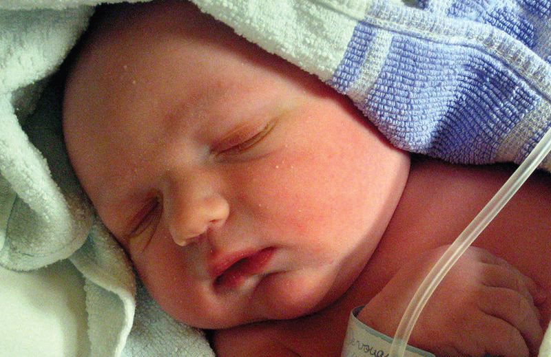
Skin forms a dynamic interface with the external environment and is a complex organisation of cell types and associated structures that performs many essential functions. Although the stratum corneum of full-term neonates is analogous to that of adult skin, structural and compositional differences of the skin renders the newborn more susceptible to bacterial colonisation. Particularly for the preterm neonate, impaired cutaneous barrier function and an immature immune system reduce the capacity to defend against bacterial pathogens. The majority of cutaneous bacterial infections are localised to the skin and are easily treated, however, systemic bacterial infection and disseminated disease in the neonatal period may be life-threatening.

Differences in neonatal skin

Newborn skin is fundamentally different from that of the adult and adapts to the extrauterine environment during the first year of life through ongoing structural and functional changes. [1] Skin is a complex, selectively permeable membrane that performs a number of roles, including protection from infection and external stressors such as ultraviolet (UV) light damage, temperature regulation, sensation, and physical appearance. Protection against the external environment is primarily due to the most superficial layer of the epidermis, the stratum corneum, and recent advances in fluorescence spectroscopy and electron microscopy have helped elucidate the differences between adult and newborn skin. [1-3] The stratum corneum is a layer of lipid-depleted, protein-rich corneocytes embedded in a matrix of extracellular lipids, resulting from the continuous proliferation of keratinocytes in the basal epidermis. [3] Compared with adult skin, newborn skin produces smaller corneocytes, a thinner epidermis, and an increased density of microrelief grooves (Table 1). [1] The corneocytes of newborns also have a higher degree of irregularity and decreased organisation in both development and subsequent desquamation phases. [2] The change in neonatal skin pH from neutral, at birth, towards a more acidic mantle is also likely to impact on stratum corneum integrity, as incomplete skin surface acidification is linked to variable rates of desquamation. [3,4] Overall, decreased corneocyte size and a thinner, less cohesive stratum corneum has negative implications for skin barrier function, as indicated by increased transepidermal water loss (TEWL) in newborn skin. [5]

Compared with full-term infants born at 37-42 weeks’ gestation, preterm newborns do not develop the same level of protection provided by the stratum corneum until 2-4 weeks after birth. [6,7] The most significant difference is an increase in the stratum corneum from two to three cell layers at 28 weeks’ gestation to the equivalent of adult skin with 15 layers by 32 weeks’ gestation. [8] Due to the role of the stratum corneum in barrier protection, the premature newborn is at considerably greater risk of cutaneous complications.

Changes in TEWL, skin pH, and sebaceous activity all lead to the creation of a skin environment that promotes colonisation of certain microbial skin flora. [9] Colonisation of resident flora commences at birth, but newborns have a unique skin microbiome profile that develops throughout the first year of life and beyond. [9] The protection offered by resident commensal and mutualistic skin flora in the adult is therefore not immediately present in the newborn, leading to different patterns of subsequent infection.

Table 1. Differences between newborn and adult skin. [1,2]

Skin defences and immune response of the newborn:

Skin has antimicrobial function afforded by the innate immune system and antigen presenting cells (APCs) of the epidermis and dermis, as well as circulating immune cells that migrate into the dermis. This innate system works together with the adaptive immune system to defend against infection. In the newborn, innate immunity is the most important mechanism of defence, as this system is present at birth as a result of pattern recognition receptors encoded by germline DNA. This system responds to biochemical structures common to a number of pathogens, producing a rapid response with no residual immunity or memory. In contrast, adaptive immunity develops slowly and involves specific antigen receptors of T- and B-lymphocytes as part of a system that develops memory for faster successive responses.

Innate immune defences comprise the physical barrier formed by the skin itself, antimicrobial peptides (AMPs), complement pathways, and immune cells including monophages, macrophages, dendritic cells, and natural killer cells. [10,11] While the keratinocytes of the skin are typically considered to be static ‘bricks’ of the physical skin barrier, they are also dynamically involved in immunity by their secretion of cytokines and chemokines, AMPs, and complement components. AMPs secreted by keratinocytes are cationic proteins termed cathelicidins and defensins, and they appear to be particularly important in neonatal immunity, with selective bactericidal activity against common cutaneous pathogens. [12,13] Cathelicidins (LL-37) and beta-defensins (BD-1, BD-2, BD-3) are attracted to negatively charged bacteria, viruses, and fungi and exert their influence by membrane insertion and pore formation. [13] Neonates display higher baseline concentrations of cutaneous AMPs than adults, suggesting a greater role for these proteins in newborn skin defences. In the absence of specific antibodies, pattern recognition receptors such as Toll-like receptors (TLRs) play a pivotal role, with subsequent cytokine production changing with increasing age and correlating to age-specific pathogen susceptibility. [14]

Figure 1. Innate immunity of the skin in the newborn

Bacterial infections

Cutaneous staphylococcal and streptococcal infections cause a variety of clinical presentations depending on site of infection, strain of organism, and neonatal immunity. Impetigo is a superficial bacterial cutaneous infection that may present with or without bulla formation, as described by the conditions non-bullous and bullous impetigo. The bullae of bullous impetigo are invariably due to infection with S. aureus and the subsequent production of epidermolytic toxin, which is also responsible for the widespread bullae and desquamation in staphylococcal scalded skin syndrome (SSSS). Development of resistant strains, overcrowding, and poor infection control have been linked to nosocomial outbreaks of S. aureus and is of particular concern in neonatal intensive care units where neonates are more susceptible to infection. [15]

Non-bullous impetigo

Both Streptococcus pyogenes and S. aureus are associated with the non-bullous form of impetigo, which presents as an erythematous macular rash before developing eroded lesions with a honey-coloured crust. [16] Isolated staphylococcal pustules and paronychia are also common in neonates. Although mild non-bullous impetigo has the capacity self-resolve, treatment with topical mupirocin and fusidic acid limits the opportunity for disease to persist. [16]

Bullous impetigo

Localised cutaneous S. aureus infection presents with an erythematous vesiculopustular rash that preferentially affects the diaper area and skin folds, coalescing to form large flaccid bullae that rupture easily and appear as honey-crusted erosions. [16,17] Bacteria are present in the lesions, and the infection usually responds to first-line systemic flucloxacillin, which may be used in conjunction with topical fusidic acid. [16,17] Certain strains of S. aureus are associated with epidermolytic toxins, which facilitate pathogen entry beneath the stratum corneum and limit disease to the superficial epidermis. [18,19] The distinct bullae present in bullous impetigo are due to the toxin-induced cleavage of desmosomal cadherin proteins in the granular layer of the epidermis, which are normally responsible for maintaining functional adhesion between keratinocytes. [18] These same toxins are produced in SSSS.

Staphylococcal scalded skin syndrome

Haematogenous spread of S. aureus is facilitated by inoculation at a distant site such as the conjunctiva, umbilicus, or perineum, and the effects of bulla formation and desquamation are the direct result of circulating epidermolytic toxins. [20] This haematogenous spread results in a widespread infection that is more severe than the localised infection of bullous impetigo. Generalised erythema and skin tenderness are the initial clinical features, with evolution into large flaccid bullae and desquamation of the entire cutaneous surface. The Nikolsky sign is present, where blistering can be elicited by light stroking of the skin. [21] Bacterial cultures of cutaneous lesions are typically negative and S. aureus is only found at the distant sites of infection. Skin biopsy is considered the gold standard of diagnosis and is particularly relevant when considering toxic epidermal necrolysis (TEN) as a differential. In contrast to SSSS, TEN results in subepidermal blisters and keratinocyte necrosis rather than epidermal cleavage and typically involves oral mucous membranes. [20,21] Although biopsy is helpful in providing a definite diagnosis, neonatal biopsies are rarely performed due to the characteristic clinical presentation of both conditions. Despite the apparent polarity of the cutaneous and haematogenous forms of S. aureus infection, a handful of mild SSSS cases have been reported, lending support to a likely clinical spectrum ranging from a mild form to the classic severe disease. [22]

Omphalitis

After birth and separation of the umbilical cord, necrosis of the stump is followed by epithelialisation. The healing stump may become colonised, with the exposed umbilical vessels forming a potential portal of entry for pathogenic bacteria. [23] Omphalitis is characterised by stump erythema and periumbilical oedema, with or without discharge, and is frequently due to S. aureus. It is more common in developing countries, and the risk is increased in cases of protracted labour, non-sterile delivery, and prematurity. [23] A recent Cochrane systematic review identified significant evidence to support the use of topical chlorhexidine on the umbilical stump to reduce omphalitis and neonatal mortality in developing countries. However, this benefit could not be demonstrated in developed countries, possibly owing to reduced risk factors for omphalitis. [24]

Necrotising fasciitis

Infection of the fascia and overlying soft tissues is a rapidly progressive neonatal emergency. Pathogens gain entry by cutaneous breaches such as omphalitis, birth trauma, and superficial skin wounds, with group A streptococci most commonly implicated as the causative organism. [25] Infection may also be polymicrobial, with a combination of organisms detected on wound cultures. [26] The infection follows the fascial plane, causing thromboses in the blood supply to overlying tissues and leading to tissue necrosis, and the skin becomes progressively more discoloured, tender, and warm. [26] While the initial presentation may not appear concerning, neonates rapidly become disproportionately tender and toxic. [26] Necrotising fasciitis has a high morbidity and mortality and requires immediate identification for surgical debridement. [25]

Ecthyma gangrenosum

Pseudomonas aeruginosa septicaemia is the most common underlying cause for this cutaneous manifestation, which typically presents with macules that progress via a necrotising vasculitis to form indurated necrotic ulcers with surrounding erythema. [27,28] Prematurity, immune deficiencies, and neutropaenia are the main predisposing factors, but lesions may develop in the absence of immunodeficiency when direct inoculation occurs through a breach in the skin barrier. [28]

Antimicrobial resistance and prevention

The treatment of neonatal bacterial infection depends on the pathogen and sensitivities to antibiotic treatments. In the Australian healthcare setting and internationally, antibiotic resistance poses a growing problem in this ‘post-antibiotic’ era. Methicillin-resistant S. aureus (MRSA) has become increasingly prevalent, particularly in the intensive care setting such as the neonatal intensive care unit (NICU). [29] As colonised neonates are continually admitted, the introduction of many unique sources and various strains over time adds to the ongoing burden and is likely to contribute to difficulties in fully eradicating MRSA from the NICU. [30] Transmission of organisms such as S. aureus most commonly occurs secondary to direct contact with colonised caregivers, and this problem is compounded when hand hygiene and barrier protection is inadequate. Premature infants in the NICU are particularly susceptible, due to their immature immune systems and the increased risk of nosocomial infection with invasive monitoring and frequent healthcare worker contact. [31] The identification of previous treatment with third-generation cephalosporins and carbapenem as independent risk factors for the development of multidrug-resistant Gram-negative bacteraemia in the NICU highlights the issue of antibiotic resistance and underscores the importance of judicious antibiotic use. [32]

Conclusion

Skin is the first line of defence against invading pathogens, and there are a number of unique cellular, functional, and immunological factors that underpin an increased susceptibility to bacterial infection in the newborn. Premature newborns are at particular risk of infection, owing to potential deficits in cutaneous barrier function. Future practice in treating bacterial infections is likely to be influenced by the emergence of multi-resistant strains and may shift the focus toward improved prevention measures.

Conflict of Interest

None declared.

Correspondence

J Read: jazlyn.read@griffithuni.edu.au

References

[1] Stamatas GN, Nikolovski J, Luedtke MA, Kollias N, Wiegand BC. Infant skin microstructure assessed in vivo differs from adult skin in organization and at the cellular level. Pediatr Dermatol. 2010;27(2):125-31.

[2] Fluhr JW, Lachmann N, Baudouin C, Msika P, Darlenski R, De Belilovsky C, et al. Development and organization of human stratum corneum after birth. Electron microscopy isotropy score and immunocytochemical corneocyte labelling as epidermal maturation’s markers in infancy. Br J Dermatol. 2014 Feb 7. DOI:10.1111/bjd.12880.

[3] Stamatas GN, Nikolovski J, Mack MC, Kollias N. Infant skin physiology and development during the first years of life: a review of recent findings based on in vivo studies. Int J Cosmet Sci. 2011;33(1):17-24.

[4] Fluhr JW, Man MQ, Hachem JP, Crumrine D, Mauro TM, Elias PM, et al. Topical peroxisome proliferator activated receptor activators accelerate postnatal stratum corneum acidification. J Invest Dermatol. 2009;129(2):365-74.

[5] Raone B, Raboni R, Rizzo N, Simonazzi G, Patrizi A. Transepidermal water loss in newborns within the first 24 hours of life: baseline values and comparison with adults. Pediatr Dermatol. 2014;31(2):191-5.

[6] Fluhr JW, Darlenski R, Taieb A, Hachem JP, Baudouin C, Msika P, et al. Functional skin adaptation in infancy — almost complete but not fully competent. Exp Dermatol. 2010;19(6):483-92.

[7] Fluhr JW, Darlenski R, Lachmann N, Baudouin C, Msika P, De Belilovsky C, et al. Infant epidermal skin physiology: adaptation after birth. Br J Dermatol. 2012;166(3):483-90.

[8] Taeusch HM, Ballard RA, Gleason CA, editors. Avery’s diseases of the newborn. 8th ed. Philadelphia: Elsevier Saunders; 2005.

[9] Capone KA, Dowd SE, Stamatas GN, Nikolovski J. Diversity of the human skin microbiome early in life. J Invest Dermatol. 2011;131(10):2026-32.

[10] Cuenca AG, Wynn JL, Moldawer LL, Levy O. Role of innate immunity in neonatal infection. Am J Perinatol. 2013;30(2):105-12.

[11] Power Coombs MR, Kronforst K, Levy O. Neonatal host defense against staphylococcal infections. Clin Dev Immunol. 2013;2013:826303. DOI:10.1155/2013/826303.

[12] Nelson A, Hultenby K, Hell E, Riedel HM, Brismar H, Flock JI, et al. Staphylococcus epidermidis isolated from newborn infants express pilus-like structures and are inhibited by the cathelicidin-derived antimicrobial peptide LL37. Pediatr Res. 2009;66(2):174-8.

[13] Yoshio H, Lagercrantz H, Gudmundsson GH, Agerberth B. First line of defense in early human life. Semin Perinatol. 2004;28(4):304-11.

[14] Kollmann TR, Levy O, Montgomery RR, Goriely S. Innate immune sensing by Toll-like receptors in newborns and the elderly. Immunity. 2012;37(5):771-83.

[15] Bertini G, Nicoletti P, Scopetti F, Manoocher P, Dani C, Orefici G. Staphylococcus aureus epidemic in a neonatal nursery: a strategy of infection control. Eur J Pediatr. 2006 Aug;165(8):530-5.

[16] Sladden MJ, Johnston GA. Current options for the treatment of impetigo in children. Expert Opin Pharmacother. 2005;6(13):2245-56.

[17] Johnston GA. Treatment of bullous impetigo and the staphylococcal scalded skin syndrome in infants. Expert Rev Anti Infect Ther. 2004;2(3):439-46.

[18] Hanakawa Y, Schechter NM, Lin C, Garza L, Li H, Yamaguchi T, et al. Molecular mechanisms of blister formation in bullous impetigo and staphylococcal scalded skin syndrome. J Clin Invest. 2002;110(1):53-60.

[19] Yamasaki O, Yamaguchi T, Sugai M, Chapuis-Cellier C, Arnaud F, Vandenesch F, et al. Clinical manifestations of staphylococcal scalded-skin syndrome depend on serotypes of exfoliative toxins. J Clin Microbiol. 2005;43(4):1890-3.

[20] Stanley JR, Amagai M. Pemphigus, bullous impetigo, and the staphylococcal scalded-skin syndrome. N Engl J Med. 2006;355(17):1800-10.

[21] Berk DR, Bayliss SJ. MRSA, staphylococcal scalded skin syndrome, and other cutaneous bacterial emergencies. Pediatr Ann. 2010;39(10):627-33.

[22] Hubiche T, Bes M, Roudiere L, Langlaude F, Etienne J, Del Giudice P. Mild staphylococcal scalded skin syndrome: an underdiagnosed clinical disorder. Br J Dermatol. 2012;166(1):213-5.

[23] Fraser N, Davies BW, Cusack J. Neonatal omphalitis: a review of its serious complications. Acta Paediatr. 2006;95(5):519-22.

[24] Imdad A, Bautista RM, Senen KA, Uy ME, Mantaring JB 3rd, Bhutta ZA. Umbilical cord antiseptics for preventing sepsis and death among newborns. Cochrane Database Syst Rev. 2013;5:CD008635.

[25] Das DK, Baker MG, Venugopal K. Increasing incidence of necrotizing fasciitis in New Zealand: a nationwide study over the period 1990 to 2006. J Infect. 2011;63(6):429-33.

[26] Jamal N, Teach SJ. Necrotizing fasciitis. Pediatr Emerg Care. 2011;27(12):1195-9.

[27] Pathak A, Singh P, Yadav Y, Dhaneria M. Ecthyma gangrenosum in a neonate: not always pseudomonas. BMJ Case Rep 2013 May 27. DOI:10.1136/bcr-2013-009287.

[28] Athappan G, Unnikrishnan A, Chandraprakasam S. Ecthyma gangrenosum: presentation in a normal neonate. Dermatol Online J. 2008;14(2):17.

[29] Isaacs D, Fraser S, Hogg G, Li HY. Staphylococcus aureus infections in Australasian neonatal nurseries. Arch Dis Child Fetal Neonatal Ed. 2004;89(4):F331-5.

[30] Gregory ML, Eichenwald EC, Puopolo KM. Seven-year experience with a surveillance program to reduce methicillin-resistant Staphylococcus aureus colonization in a neonatal intensive care unit. Pediatrics. 2009;123(5):e790-6.

[31] Cipolla D, Giuffre M, Mammina C, Corsello G. Prevention of nosocomial infections and surveillance of emerging resistances in NICU. J Matern Fetal Neonatal Med. 2011;24 Suppl 1:23-6.

[32] Tsai MH, Chu SM, Hsu JF, Lien R, Huang HR, Chiang MC, et al. Risk factors and outcomes for multidrug-resistant Gram-negative bacteremia in the NICU. Pediatrics. 2014;133(2):e322-9.

Categories
Feature Articles

The history of modern general anaesthesia

Safe and effective anaesthesia is among the greatest advances in medical history. Modern surgery and the considerable benefits it brings would be impossible without the significant academic, pharmacological, and practical advances in anaesthesia over the past 200 years. At the forefront of these are the major developments in general anaesthesia and airway management. This article aims to provide a basic framework to understand the development of modern general anaesthesia.

A brief history of general anaesthesia

v5_i1_a21
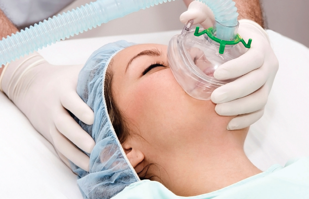
Anaesthesia is a relatively new field in modern medicine. Prior to its development, most surgical procedures were either minor or emergency operations. [1] It is clear that modern surgery and the considerable benefits it brings would be impossible without the significant academic, pharmacological, and practical advances in anaesthesia during the 19th and 20th centuries. First and foremost among these is the development of safe and effective general anaesthesia.

Carbon dioxide was first explored as an anaesthetic in the 1820s by the English physician Henry H. Hickman. By inducing partial asphyxiation, Hickman demonstrated that animals could be rendered unconscious for a prolonged period, enabling surgical procedures to be performed. [2] This was a major breakthrough, however the risks associated with hypoxic anaesthesia were too great to see the widespread adoption of carbon dioxide as an anaesthetic.

Diethyl ether, a solvent commonly referred to simply as ‘ether’, was first used clinically by American physician William E. Clarke for a tooth extraction in January 1842. [3, 4] Several months later Crawford W. Long, an American surgeon and pharmacist, famously used ether as a surgical anaesthetic to remove a growth on a young man’s neck. He published his findings after seven years, revealing that the patient felt nothing throughout the procedure. [5] The discovery of ether’s clinical utility represented a significant advance in effective general anaesthesia, spurring a flurry of interest in potential anaesthetic agents.

Still used today for its anaesthetic properties, nitrous oxide was experimented with during the 19th century. Positive experiences by chemist Humphrey Davy lead to public gatherings, in which members of the public would inhale nitrous oxide for its exhilarating and pleasurable effects. [2] A medical student, Gardner Quincy Colton, made over $400 profit from one such affair that attracted three to four thousand attendees. [6] These events were similar to those held for ether, known as ‘ether frolics.’ [2] Following the observation in these gatherings of nitrous oxide’s analgesic and anaesthetic effects, it was formally tested in December 1844. Horace Wells, a dentist who had attended one of Colton’s exhibitions, persuaded a colleague to extract one of Wells’ teeth while Colton administered nitrous oxide gas. [7] The procedure was performed successfully, reportedly the first tooth ever removed painlessly. [8]

A former student of Wells, William T. G. Morton, was instrumental in the popularisation of ether as an anaesthetic. Morton performed a successful public demonstration of the anaesthetic capabilities of ether in October 1846 at Massachusetts General Hospital. [1] This event is often considered to mark the birth of modern anaesthesia, following which ether was widely adopted around the world. Later that year, Oliver W. Holmes, a writer and professor of anatomy, named the process which Morton demonstrated anaesthesia, derived from the Greek for ‘without sensation.’ [2]

Scottish obstetrician James Y. Simpson was the first to adopt the organic compound chloroform to relieve the pain of childbirth in 1847. Chloroform anaesthesia grew in popularity around the world and was in wide use when Queen Victoria gave birth to Prince Leopold under its influence in 1853. The chloroform was administered by the famous physician and epidemiologist John Snow. [2] In the early 20th century, chloroform came to supersede ether as a general anaesthetic in light of its less offensive odour, and rapid induction and emergence.

Though the first intravenous injections took place in 1656, [9] the first intravenous anaesthetic, sodium thiopental (thiopentone), was not synthesised until 1934. [10] Thiopentone is a short-acting, rapid-onset barbiturate sometimes used for anaesthetic induction. Its earliest documented use in humans was later in 1934 by Ralph Waters, an American anaesthetist. [2] Intravenous anaesthesia allowed more precise dosing and a less confrontational experience for the patient, and thiopentone rapidly entered common usage. Although it remained popular for many years, thiopentone was gradually replaced by propofol as the preferred induction agent. Introduced in the late 1980s, propofol allowed rapid induction and emergence, reliable hypnosis, and has antiemetic properties. [1]

Significant advances were also made in the 20th century in developing better halogenated inhaled agents. The advent of improved volatile agents, in parallel with a rising interest and focus on patient safety, saw a shift from ether and chloroform anaesthesia to the use of newer intravenous and inhalational agents with more favourable characteristics. Routinely used today, these agents provide fast induction and emergence, and are ideally suited for maintenance of anaesthesia. After halothane and enflurane came isoflurane, then sevoflurane, and finally desflurane in the early 1990s. [1] These new volatile agents had a number of desirable properties including low solubility, minimal cardiorespiratory depression, and unlike ether, are non-flammable. In contrast to ether and chloroform however, they lack analgesic effects, necessitating the use of other agents such as opioids, local anaesthetics, or nitrous oxide to ensure adequate pain relief. Nitrous oxide saw a steady decline in use over the following years, in part due to the availability of these newer agents, but also because of concerns about potential toxicity and its link with postoperative nausea and vomiting. [1]

The introduction of muscle relaxants to clinical practice in the early 1950s allowed for major advances in anaesthetic techniques and thereby surgery. Curare, a natural alkaloid historically used on poison darts and arrows by aboriginal people across Africa, Asia, and the Americas, [11] was the first non-depolarising muscle relaxant used. Through the late 1970s to 1990s, quaternary ammonium muscle relaxants were developed, including vecuronium, atracurium, and rocuronium. These compounds brought several advantages, including more favourable cardiovascular effects and minimal release of histamine. [12] Due to their clearance by Hoffman elimination rather than renal excretion, atracrurium and subsequently cisatracurium also possess the additional benefit of predictably rapid recovery with little cumulative effect following repeated administration. Suxamethonium, still in use today, was also developed in the 1950s. It is a depolarising neuromuscular blocking agent with fast onset and offset of action. It is considered by many as the agent of choice for rapid-onset neuromuscular blockade and has a short duration of action, although its side-effects of potassium release and increased intra-thoracic, intra-abdominal, and intra-cranial pressures will sometimes contraindicate its use.

Advances in monitoring have significantly impacted upon the practice of anaesthesia including the introduction of pulse oximetry and capnography in the 1980s. [1] The routinely used combination of these has contributed to a reduction in the proportion of anaesthesia-related complications that are preventable by monitoring from 39% in the 1970s to only 9% in the 1990s. [13] Other advances have included the measurement of inspired and end-tidal gases, including oxygen, nitrous oxide, and the volatile agents. The advent of ‘depth of anaesthesia’ monitors such as bispectral index (BIS) monitors has advanced our understanding of anaesthetic practice. Modern anaesthesia has achieved such an impressive degree of safety that the anaesthesia-related mortality in Australia is less than 3 deaths per million annually. [14]

A brief history of endotracheal intubation
In the late 19th century great advances were made in airway management for patients undergoing general anaesthesia. Without advanced airway support, the great safety and efficacy of modern anaesthesia would be impossible. The laryngeal tube had reportedly existed since at least 1791, and was used for a range of purposes including to facilitate breathing in oedema of the glottis, for direct delivery of medications to lung tissue, and for artificial respiration. [15]

Charles Trueheart of Texas published an account in 1869 describing a biphasic artificial respiration device, which included a laryngeal airway. However, the first successful delivery of endotracheal general anaesthesia was performed through tracheotomy by German surgeon Friedrich Trendelenburg in 1871. [16] Over the following decades, this technique was adapted in multiple settings to be delivered by oro-tracheal intubation and thus avoid the need for a surgical airway. [16, 17]

A further breakthrough in intubation came in 1895, when German physician Alfred Kirstein performed the first laryngoscopy with direct visualisation of the vocal cords. [18] Previously, direct visualisation was thought impossible, and the glottis and larynx had been visible only by indirect vision using mirrors. Kirstein called his device the autoscope, now known as a laryngoscope, and in the process of its development he established many of the principles of laryngoscopy which continue to be used in clinical practice. [18]

In 1913, Chevalier Jackson introduced a new laryngoscope blade with a light source at the distal tip, rather than the proximal light source used by Kirstein. [19] That same year, Henry Janeway expanded upon this, also including batteries in the handle, a central notch for maintaining the tracheal tube in the midline of the oropharynx, and a slight curve to the tip of the blade. [20] These changes were instrumental in popularising the use of direct laryngoscopy and tracheal intubation in anaesthesia, and the use of endotracheal intubation spread greatly following the First World War. [15]

Sir Ivan Magill went further with his invention, the Magill laryngoscope blade. The most significant features of this blade included a flat and wide distal end of the speculum, improving control of the epiglottis, and a slot on the side allowing the passage of catheters and tubes without obscuring vision. [21, 22] He also developed the technique of awake blind nasotracheal intubation in 1928, along with a new type of angulated forceps (the Magill forceps) for nasotracheal intubation, and a new endotracheal tube. [21] The Magill laryngoscope blade remains in use today, however in 1943 Sir Robert Macintosh introduced the Macintosh blade, a curved model which is currently the most widely used laryngoscope blade. [23] Other specific blades may be used for certain patient subsets, such as straight laryngoscope blades in infants or the McCoy laryngoscope blade for difficult intubations.

The laryngeal mask airway (LMA) was first used in 1981 before being officially released in 1988. [24] The LMA revolutionised airway management – it provides a clear airway, forms an effective seal at the glottic inlet, and largely avoids the risk of trauma associated with intubation. [24] Endotracheal intubation remains an indispensable skill for the anaesthetist, with both LMAs and endotracheal tubes widely used.

A range of equipment for intubation is now available, including video laryngoscopes and fibre-optic bronchoscopes to aid in visualising the difficult airway, tubes with and without cuffs, reinforced tubes, and double-lumen tubes. Measurement of end-tidal carbon dioxide by capnometry has also provided a useful adjunct to direct visualisation for confirming correct placement of the endotracheal tube. [25, 26] Despite these advances, the modern endotracheal intubation still relies heavily on the principles laid down by Kirstein and his successors.

Conclusion
Considerable progress has been made in the field of anaesthesia over the past two centuries. The development of safe, effective general anaesthesia is one of the most important advances in medical history, allowing the widespread expansion of surgery and the considerable benefits it brings. Significant advances beyond the scope of this article include the developments of local anaesthesia, regional anaesthesia, conscious sedation, and analgesia.

Conflict of interest

None declared.

References

[1] Urman RD, Desai SP. History of anesthesia for ambulatory surgery. Curr Opin Anaesthesiol. 2012 Dec;25(6):641-7.

[2] Miller R. Anesthesia. Philadelphia: Churchill Livingstone; 2000.

[3] Lyman H. Artificial anaesthesia and anaesthetics. New York: William Wood and Co.; 1881.

[4] Keys T. The history of surgical anesthesia. Huntington: Robert E. Krieger Publishing Company; 1978.

[5] Long C. An account of the first use of sulphuric ether by inhalation as an anaesthetic in surgical operations. South Med Surg J. 1849;5:705-13.

[6] Smith GB, Hirsch NP. Gardner Quincy Colton: pioneer of nitrous oxide anesthesia. Anesth Analg. 1991 Mar;72(3):382-91.

[7] LeVasseur R, Desai SP. Ebenezer Hopkins Frost (1824-1866): William T.G. Morton’s first identified patient and why he was invited to the ether demonstration of October 16, 1846. Anesthesiology. 2012 Aug;117(2):238-42.

[8] Colton G. Boyhood and manhood recollections. The story of a busy life. New York: A. G. Sherwood; 1897.

[9.] Dagnino J. Wren, Boyle, and the origins of intravenous injections and the Royal Society of London. Anesthesiology. 2009 Oct;111(4):923-4.

[10] Tabern D, Volwiler E. Sulfur-containing barbiturate hypnotics. J Am Chem Soc. 1935;57(10):1961–3.

[11] Dorkins HR. Suxamethonium-the development of a modern drug from 1906 to the present day. Med Hist. 1982 Apr;26(2):145-68.

[12] Ball C, Westhorpe RN. Muscle relaxants: pancuronium and vecuronium. Anaesth Intensive Care. 2006 Apr;34(2):137.

[13] Lee LA, Domino KB. The closed claims project: has it influenced anesthetic practice and outcome? Anesth Clin N Am. 2002 Sep;20(3):485-501.

[14] Gibbs N, editor. Safety of anaesthesia: a review of anaesthesia-related mortality reporting in Australia and New Zealand 2006-2008. Melbourne: Australian and New Zealand College of Anaesthetists; 2009.

[15] Waters R, Rovenstine E, Guedel A. Endotracheal anesthesia and its historical development. Anesth Analg. 1933;12:196-203.

[16] Trubuhovich RV. Early artificial ventilation: the mystery of “Truehead of Galveston” – was he Dr Charles William Trueheart? Crit Care Resusc. 2008 Dec;10(4):338.

[17] Macewen W. General observations on the introduction of tracheal tubes by the mouth, instead of performing tracheotomy or laryngotomy. Br Med J. 1880 Jul 24;2(1021):122-4.

[18] Hirsch NP, Smith GB, Hirsch PO. Alfred Kirstein: pioneer of direct laryngoscopy. Anaesthesia. 1986 Jan;41(1):42-5.

[19] Zeitels SM. Chevalier Jackson’s contributions to direct laryngoscopy. J Voice. 1998 Mar;12(1):1-6.

[20] Burkle CM, Zepeda FA, Bacon DR, Rose SH. A historical perspective on use of the laryngoscope as a tool in anesthesiology. Anesthesiology. 2004 Apr;100(4):1003-6.

[21] McLachlan G. Sir Ivan Magill KCVO, DSc, MB, BCh, BAO, FRCS, FFARCS (Hon), FFARCSI (Hon), DA, (1888-1986). Ulster Med J. 2008 Sep;77(3):146-52.

[22] Magill I. An improved laryngoscope for anaesthetists. Lancet. 1926;207(5349):500.

[23] Scott J, Baker PA. How did the Macintosh laryngoscope become so popular? Paediatr Anaesth. 2009 Jul;19 Suppl 1:24-9.

[24] van Zundert TC, Brimacombe JR, Ferson DZ, Bacon DR, Wilkinson DJ. Archie Brain: celebrating 30 years of development in laryngeal mask airways. Anaesthesia. 2012 Dec;67(12):1375-85.

[25] Grmec S. Comparison of three different methods to confirm tracheal tube placement in emergency intubation. Intensive Care Med. 2002 Jun;28(6):701-4.

[26] Rudraraju P, Eisen LA. Confirmation of endotracheal tube position: a narrative review. J Intensive Care Med. 2009 Sep-Oct;24(5):283-92.

Categories
Feature Articles

Penicillin allergies: facts, fiction and development of a protocol

Penicillins, a member of the beta-lactam family, are the most commonly prescribed antibiotic class in Australia. Beta-lactam agents are used in a sexual health setting for the management of syphilis, uncomplicated gonococcal infections and pelvic inflammatory disease. Patients frequently report allergies to penicillin, which can be protective but also counterproductive if it does not represent a ‘true’ allergy. Features of a reported reaction may be stratified as either high or low risk, which has implications for both re-exposure to penicillins; but also cross-reactivity to other members of the beta-lactam family such as cephalosporins. We reviewed the evidence surrounding penicillin allergies, in the context of developing a local protocol for penicillin-allergic patients at a sexual health clinic.

Case scenario

v5_i1_a20
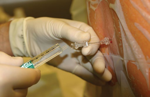
A 27-year-old male is referred to a sexual health clinic by a general practitioner (GP). He presents with a widespread maculopapular rash, fever and malaise for the past four days. Whilst he does not describe any other symptoms, he did notice a painless genital ulcer approximately four weeks ago. The ulcer resolved spontaneously; hence he initially did not seek medical advice. He does not have a stable sexual partner and mentions engaging in several episodes of unprotected sex with both women and men in the previous three months. Secondary syphilis is the suspected diagnosis given the widespread rash and preceding chancre, and testing confirms this with a positive syphilis enzyme-linked immunoassay (EIA) screening test, Treponema pallidum particle agglutination assay (TPPA), and an rapid plasma reagin (RPR) of 1:32. As part of a sexual health screen, he tested positive for rectal gonorrhoea by culture. The treatment regime includes 1.8 g intramuscular benzathine penicillin for syphilis, in addition to 500 mg intramuscular ceftriaxone and 1g oral azithromycin for gonorrhoea, all given as stat doses. Before signing the drug order, the clinician questions about any allergies. The patient mentions having an allergic reaction to penicillin when he was six years old but cannot remember any particular details. What is the plan now?

Introduction

This case presents a challenging scenario for the clinician. In this article, we hope to outline some of the facts surrounding penicillin allergies, dismiss some myths and provide a systematic approach to aid decision-making, especially in the sexual health setting where there are limited treatment options for gonorrhoea and syphilis.

The beta-lactam family of antibiotics are one of the most commonly prescribed antibiotic classes in medicine. The beta-lactam ring forms the structural commonality between different types of penicillins and this is also shared with other drug classes, such as the cephalosporins and carbapenems.

Penicillin allergy is the most commonly reported medication allergy, either by the patient or medical providers. [1] The implications of this ‘label’ can be either protective or counterproductive. For those patients with a severe previous reaction, such as anaphylaxis, this allergy is important and re-exposure can prove disastrous and potentially fatal. However, for other patients with a minor or inconclusive reaction, not administering penicillin may be denying the patient first-line, efficacious treatment. Additionally, there are concerns that treating patients with alternative agents in this context contribute to the development of resistance, which is of public health concern. [2]

Whilst the use of beta-lactam antibiotics crosses many realms of medicine, this article is written in the context of developing a protocol for the management of patients with penicillin allergy in an urban sexual health clinic. It is not designed to provide guidance outside of this setting, nor to replace existing protocols in other clinical units.

Rationale for a protocol

Management of patients with penicillin allergy requiring beta-lactam treatment was reviewed as part of an overall revision of internal treatment guidelines. Sexually transmitted infections pose treatment challenges whereby certain conditions or patient sub-groups (e.g. pregnant women) have no equally efficacious or appropriate alternatives to penicillins. [3] For example, one acceptable alternative to penicillin for syphilis treatment is to administer oral doxycycline, however the potential harm associated with this treatment (permanent dental staining) contraindicates its use in pregnancy. [4]

Development process

Senior clinicians at the sexual health clinic provided the protocol brief in May 2013. This included, but was not limited to: reviewing existing guidelines for the management of penicillin-allergic patients both at a national and international level; reviewing literature about the incidence of penicillin allergy, and cross-reactivity rates in patients with a documented history of penicillin allergy; formulating a protocol, based on existing protocols and evidence which would be applicable for managing patients with a penicillin allergy; designing a flowchart which summarises the protocol in a clear manner, including clear decision making and referral points in addition to an estimation of risk; engaging senior nursing staff and the director to assess the usability and practicality of the protocol; and presenting a draft for consideration to medical and nursing personnel, with subsequent review, endorsement and implementation.

Use of penicillins and cephalosporins for treatment of sexually transmitted infections

Current protocols in the clinic suggest the use of beta-lactam antibiotics as first line treatment for the following conditions, in accordance with national guidelines [5,6]:

Syphilis

IM benzathine penicillin 1.8 g stat single dose for early syphilis (including early latent syphilis), 3 weekly doses for late latent syphilis

Uncomplicated gonococcal infections

IM ceftriaxone 500 mg stat (in conjunction with azithromycin 1 g orally)

Pelvic inflammatory disease (PID)

IM ceftriaxone 500 mg stat (in conjunction with doxycycline 100 mg BD for 14 days and metronidazole 400 mg BD for 14 days)

Mechanism of penicillin allergy and associated reactions

This article focuses on the main concern with penicillin allergy, which is the possibility of anaphylaxis, an IgE-mediated (type I) hypersensitivity reaction. However, the clinician should be aware that delayed type hypersensitivity reactions (type IV) can also occur, causing exanthema or other skin eruptions, such as morbilliform reactions. These are not determined by the beta-lactam ring or side chains of the antibiotics, but rather, the ability for a drug to act independently as a hapten and become antigenic in nature. [7] This antigenicity triggers an immune response through interaction with antigen-presenting cells (APC) and T-cells. [8]

The major determinant of anaphylactic reactions to beta-lactam antibiotics is the beta-lactam ring, which is shared amongst the penicillin class, as this binds to endogenous lysine proteins to form a hapten. [9] However, there is also evidence to suggest that an IgE-mediated reaction can occur with the minor determinants of the molecule, which is the R chain (acyl) side group of individual penicillins (Figure 1). IgE binding results in mast cell activation and histamine release, in addition to the release of inflammatory mediators.

In patients who develop an IgE-mediated reaction, there is subsequent risk of a more severe reaction on re-exposure. IgE-mediated reactions can have effects on the following body systems: dermatologic (urticarial rashes, angioedema, macroglossia), respiratory (asthma, bronchospasm, wheezing, laryngeal swelling), gastrointestinal (abdominal pain, vomiting, diarrhoea, cramping), and cardiovascular (hypotension, vascular collapse, altered consciousness, shock).

Whilst no universal definition exists for anaphylaxis, two commonly accepted definitions in Australia are: (1) the acute onset of illness, with typical skin features (urticarial rash, erythema/flushing, angioedema) plus involvement of one other body system; or (2) the acute onset of hypotension, bronchospasm or upper airway obstruction with or without skin features. [10]

Figure 1.The beta-lactam ring is found in penicillins and several other closely related drug classes. Variants in the R chain are found within each drug class.
Figure 1.The beta-lactam ring is found in penicillins and several other closely
related drug classes. Variants in the R chain are found within each drug class.

Incidence of penicillin allergy and cross reactivity with cephalosporins
Various early studies suggested the incidence of penicillin allergy to be approximately 2% per course, with anaphylaxis estimated in 0.05% of all penicillin courses. [1] However, a large retrospective cohort study in the UK, which looked at 3,375,162 patients prescribed subsequent courses of penicillin, found a much lower incidence of only 0.18%. [11] It must be noted when quoting these figures that the definition of an ‘event’ in this study did not include asthma or eczema; however, when included, the incidence increased from 0.18% to 9% which makes interpretation difficult as we consider asthma a feature of allergy in Australian definitions. [11]

It is difficult to accurately identify if a trend over time exists in patients having a penicillin allergy. Multiple protocols exist internationally about the diagnosis of penicillin allergy and subsequent testing. [12] Furthermore, an element of bias may be present from both the patient and the clinician as the label of an ‘allergy’ can be highly subjective, and a permanent feature on a health record without subsequent confirmation.

Many early studies have quoted 10% cross-reactivity between penicillins and cephalosporins. [13,14] Unfortunately, these original studies assessing cross-reactivity over three decades ago were flawed, poorly designed open studies, lacking control groups, and have consequently overestimated this figure. [15] Furthermore, it is also postulated that the original manufacturing processes of cephalosporins contributed to inflated allergy rates, due to cross-contamination with penicillin compounds. [15] Since manufacturing processes have been refined, there has been a reduced incidence in cross-reactivity. If studies past 1980 are exclusively considered, a patient with a confirmed penicillin allergy (by positive skin test) will react with a cephalosporin in less than 2% of occasions. [15]

There is further evidence to suggest that there is less cross-reactivity with newer cephalosporins (second generation and onwards). A recent review and meta-analyses have found that third generation cephalosporins, such as ceftriaxone, have a cross reactivity rate of only 0.8% in those patients who are confirmed to be penicillin-allergic by skin testing, compared to 2.9% with older cephalosporins such as cephalexin. [16,17] Furthermore, these papers established that the risk of anaphylaxis due to cephalosporin cross-reactivity is quite small, as there was a higher incidence of anaphylactic reactions to cephalosporins with a negative penicillin skin test, compared to positive skin test patients. [16] This demonstrates that anaphylaxis reactions are often unpredictable.

Assessing the type and severity of penicillin allergy

Evidence suggests that history from the patient, especially when vague or not documented, is insufficient for assessing the degree of penicillin allergy. [18] Furthermore, the potential for allergy changes over time, with 80% of individuals with a documented IgE-mediated reaction having no evidence of reactive IgE after 10 years from initial reaction. [19] Most IgE-mediated reactions occur within seconds (IV administration) or up to an hour if administered orally with food. [20] Reactions outside of this timeframe are less likely to be IgE-mediated.

Hence, when taking a history, specific information should be sought to identify the presence of low- or high-risk features of the previous penicillin-related reaction, and consequently stratify the risk of future allergic reactions to a penicillin or cephalosporin.

High-risk features include: reaction occurred within one hour of administration; reaction occurred within the last 10 years; well documented history of features suggestive of anaphylaxis; required hospitalisation; any features suggestive of anaphylaxis as defined previously; and features of type IV hypersensitivity reactions including blistering, mucosal involvement, early onset desquamation (peeling), blood abnormalities such as derangements in liver function, renal function, or eosinophils. [15]

Low-risk features include: reaction occurred more than one hour after administration; reaction occurred more than 10 years ago; history is vague, unclear or poorly documented; localised reaction of mild severity involving one system only (rash not displaying any ‘high-risk features’ or stomach cramping); and a reaction that is not a true allergy; for example, an amoxicillin-Epstein Barr virus reaction. [10,15]

Role of the radioallergosorbent test, skin testing and desensitisation for penicillin allergy

In any patient with features of penicillin allergy, there should be consideration of referral for immunological skin testing and/or desensitisation, which can be performed quickly and is cost effective. The radioallergosorbent test (RAST) is more expensive, takes time to analyse and has poor positive predictive value. Desensitisation is performed in a supervised setting, can take 4-12 hours to complete in an acute setting, and results in a temporary reduction in immunogenic potential towards penicillins or associated medications. [21] Immediately following desensitisation, the first dose of penicillin is usually given. It must be noted that penicillin skin testing should also include testing against related beta-lactams such as cephalosporins, as a positive penicillin skin test cannot accurately predict cross-reactivity. [17]

Recommended drug choice in penicillin allergic patients

Based on the information outlined, and current existing guidelines, the following recommendations have been derived:

  1. Any patient who has high-risk features on history should not receive a beta-lactam agent. [22] An alternative efficacious drug should be prescribed. If there is no efficacious alternative, or a cephalosporin is required, referral to immunology should occur for skin testing and desensitisation if needed.
  2. 2. In those patients who have only low-risk features on history, the following question must be addressed: ‘which antibiotic is required?’ If a penicillin-based compound (i.e., benthazine penicillin) is required, the same precautions should be taken as mentioned above. However, if a cephalosporin such as ceftriaxone is required, the medication may be administered as the risk is less than 1% for third-generation cephalosporins. In this setting, the patient should be advised of the small but possible risk of an allergic reaction. [16]

Routine monitoring

Regardless of what antibiotic is prescribed, routine monitoring is advised as all serious allergic reactions need appropriate medical care. Observation facilities in addition to life support equipment and staff trained in first aid are essential in administering stat doses of antibiotics for the treatment of sexually transmitted infections. Signs and symptoms to look for during the observation period include the following: rash, swelling around the face/tongue/eyes, breathing difficulty, wheeze, vomiting, diarrhoea, abdominal pain, syncope or pre-syncope (low blood pressure), altered consciousness or shock. If any of these features are present or there is concern, refer to the local anaphylaxis and emergency protocols.

Case outcome

Although specific details of the previous allergic reaction could not be recalled by the patient, collateral history from his family suggested an urticarial reaction at the age of seven, with no other systemic features and not requiring hospitalisation. Consequently, the patient was deemed low-risk for a cross-reactivity allergic reaction towards ceftriaxone. He received the original prescribed treatment of 500 mg IM ceftriaxone and 1 g oral azithromycin without any adverse effects, or features of an allergic reaction. To treat his syphilis, he underwent a rapid desensitisation and subsequently received 1.8 g intramuscular benzathine penicillin, with no adverse effects.

Conflicts of interest

None declared.

Correspondence

J Floridis: john.floridis@nt.gov.au

References

[1] Solensky R. Allergy to penicillins. In: UpToDate. [Online].; 2012 [cited 2013 June 1] Available from: http://www.uptodate.com/contents/allergy-to-penicillins?source=search_result&search=allergy+to+penicillins&selectedTitle=1~150.

[2] Solensky R. Penicillin allergy as a public health measure. J Allergy Clin Immun. 2014;133(3):797-8.

[3] Emerson C. Syphilis: A review of the diagnosis and treatment. Open Infect Dis J. 2009;3:143-7.

[4] Australian Medicines Handbook Pty Ltd. Australian Medicines Handbook Rossi S, editor. Adelaide; 2013.

[5] STD Services. Diagnosis and Management of STDs (including HIV infection). 7th ed. Adelaide: Royal Adelaide Hospital; 2013.

[6] Antibiotic Expert Group. Therapeutic Guidelines: Antibiotics. Version 14. Melbourne: Therapeutic Guidelines Limited; 2010.

[7] Adam J, Pichler W, Yerly D. Delayed drug hypersensitivity: models of T-cell stimulation. Brit J Clin Pharmaco. 2011;71(5):701-7.

[8] Friedmann P, Pickard C, Ardern-Jones M, Bircher A. Drug-induced exanthemata: a source of clinical and intellectual confusion. Eur J Dermatol. 2010;20(3):255-9.

[9] Arroliga M, Pien L. Penicillin allergy: Consider trying penicillin again. Clev Clin J Med. 2003;70(4):313-26.

[10] Australasian Society of Clinical Immunology and Allergy. ASCIA. [Online].; 2012 [cited 2013 May 28] Available from: www.allergy.org.au/health-professionals/anaphylaxis-resources/adrenaline-autoinjector-prescription.

[11] Apter A, Kinman J, Bilker W, Herlim M, Margolis D, Lautenbach E, et al. Represcription of penicillin after allergic-like events. J Allergy Clin Immun. 2004;113(4):764-70.

[12] Macy E. The clinical evaluation of penicillin allergy: what is necessary, sufficient and safe given the materials currently available? Clin Exp Allergy. 2011;41(11):1498-1501.

[13] Dash C. Penicillin allergy and the cephalosporins. J Antimicrob Chemoth. 1975;1(3):107-18.

[14] Petz L. Immunologic cross-reactivity between penicillins and cephalosporins: a review. J Infect Dis. 1978;137 Suppl:S74-S79.

[15] Solensky R. Penicillin-allergic patients: Use of cephalosporins, carbapenems and monobactams. In: UpToDate. [Online].; 2013 [cited 2013 June 1] Available from: http://www.uptodate.com/contents/penicillin-allergic-patients-use-of-cephalosporins-carbapenems-and-monobactams?source=search_result&search=penicillin+allergic+patients&selectedTitle=1~150.

[16] Pichichero M. A review of evidence supporting the American Academy of Pediatrics recommendation for prescribing cephalosporin antibiotics for penicillin-allergic patients. Pediatrics. 2005;115(4):1048-57.

[17] Pichichero M, Casey J. Safe use of selected cephalosporins in penicillin-allergic patients: a meta-analysis. Otolaryngol Head Neck Surg. 2007;136(3):340-7.

[18] Wong B, Keith P, Waserman S. Clinical history as a predictor of penicillin skin test outcome. Ann Allergy Asthma Im. 2006;97(2):169-74.

[19] Blanca M, Torres M, Garcia J. Natural evolution of skin test sensitivity in patients allergic to beta-lactam antibiotics. J Allergy Clin Immun. 1999;103(5 Pt 1):918-24.

[20] Pichler W. Drug allergy: Classification and clinical features. In: UpToDate. [Online].; 2013 [cited 2013 May 28] Available from: http://www.uptodate.com/contents/drug-allergy-classification-and-clinical-features?source=search_result&search=drug+allergy%3A+classification&selectedTitle=1~150.

[21] Centers for Disease Control and Prevention. Sexually Transmitted Diseases Treatment Guidelines. Management of persons who have a history of penicillin allergy. 2011 January 28.

[22] Smith W. Adverse drug reactions. Allergy? Side-effect? Intolerance? Aust Fam Physician. 2013;42(1-2):12-6.

Categories
Feature Articles

Exercising patient-centred care: A review of structured physical activity, depression and medical student engagement

Structured physical activity has a wide range of benefits that include improving mood and preventing chronic disease. Recently, there has been an explosion of research aimed at treating diseases such as depression using nothing more than exercise. This article presents an overview of research conducted into the use of exercise to treat depression. As a body of work, the literature finds it to be a practice that has significant clinical benefits; however, its implementation is not straightforward. Issues concerning exercise adherence have hampered studies and force us to ask whether prescribing exercise for sufferers of depression is indeed appropriate. Nonetheless, is there a role for medical students in encouraging physical activity as treatment? If we re-examine the use of exercise from a patient-centred perspective, medical students have an opportunity to engage with patients, promote exercise and possibly prevent depression.

Introduction

v5_i1_a19Structured physical activity has a wide range of health benefits that include the prevention of chronic diseases such as cancer, cardiovascular disease, diabetes and obesity. [1] There is also consensus that exercise has a short-term ‘feel good’ effect that improves mood and wellbeing. [2-4] Recently, converging interest in these two areas has spawned an explosion of research aimed at treating diseases such as depression using nothing more than exercise. [5-7] A 2012 meta-analysis of over 25 trials found that prescribing exercise to treat depression is on par with pharmacological and physiological interventions, and that exercise alone is moderately more effective than no therapy. [8] The study also highlights that prescribing exercise for depression is not straightforward. Poor patient attendance rates, along with issues including exercise adherence and the type, duration and intensity of exercise, all question whether prescribing exercise for sufferers of depression is indeed appropriate. The authors of the study admit the implementation is complex and that further study is required. Nonetheless, is there a role for medical students in encouraging physical activity as treatment? Can our skills in motivational interviewing and goal setting play a role? If we re-examine the use of exercise from a patient-centred perspective, medical students can promote exercise adherence and support those who are already exercising to stay exercising. In doing so, we can facilitate the prescription of exercise and possibly prevent depression. [5,7,9]

A background to depression

Depression affects a staggering 350 million people globally, with sufferers commonly reporting changes in emotional, cognitive and physical behaviour. [10] Depression also presents with high rates of comorbidity (the occurrence of more than one condition or disease). [11] More locally, a 2007 National Survey of Mental Health and Wellbeing found that almost half of the Australian population aged 18-85 (7.3 million people) had experienced a mental illness at some point in their lifetime. [3] A study by the Australian Bureau of Statistics (ABS) in the same year revealed that of those people suffering from mental illness, only 35% actually sought treatment, suggesting that within Australia there are 2.1 million potential patients going without much-needed assistance. [12]

Mental illness, while indiscriminate, has a higher incidence within certain sub-populations. Perhaps surprisingly, doctors and medical students experience higher rates of depression and stress than the general population. Medical students report increased depressive symptoms as a result of medical school while a significant number of doctors report that they are less likely to seek treatment for depression despite their awareness of the condition. [13,14] With a reported 3668 students admitted to medical schools in Australia during 2013, these statistics highlight the importance for medical students to identify and understand depression. [15]

Pathophysiology and current treatment options

Therapeutic treatment options for patients who are depressed fall into two broad categories: psychological and pharmacological. Typically, people are treated using cognitive behavioural therapy (CBT), anti-depressant medication, or a combination of both. [4,16,17] Despite being able to treat depression, a simple pathogenesis is yet to be found. Current opinion centres on depression being a result of chemical imbalances within the brain, specifically the action of monoamine neurotransmitters, including dopamine, serotonin, and norepinephrine. [17] This approach has allowed the pharmaceutical industry to develop medications including selective serotonin reuptake inhibitors (SSRIs), which target neurotransmitter reuptake to help restore their usual balance and so reduce symptoms. [18]

One promising new development in the quest to understand the pathophysiology of depression concerns the emergence of inflammation as a mediator of depression. A recent meta-analysis of 24 separate studies found that depression is accompanied by immune dysregulation and activation of the inflammatory response system (IRS). [19] Specifically, when compared to non-depressed patients, sufferers of major depression were found to have significantly higher (p < 0.001) concentrations of the pro-inflammatory cytokines tumour necrosis factor-α and interleukin-6 in their blood. Once the increased cytokine signal reaches the brain, it is able to down-regulate the synthesis, release and reuptake of the very same monoamines targeted by antidepressant medications. [20] Understanding and preventing this interaction from occurring could lead to promising new treatments for depression. Despite widespread prescription, the use of antidepressant medication is not without its drawbacks. Many patients experience unwanted side effects and stop taking their medication, while others simply do not want to take medicine that will make them feel worse. [21] Additionally, a 2012 report on Australia’s overall health revealed that some patients who experience depression suffer worse health outcomes due to the social stigma associated with taking antidepressants. [22] As a result, many patients choose to shun medication altogether, opting for alternative therapies such as acupuncture and yoga to help manage depression. [21] Viewed from the patient’s perspective, the use of antidepressants can be seen as a choice between the lesser of two evils: treatment or depression. Exercise and depression

The link between exercise and relieving depression has been a difficult one to make. A 2009 meta-analysis published by The Cochrane Collaboration evaluated the use of exercise, defined as “repetitive bodily movement done to improve or maintain … physical fitness”, in treating depression. [23] Initially, the Cochrane review pooled data from 25 trials and found a large clinical effect in the reduction of depressive symptoms when compared to a placebo or no treatment at all. However, in 2012, when the authors repeated the study, correcting for what they saw as errors and bias in the previous analysis, the data found only a moderate effect in relieving depressive symptoms. [8] Interestingly, in both studies, the data concerned with treating depression with exercise was found to be on par with the use of antidepressant medication. Nonetheless, despite the downgraded clinical effect, the authors of the study continue to find it reasonable to prescribe exercise to people with depressive symptoms; however, they caution to expect only a moderate effect at best.

Research into a preventative, rather than curative approach to depression is also being conducted. In a large-scale longitudinal study of depression, researchers hypothesised that an inverse relationship existed between the level of physical activity and depressive symptoms: those who were more active should not become depressed. [9] Researchers grouped exercise into three categories: light activity such as ‘necessary household chores’, moderate activity which included regular walks, and strenuous activity such as ‘participation in competitive sports’. The study revealed that participants in the ‘competitive sports’ category reported less depressive symptoms than those in the ‘necessary household chores’ category – an unsurprising finding given the ability of exercise to lift mood. But perhaps more revealing is the finding that participants who decreased their activity from moderate to light, and from strenuous to light, reported the greatest increase in depressive symptoms, implying that exercise may act as a buffer against depression.

Exercising patient-centred care

While scientific analysis and treatment forms the foundation of modern health care, a focus on the patient as a person should be paramount. In 2010, the Australian government endorsed patient-centred care, a framework that enshrines the values and individual needs of the patient. Since then, patient-centred care has broadened to encompass ‘an approach to the planning, delivery, and evaluation of health care that is grounded in mutually beneficial partnerships among healthcare providers, patients, and families’. [24] While relatively new, patient-centred care now shapes the delivery of health care in Australia. It has also reaffirmed the rights of the individual in developing and delivering health care.

Patient-centred care forces us to examine whether exercise as an intervention is at all appropriate for the depressed. While there may be some clinical benefit, is it really patient-centred? The Cochrane Collaboration study of exercise as a treatment for depression highlights low exercise adherence and high dropout rates amongst participants. [8] While the reasoning behind the dropouts was absent from the report, one could easily imagine the potential challenges in asking a person who is already lacking in drive and motivation to participate in an exercise program. Is it possible that in considering the prescription of exercise to the depressed we are violating key Hippocratic notions including that of non-maleficence? Are we exposing an already vulnerable individual to a situation where they are likely to fail and experience a decline in their mental health as a result? [1] It would seem that when it comes to exercise and depression, the patient-centred perspective would advise against such risks. [24]

While it is clear that exercise is good for us, it is also clear that its prescription for depression is fraught with ethical issues. As medical students, how then can we engage in such an uncertain and potentially lethal landscape when we do not possess the skills to interact with depression in any therapeutic manner? If we re-examine the issue from a patient-centred perspective, we are able to view exercise as a preventative, rather than curative, approach to depression. This opens the door to medical student engagement. For example, we can use the motivational and goal-setting skills taught as part of the preclinical curriculum to help patients achieve and maintain their exercise goals. This could involve scheduling a progress telephone call to maintain the patient’s motivation, a periodic home visit in an effort to reduce recidivism, or the discussion and identification of barriers that may prevent them from achieving their goals. Placements are the ideal environment for us to develop these mutually beneficial partnerships. Whatever the effort, promoting a healthier and more active lifestyle is a patient-centred perspective that all medical students should feel comfortable in advocating.

Conclusion
Re-examining the issue from a patient-centred perspective sees exercise as a multi-benefit, primary prevention tool that may also safeguard against developing depression. Moreover, this is wholly within our advocacy as medical students. Not only does this approach echo recommendations supported by the Australian Government to include collaborative, patient-centred care programs in undergraduate health programs, it also provides many practical opportunities for medical student engagement. For example, during placements in rural and remote areas, students often participate in community-based activities where we leverage the associative influence of our medical profession to promote the benefits of a healthier and more active lifestyle.

Despite reviews of studies inferring the protective effect of exercise against developing depressive symptoms, prescribing exercise as a treatment option requires skill and experiences beyond the scope of medical students. However, medical students do have skills in motivational interviewing and goal-setting strategies that enable us to promote exercise adherence. Therefore, if we consider exercise as a tool for disease prevention that may also safeguard against depression, patients will experience greater health outcomes and medical students can be active in its prescription.

If you or someone you care about is in crisis and you think immediate action is needed, call emergency services (triple zero – 000) or contact your doctor or local mental health crisis service, such as Lifeline (13 11 14).

Correspondence

D Lowden: danlowden@gmail.com

Acknowledgements

I’d like to thank the James Cook University Ecology of Health subject coordinators and Dominic Lopez for the valuable feedback provided in preparing this article.  

Conflict of interest declaration

None declared. 

References

[1] Allen K, Morey M. Physical activity and adherence. In: Bosworth H, editor. Improving patient treatment adherence. New York: Springer; 2010. p. 9-38.

[2] Ströhle A. Physical activity, exercise, depression and anxiety disorders. J Neural Transm. 2009;116(6):777-84.

[3] Australian Institute of Health and Welfare (AIHW). Comorbidity of mental disorders and physical conditions 2007 [Internet]. 2012 [cited 2014 May 30]. Available from: http://www.aihw.gov.au/publication-detail/?id=10737421146.

[4] Carlson VB. Mental health nursing: the nurse-patient journey. Philadelphia: W B Saunders Co.; 2000.

[5] Blake H. How effective are physical activity interventions for alleviating depressive symptoms in older people? A systematic review. Clin Rehabil. 2009;23(10):873-87.

[6] Carlson DL. The effects of exercise on depression: a review and meta-regression analysis [dissertation]. Milwaukee: University of Wisonin; 1991.

[7] Pinquart M DP, Lyness JM. Effects of psychotherapy and other behavioral interventions on clinically depressed older adults: a meta-analysis. Aging Ment Health. 2007;11(6):645-57.

[8] Rimer J DK, Lawlor DA, Greig CA, McMurdo M, Morley W, Mead GE. Exercise for depression. Cochrane Database Syst Rev. 2012;7:CD004366.

[9] Graddy JT, Neimeyer GJ. Effects of exercise on the prevention and treatment of depression. Journal of Clinical Activities, Assignments & Handouts in Psychotherapy Practice. 2002;2(3):63-76.

[10] World Health Organization. Depression: Fact sheet no. 369 [Internet]. 2012 [cited 2014 March 14]. Available from: http://www.who.int/mediacentre/factsheets/fs369/en/.

[11] World Health Organization. The world health report 2001: mental health – new understanding, new hope [Internet]. 2001 [cited 2014 May 24]. Available from: http://www.who.int/whr/2001/en/.

[12] Department of Health and Ageing. National mental health report 2010: Summary of 15 years of reform in Australia’s mental health services under the National Mental Health Strategy 1993-2008. Canberra: Commonwealth of Australia; 2010.

[13] beyondblue. Doctors’ mental health program [Internet]. 2014 [cited 2014 March 30]. Available from: http://www.beyondblue.org.au/about-us/programs/workplace-and-workforce-program/programs-resources-and-tools/doctors-mental-health-program.

[14] Rosal MC, Ockene IS, Ockene JK, Barrett SV, Ma Y, Hebert JR. A longitudinal study of students’ depression at one medical school. Acad Med. 1997;72(6):542-6.

[15] Medical Deans Australia and New Zealand Inc. Annual tables [Internet]. 2013 [30 March 2014]. Available from: http://www.medicaldeans.org.au/statistics/annualtables.

[16] Imel ZE, Malterer MB, McKay KM, Wampold BE. A meta-analysis of psychotherapy and medication in unipolar depression and dysthymia. J Affect Disord. 2008 Oct;110(3):197-206.

[17] Carlson NR. Foundations of physiological psychology. 7th ed. Boston, MA: Allyn & Bacon; 2008. p. 108-22.

[18] Nutt DJ. Relationship of neurotransmitters to the symptoms of major depressive disorder. J Clin Psychiatry. 2008;69 Suppl E1:4-7.

[19] Dowlati Y, Herrmann N, Swardfager W, Liu H, Sham L, Reim EK, et al. A meta-analysis of cytokines in major depression. Biol Psychiatry. 2010;67(5):446-57.

[20] Miller AH, Maletic V, Raison CL. Inflammation and its discontents: the role of cytokines in the pathophysiology of major depression. Biol Psychiatry. 2009;65(9):732-41.

[21] Sarris JK, Newton DJ. Depression and Exercise. J Comp Med. 2008;7(3):48-50.

[22] Australian Institute of Health and Welfare (AIHW). Australia’s health. Canberra: AIHW; 2012.

[23] Mead GE, Morley W, Campbell P, Greig CA, McMurdo M, Lawlor DA. Exercise for depression. Cochrane Database Syst Rev. 2009 Jul 8;(3):CD004366.

[24] Australian Commission on Safety and Quality in Health Care (ACSQHC). Patient-centred care: Improving quality and safety through partnerships with patients and consumers. Canberra: ACSQHC; 2011.