Categories
Case Reports

Efficacy of mirtazapine as adjunct therapy to antipsychotics in the treatment of chronic schizophrenia

Aim: The aim of this article was to review the literature and evaluate the evidence that is available on the effectiveness of mirtazapine as adjunct therapy to antipsychotics for chronic schizophrenia. Case Study: SC, a 44 year old male with a previous psychiatric history of chronic paranoid schizophrenia, voluntarily presented to an acute mental health service with insomnia, delusional ideations, and negative symptoms. He was subsequently diagnosed with relapse of his schizophrenia and prescribed olanzapine. He responded poorly and slowly, which then prompted the addition of mirtazapine as an augmenting agent to the regimen. His insomnia resolved shortly after and significant improvement of his negative symptoms was observed. Methods: A literature search was conducted using the ScienceDirect and Pubmed databases. The search terms mirtazapine AND chronic schizophrenia; mirtazapine AND antipsychotics AND chronic schizophrenia AND efficacy  were  used.  Results:  Four  randomised  controlled  trials and one open-label trial were identified. Two of the randomised trials demonstrated substantial reduction in the total scores of the Positive and Negative Syndrome Scale (PANSS) and the Scale for the Assessment of Negative Symptoms (SANS) when mirtazapine was combined with the antipsychotics, risperidone and clozapine, respectively.  The  remaining  studies  showed  that  mirtazapine in combination with risperidone yields greater improvement in neurocognition. There were no studies identified that directly investigated the efficacy of a combined olanzapine and mirtazapine treatment strategy. Conclusion: Current level II evidence suggests that mirtazapine may be beneficial as an adjunct agent in patients with chronic schizophrenia. However, this evidence is limited to a select number of primary therapies and the mechanism and long term effects are currently unclear.

v6_i1_a15

Case Report

SC, a 44 year old Caucasian male with a background of chronic paranoid schizophrenia, was brought in by his sister to an Acute Mental Health Service with a 12 month history of insomnia which he believed was a consequence of the depot (Risperidone Consta) he was given a year ago. He averaged 2-3 hours of sleep most nights and had delusional ideations about needing 2-3 blood transfusions to remove the “chemicals from the depot” from his blood stream. He also appeared to have somatic delusions as he believed that the contents of the depot were slowly being leached out through his sweat and feet as “aqua ammonia”.

SC was previously working as a banana farmer, but is currently unemployed and lives alone. Collateral from the sister further revealed that SC was socially withdrawn from his family and friends, lacked motivation to resume his job as a banana farmer or any job for that matter and failed to look after his personal hygiene. He had no other past medical history and his only medication prior to admission was Olanzapine, of which he had poor compliance with, as reported by his sister.

On assessment, SC looked unkempt with long dry, frizzy hair and a long, scraggly beard. He had a lean build and was dressed in worn-out jeans and a faded, dirty t-shirt. He had downcast eyes but was passively cooperative. His speech was slow with low volume and he needed to be prompted repeatedly.  He said he always had a frustrated mood due to his lack of sleep and rated it “0/10”. His affect was stable and blunted. He had delusional thought processes and showed aspects of paranoia. Both his insight and judgment were poor and he was assessed to have a moderate risk for suicide/self harm.

SC was diagnosed with relapse of his chronic paranoid schizophrenia. He was continued on Olanzapine, with an increased dose, which saw a reduction in his delusional thought processes and an improvement in his insight and judgment. However he continued to suffer from insomnia and his avolition, reduced socialization and diminished emotional responsiveness remained unchanged. Mirtazapine was added to the regimen and improvement in all these domains was seen within 1-2 weeks.

Introduction

Schizophrenia, as defined by the Diagnostic and Statistical Manual of Mental Disorders, 5th Edition, is characterized by a mixture of both positive and negative symptoms that have been present for a substantial amount of time during a 1-month period, with some symptoms persisting for at least 6 months.[1] Positive symptoms include delusions, hallucinations, disorganized speech or grossly disorganized or catatonic behavior, whilst negative symptoms include affective flattening, alogia or avolition. These symptoms are further associated with social/occupational dysfunction and are not accounted for by another disorder.[1] Whilst DSM-V does not specifically classify schizophrenia into acute or chronic forms, it indicates that the course of schizophrenia varies, with some patients showing exacerbations and remissions, whilst others remain chronically ill with symptoms lasting greater than 1 year.[1] The pharmacological management of schizophrenia primarily addresses the positive symptoms of the disorder as they are particularly responsive to the current recommended second generation antipsychotics such as risperidone, olanzapine, aripiprazole, quetiapine and clozapine. Negative symptoms only respond modestly at best to these antipsychotics.[2] This is of particular concern in patients with chronic schizophrenia as this form of the illness is usually characterized by an increasing prominence of negative symptoms throughout its course, leading to poor functional outcomes and quality of life for these patients.[2] Literature suggests that certain antidepressants may have a positive impact on negative symptoms.[2] In the above case, SC was first given olanzapine but responded only partially which in turn prompted the addition of mirtazapine. This makes us question whether the use of mirtazapine as add-on therapy to antipsychotics is efficacious in the treatment of chronic schizophrenia.

Objective

The objective of this article was to evaluate the evidence that is available on the effectiveness of mirtazapine as adjunct therapy to antipsychotics for chronic schizophrenia.

Data Collection

To address the objective identified above, a literature search of the ScienceDirect and NCBI Pubmed databases was done with limits set to include articles that were written between the year 2000 and the present time. References from retrieved articles were also reviewed for relevance and inclusion in the review. The search terms were mirtazapine AND chronic schizophrenia; mirtazapine AND antipsychotics AND chronic schizophrenia AND efficacy. The search identified five studies: four randomized, double-blind, placebo-controlled trials (Level II Evidence) and one open-label trial (Level III-3 Evidence). Of these studies, none specifically investigated the combination therapy of olanzapine and mirtazapine (that which is relevant to the patient described in the case report). They did however investigate the efficacy of mirtazapine with other related second-generation antipsychotics.

Discussion

Effects of mirtazapine on the negative symptoms of chronic schizophrenia

 One study was identified that evaluated the efficacy of mirtazapine as add-on therapy to risperidone in patients with chronic schizophrenia and prominent negative symptoms. It was an 8 week, randomized, double-blind, placebo-controlled trial involving a sample of 40 in-patients who met the DSM-V criteria for schizophrenia with 20 assigned to risperidone 6mg/day + mirtazapine 30mg/day and 20 to risperidone 6mg/day + placebo.[3] Both treatment groups had a chronic background of schizophrenia but were in the active phase of their illness with similar baseline characteristics. Patients were assessed at baseline and at the end of the study and the Positive and Negative Syndrome Scale (PANSS) was used as the primary outcome measure.[3]

The study found that the mirtazapine group had a greater mean improvement in the negative symptoms and PANSS total scores over the eight-week period.[3] Furthermore, clinical response (characterized by a 50% or more reduction in the PANSS total score) was seen in 68.18% of patients receiving mirtazapine compared to 31.81% of those assigned to placebo. Other measures such as the negative subscale score (p<0.001) also saw larger score reductions in the mirtazapine group.[3]

This study showed the superior efficacy of mirtazapine in the treatment of chronic schizophrenia in comparison to placebo. Given that no significant adverse effects were observed with the administered dose of mirtazapine[3], the study further suggests its use as a potential combination treatment strategy particularly when negative symptoms prevail.

A similar 8 week, randomized, double-blind, placebo-controlled trial was identified that tested the role of mirtazapine in augmenting clozapine therapy for chronic schizophrenic patients.[4] Its methodology and criteria for inclusion were similar to that of the aforementioned study. The study involved 48 in-patients, half of whom were assigned to mirtazapine 30mg/day and the other half administered placebo.[4] Each patient was on a stable dose of clozapine monotherapy for at least 1 month prior to the study. Their doses ranged from 150-650mg daily and did not change throughout the study.[4] Its primary efficacy measure was the Scale for the Assessment of Negative Symptoms (SANS) total scores which saw a substantial reduction in the mirtazapine group compared to the placebo group with particular improvements on the SANS subscales avolition/apathy and anhedonia/asociality.[4] Mirtazapine also showed greater superiority over placebo in the Brief Psychiatric Rate Scale (BPRS) total score at the end of the trial.[4]

The evidence from both studies indicates that the combination of antipsychotics and mirtazapine may be more effective for the treatment of negative symptoms in chronic schizophrenia than antipsychotics alone. However, both studies had limitations, namely the small sample sizes and the short treatment period, given the long-term nature of the illness. Furthermore, whether these findings can be generalized to all common second-generation antipsychotics such as olanzapine is also worthwhile questioning.

Effects of mirtazapine on neurocognition

 The efficacy of adjunctive mirtazapine in chronic schizophrenia does not appear to be limited to improving the negative symptoms of the illness. The literature suggests that add-on mirtazapine may also have desirable effects on neurocognition.[5], [6] An 8 week, double-blind clinical trial was conducted whereby 21 patients with chronic schizophrenia and stabilized on risperidone were randomly assigned to adjunctive treatment with either mirtazapine or a placebo. Cognitive performance was measured by the Repeatable Battery for the Assessment of Neuropsychological Status (RBANS).[5] Unlike the placebo group, the mirtazapine group saw statistically significant improvements in the RBANS total scores, and also in the subscales for immediate and delayed memory.[5] Like all other studies discussed so far, the short treatment period was a major limitation.

This shortcoming was addressed in another study with a similar methodology and criteria for inclusion. It was a 6-week double-blind, randomized trial with a 6 week open label extension phase, designed to explore the effects of prolonged mirtazapine treatment.[7], [8]  During the extension phase, the 12 week mirtazapine exposure group (i.e. those who received mirtazapine from the beginning) and the 6 week mirtazapine exposure group (i.e. those who received placebo initially and were then shifted to mirtazapine at the extension phase) both showed advancements in various neuropsychological tests particularly in the areas of visual-spatial functions, verbal/visual memory, executive functions, verbal fluency, and general mental and psychomotor speed.[7], [8] However, the 12 week mirtazapine exposure group was found to convey neurocognitive superiority over the 6 week mirtazapine exposure group[7], [8], suggesting that additional benefits may be yielded with prolonged treatment.

Conclusion

Chronic schizophrenia is a complex illness that is characterized by a combination of positive and negative symptoms.[1] Whilst antipsychotics are the recommended first-line treatment, the prolonged nature of the illness often results in residual negative symptoms and sustained neurocognitive deficits that tend to have a poor response to antipsychotics.[2] The literature suggests that the use of adjunct mirtazapine to antipsychotics may augment the treatment of chronic schizophrenia. However, the evidence base for this is quite limited and the current findings need to be corroborated by further large trials. Given that no studies were identified that specifically addressed the efficacy of a combined treatment strategy of olanzapine and mirtazapine, it is difficult to determine the appropriateness of the management approach taken for SC’s illness.

Before the use of mirtazapine as an adjuvant to antipsychotics in chronic schizophrenia can be recommended for clinical practice, it is important to conduct large-scale, placebo-controlled studies that are lengthy in duration, so that the full efficacy and potential side effects of mirtazapine can be properly explored. It may also be worthwhile to determine whether mirtazapine is synergistic with most or only selective antipsychotics.

Nonetheless, SC’s considerable improvement upon administration of mirtazapine provides the grounds for questioning what treatment approach is best for a patient with chronic schizophrenia.


Acknowledgements

None.

Consent declaration

Informed consent was obtained from the patient for publication of this case report.

Conflict of interest

None declared.

Correspondence

K A Mathew: karen.mathew@my.jcu.edu.au

[1] American Psychiatric Association. Diagnostic and statistical manual of mental disorders [Internet]. 5th Arlington: American Psychiatric Publishing Inc.; c2000 [cited 2014 Feb 8]. Available from: http://dsm.psychiatryonline.org.elibrary.jcu.edu.au/content.aspx?bookid=556&sectionid=41101758#103437013

[2] Velligan D, Alphs LD. Negative symptoms in schizophrenia: the importance of identification and treatment [Internet]. London: UBM Medica; 2008 Mar [cited 2014 Feb 8]. Available from: http://www.psychiatrictimes.com/schizophrenia/negative-symptoms-schizophrenia-importance-identification-and-treatment?pageNumber=2

[3] Abbasi SH, Behpournia H, Ghoreshi A, Salehi B, Raznahan M, Rezazadeh SA, Rezaei F, Akhondzadeh S. The effect of mirtazapine add on therapy to risperidone in the treatment of schizophrenia: a double-blind randomized placebo-controlled trial. Schizophr Res. 2010 Feb;116(2-3):101-06.

[4] Zoccali R, Muscatello MR, Cedro C, Neri P, La Torre D, Spina E, Di Rosa AE, Meduri M. The effect of mirtazapine augmentation of clozapine in the treatment of negative symptoms of schizophrenia: a double-blind placebo-controlled study. Int Clin Psychopharmacol. 2004 Mar;19(2):71-6.

[5] Sung JC, Keunyoung Y, Borah K, Tai KC, Kang SL, Yong WK, Ji Eun L, Shin Young S, Ki Hwan Y, Sang-Hyuk L. Mirtazapine augmentation enhances cognitive and reduces negative symptoms in schizophrenia patients treated with risperidone: a randomized controlled trial. Pro Neuropsychopharmacol Biol Psychiatry. 2011 Jan 15;35(1):208-11.

[6] Delle CR, Salviati M, Fiorentini S, Biondi M. Add-on mirtazapine enhances effects on cognition in schizophrenic patients under stabilized treatment with clozapine. Exp Clin Psychopharmacol. 2007 Dec;15(6):563-68.

[7] Stenberg JH, Terevnikov V, Joffe M, Tiihonen J, Tchoukhine E, Burkin M, Joffe G. Effects of add-on mirtazapine on neurocognition in schizophrenia: an open label extension phase of a double blind randomized placebo controlled study and both phases. Schizophr Res. 2010 Apr;117(2-3):377.

[8] Stenberg JH, Terevnikov V, Joffe M, Tiihonen J, Tchoukhine E, Burkin M, Joffe G. More evidence on proneurocognitive effects of add-on mirtazapine in schizophrenia. Pro Neuropsychopharmacol Biol Psychiatry. 2011 June 1;35(4):1080-86.

Categories
Review Articles

Pathogenesis of severe allergic asthma and the therapeutic use of anti- immunoglobulin E antibody

Allergic asthma involves type 1 hypersensitivity, which is driven by immunoglobulin E (IgE) dependent immunological mechanisms. Severe asthma is associated with chronically persisting inflammation and is often relatively unresponsive to conventional treatment with corticosteroids. This review article summarises the best treatment for severe persistent asthmatics based on current understanding of its pathogenesis. The efficacy and need for the recent therapeutic intervention of anti-immunoglobulin E (anti- IgE) monoclonal antibodies is explored. Further discussion includes drug efficacy and limitations, a summary of cost–benefit analyses, and  comparison  of  anti-IgE  to  alternative  treatment  options for  asthma.  Literature  was  searched  using  MEDLINE  database to obtain relevant articles. Currently, there is glucocorticoid resistance in certain cases of severe asthma. Hence the viability and safety of anti-IgE antibodies in the treatment of severe asthma was a significant breakthrough. Anti-IgE therapy enhances lung function whilst it reduces number of hospitalisations, frequency of exacerbations and need for inhaled corticosteroids (ICSs). Potential future therapies include monoclonal antibodies against interleukins 5 and 13 (IL-5 and IL-13) for severe asthmatics with persisting eosinophilia. Patients with severe asthma who have become unresponsive to high dose inhaled corticosteroids and who are above the age of six should be prescribed anti-IgE therapy – an effective treatment option that is currently available under the Pharmaceutical Benefits Scheme (PBS).

v6_i1_a14a

Introduction

Allergic asthma is a type 1 hypersensitivity reaction that occurs in response to an antigen which would not normally trigger an immune response. Only a small proportion of asthmatics are classified as being severe, yet they contribute to a disproportionately high percentage of health care costs in comparison to mild–moderate asthmatics whose diseases are well-controlled. [1] A cross-sectional study in Barcelona identified that severe asthmatics contributed towards 41% of their total asthma-derived healthcare costs. [1] This review focuses on the pathogenesis of severe allergic asthma, especially the important role of IgE antibodies in the degranulation of mast cells and eosinophils, leading to severe inflammation and contributing to airway remodelling. It also focuses on the mechanism of action and side effects of corticosteroids and anti-IgE antibodies. Glucocorticoid resistance is an important issue in severe asthma and understanding anti-IgE therapy involves having insight into the pathogenesis of allergic asthma. This article also explores the efficacy and limitations of anti-IgE therapy, including a summary of cost–benefit analyses, and a comparison to other options for treatment of asthma.

Pathogenesis

The pathogenesis of asthma involves three major phases (Figure 1): initial sensitisation, and subsequent early and late phase reactions. The initial contact with a particular inhaled allergen is recognised by antigen presenting cells (APCs) such as dendritic cells in the airway tissue. These APCs migrate to bronchus associated lymphoid tissues and lymph nodes and interact with naïve T cells to induce a type 2 T helper (Th2) cell response. Th2 cells stimulate B cell proliferation and isotype switching within germinal centres, resulting in plasma cells switching from producing IgM to IgE antibodies specific to the allergen. [2-4] The Fc portions of IgE antibodies bind to high affinity FcεRI receptors on mast cells and basophils in the submucosa of bronchial tissues, hence completing the sensitisation process. Subsequent contact, when the same allergen binds to and cross-links adjacent Fab portions of IgE molecules present on the surface of mast cells, results in degranulation of the mast cells and release of their mediators. The immediate early phase response involves pre-formed mediators in granules such as histamine being released, which causes bronchoconstriction as well as greater vascular permeability and hence oedema of the bronchial walls and narrowing of the airways. The late phase allergic reaction occurs due to newly synthesised mediators from mast cells such as interleukin 4 (IL-4), prostaglandins, leukotrienes and tumour necrosis factor alpha (TNF-α), which cause infiltration of the bronchial walls with inflammatory cells, especially Th2 cells and eosinophils, leading to increased oedema and airway narrowing. Furthermore, the release of IL-5, granulocyte-macrophage colony-stimulating factor (GM-CSF) and IL-13 from mast cells induces the degranulation of eosinophils. These release even more mediators that further perpetuate the condition and can lead to chronic inflammation as seen in severe asthmatics who suffer from frequent exacerbations. Evidently there are serious consequences to the downstream effects of an IgE-mediated response. [5,6]

v6_i1_a14b

In the pathogenesis of severe persistent allergic asthma, the activation of  IgE that results  in  Th2-driven  chronic inflammation is linked to the development of fibrosis and airway remodelling. [7, 8] The Th2 cytokines, IL-4 and IL-13 along with transforming growth factor beta (TGF-β) increase collagen synthesis and the synthesis of eotaxin which chemically attracts eosinophils. Studies on murine models demonstrate that IL-13 plays a direct role in mucus production. [9] Myofibroblasts that synthesise collagen are responsible for the fibrotic changes seen in airway remodelling in chronic asthma. Other changes seen in airway remodelling include goblet cell metaplasia of airway epithelium and hence increased mucus synthesis and secretion. One possible reason for this could be stimulation by TGF-β. [7] Overall, the resulting disease profile for severe asthma involves persisting symptoms of dyspnoea, coughing and chest tightness, greatly compromised airflow, high eosinophil and Th2 cell differential counts within their full blood count profiles, as well as repeated hospitalisations for severe exacerbations. [10-13]

Management

Inhaled corticosteroids (ICSs)

The current standard treatment for asthma is ICSs, an anti-inflammatory medication, which is often combined with a bronchodilator for symptomatic relief. [10] The mechanism of action of corticosteroids involves binding to the glucocorticoid receptor in the cytosol, which stimulates the receptor to translocate and bind to DNA in the nucleus in order to alter the expression of a variety of genes. [13,14] For instance, corticosteroids inhibit nuclear transcription factor NF-κB and activator protein 1 (AP-1) complex, resulting in decreased production of Th2 pro-inflammatory cytokines. [13-15] Overall, ICSs prevent excessive inflammation involving infiltration by eosinophils and other leukocytes, as well as release of pro-inflammatory mediators that lead to airway remodelling. [13,16]

Even though corticosteroids successfully address the inflammatory consequences of the hypersensitivity reaction, severe asthmatics can become unresponsive to even high doses of ICSs, as well as to oral corticosteroids. The development of glucocorticoid resistance in severe asthmatics is relatively rare but requires appropriate management. [13,17] There are many theories to explain the development of glucocorticoid resistance, including an abnormal interaction between the large amounts of pro-inflammatory mediators and glucocorticoid receptors. [9,18] Hence, in June 2003 the Federal Drug Administration approved the use of omalizumab, the only recombinant human anti- IgE monoclonal antibody (mAb) currently available. [2,16]

v6_i1_a14c

Anti-IgE therapy

As previously illustrated, IgE antibodies play a crucial role in the pathophysiology of allergic asthma. The synthesis of therapeutically viable anti-IgE mAbs that can target the specific mechanisms of disease pathogenesis is an important breakthrough. [4] Anti-IgE mAb binds to the site on the Fc portion of IgE antibodies that normally binds to FcεRI receptors on mast cells and basophils. [4] Hence once anti-IgE binds to unbound IgE molecules, these IgE antibodies are unable to attach onto mast cells and hence no degranulation and release of inflammatory mediators occurs upon allergen exposure. However anti-IgE is unable to bind to IgE molecules that are already attached to mast cells or basophils due to a conformational change of the Fc portion of IgE once it is bound to the FcεRI receptor on mast cells. [2,4] Consequently, anti- IgE mAb are not able to cross-link IgE on mast cells and basophils and are fortunately non-anaphylactic. [4,8] Circulating anti-IgE:IgE immune complexes  are  removed  by  the  reticuloendothelial system  and  do not accumulate in the kidneys, and hence omalizumab has no renal toxicity. [3,16]

Furthermore, complement components do not bind to these immune complexes, no antibodies are produced against anti-IgE, and hence no serum sickness or anaphylaxis occurs. [2,3] This is because the mAb has been carefully manipulated to become “humanised” through the removal of murine components. [4,16] The emphasised safety of the drug is supported by multiple-double blind, randomised control trials (RCT) consisting of greater than 300 participants to compare the effects of omalizumab to placebos in moderate to severe asthmatics. [4,16,19] The reported significant adverse events were primarily injection site reactions. [19,20,21] The drug has only been used for the last eleven years since its approval in the United States and hence the long term side effects are unknown.

Overall, there are many therapeutic benefits of anti-IgE antibodies. These include serum IgE levels diminishing by greater than 95% compared  to  before  treatment and  consequently  weakening  early and late phase reactions. [2,16] Clinical outcomes commonly assessed in trials include the rate of exacerbations, unscheduled healthcare use, asthma-related mortality and quality of life. The 2013 Cochrane review and other systematic reviews identify that omalizumab significantly reduces asthma exacerbations, and specifically that there was a reduction in the rate of exacerbations from 26% to 16% when comparing patients given a placebo to patients receiving omalizumab. [21-23] Similarly, there was a reduction in hospitalisations from 3% to 0.5% when moderate to severe asthmatics were treated with anti- IgE therapy. Furthermore, once treatment with omalizumab begins, patients are more likely to reduce or completely withdraw their use of ICSs, which is further supported by individual RCTs. [19] The RCT conducted by Busse et. al. showed a significant reduction in the number of days with asthma symptoms in comparison to placebo group, a reduction in the need for ICSs as well as a reduction in exacerbations from 48.8% to 30.3% when participants were given omalizumab. [20] Overall, anti-IgE therapy enhances lung function whilst it decreases bronchoconstriction, sputum eosinophilia, hospitalisations, frequency of exacerbations and the need for ICSs. This occurs due to inhibition of the downstream effects of IgE antibodies. [24] Hence anti-IgE mAb provides effective symptom control and improves quality of life.

Current guidelines provided by the National Institute for Health and Care Excellence (NICE) clearly state that omalizumab is recommended as an add-on therapy only for severe persistent asthmatics in individuals above the age of six years old, who are commonly already on high dose ICSs and possibly oral corticosteroids. [25] Safety of omalizumab use among young children has not been determined and therefore there is an age restriction. [20] The pooled analysis of two RCTs involving 1070 moderate–severe asthmatics by Bousquet et al. interestingly showed that patients who had lower lung function or were taking high doses of corticosteroids, or patients who had been hospitalised for asthma treatment in the past year before beginning omalizumab therapy, all displayed the greatest benefit from treatment. [15] Mild to moderate asthmatics would still benefit from anti-IgE therapy; however its use is limited to severe asthmatics primarily due to the large cost of the drug.

The incremental cost effectiveness ratio (ICER) per quality adjusted life year gained for omalizumab is above conventional thresholds – the average annual cost of treatment per patient is £8056 in the UK. [22,25] However the cost effectiveness of omalizumab is justified in severe asthmatics due to their high risk of asthma-related mortality and hence the considerable improvement in quality of life provided by omalizumab. [21,22] Specifically it is reported that severe asthmatics cost the National Health Service (NHS) in the UK approximately greater than £680 million annually. Hence it is subsidised in the UK under the NHS. [25] Initially omalizumab was not PBS-listed until adequate cost–benefit analysis had been conducted. Currently, under Medicare Australia, omalizumab is available under the PBS. [26] However there are strict criteria for satisfying requirements to obtain omalizumab under the PBS. These include having a formal assessment, a corrected inhaler technique, a completed Asthma Control Questionnaire five- item (ACQ-5) and an IgE pathology report. Omalizumab is administered subcutaneously either every two or four weeks, depending on the baseline serum total IgE levels and the patient’s body weight. [26]

Potential future treatment options

Following the successful use and implementation of anti-IgE, there is significant investigation into the efficacy of other mAbs targeting specific inflammatory mediators involved in the pathogenesis of severe allergic asthma. Experimental trials involving monoclonal antibodies against TNF-α and interleukins 4, 17 and 9 (IL-4, IL-17 and IL-9) have not  been  successful  in  treating  severe  allergic  asthma.  [23,27-29] However mAbs against IL-5 and IL-13 are promising due to their success in trials with reducing frequency of severe exacerbations in patients with severe asthma with persistent eosinophilia. [23,27,30,31] Similar to omalizumab, mAbs against IL-5 such as mepolizumab, relizumab or  benzalizumab  reduce  rate  of  exacerbations,  reduce  need  for corticosteroids, and improve lung function and asthma control. Clinical trials for use of such immune-modulators have only occurred recently, however it is likely to become a therapeutic option for patients with the  specific  phenotype  of  severe  asthma  with  persisting  airway eosinophilia. [23,30] Furthermore, several recent studies identify the importance of phenotyping severe asthmatics in order to tailor the most appropriate treatment to each patient. [27,32,33] Personalised treatment will be greatly beneficial for severe asthmatics, however the cost of such endeavours must be considered simultaneously.

It is also important to consider whether there are any currently available alternative treatment options for severe asthmatics. Hence, we shall quickly consider leukotriene receptor antagonists (LTRAs), such as montelukast, and mast cell stabilisers, such as nedocromil. There are several types of leukotrienes (LTs), such as cysteinyl LTs

(CysLTs) and LTB4, and their release plays an important role in the pathogenesis of asthma. Montelukast is specifically a CysLT1 receptor antagonist which does not affect LTB4, an important inflammatory LT in the pathogenesis of airway inflammation in severe asthma. Evidently this drug is not effective in the treatment of severe asthma. [34] As montelukast provides some asthma symptom control, treatment guidelines from the Global Initiative of Asthma (GINA) and the US National Asthma Education and Prevention Program (NAEPP) recommend LTRAs as second-line treatment to ICSs for mild persistent asthma only. [35,36] Nedocromil is a G-protein coupled receptor 35 agonist, which is expressed on human mast cells, and hence causes mast cell stabilisation. [37] This leads to an improvement in lung function and reduces asthma symptoms. Similarly to montelukast, nedocromil only plays a role in mild asthmatics as an alternative treatment to ICSs and there is no current evidence for its role in the treatment of severe asthma. [35,38]

Conclusion

Evidently, severe uncontrollable asthma requires new treatment options other than corticosteroid anti-inflammatory medication due to some patients developing glucocorticoid resistance. Fortunately, numerous randomised control trials have proved the efficacy of anti- IgE therapy for severe asthmatics and now omalizumab is being used clinically. Anti-IgE therapy is particularly effective as it specifically inhibits the IgE-mediated severe inflammatory response which is a critical process in the pathogenesis of allergic asthma. Anti-IgE therapy enhances lung function whilst it reduces number of hospitalisations, frequency of exacerbations and need for ICSs, and greatly improves patient quality of life. It is an effective treatment option that is currently available under the PBS. Potential therapies that may be used in the near future in severe asthmatics with persisting eosinophilia include monoclonal antibodies against IL-5 and IL-13. Future research into reducing the cost of omalizumab and consequently expanding its use for mild–moderate asthmatics would be beneficial.

Acknowledgements

None.

Conflict of interest

None declared.

Correspondence

R Malik: rukaiya.malik@my.jcu.edu.au

References

[1]Serra-Batlles J, Plaza V, Morejon E, Comella A, Brugues J. Costs of asthma according to the degree of severity. European Respiratory Journal. 1998;12(6):1322-6.

[2] Fahy JV, Fleming HE, Wong HH, Liu JT, Su JQ, Reimann J, et al. The effect of an anti-IgE monoclonal antibody on the early-and late-phase responses to allergen inhalation in asthmatic subjects. American journal of respiratory and critical care medicine. 1997;155(6):1828-34.

[3] Brownell J, Casale TB. Anti-IgE therapy. Immunology and Allergy Clinics of North America. 2004;24(4):551-68.

[4] Schulman ES. Development of a monoclonal anti-immunoglobulin E antibody (omalizumab) for the treatment of allergic respiratory disorders. American journal of respiratory and critical care medicine. 2001;164(8 Pt 2):S6-S11.

[5] Wills-Karp M. IMMUNOLOGIC BASIS OF ANTIGEN-INDUCED AIRWAY HYPERRESPONSIVENESS. Annual Review of Immunology. 1999;17(1):255-81.

[6] Galli SJ, Tsai M. IgE and mast cells in allergic disease. Nature medicine. 2012;18(5):693-704.

[7] Gonzalo JA, Lloyd CM, Kremer L, Finger E, Martinez-A C, Siegelman MH, et al. Eosinophil recruitment to the lung in a murine model of allergic inflammation. The role of T cells, chemokines, and adhesion receptors. The Journal of clinical investigation. 1996;98(10):2332-45.

[8] Coyle AJ, Wagner K, Bertrand C, Tsuyuki S, Bews J, Heusser C. Central role of immunoglobulin (Ig) E in the induction of lung eosinophil infiltration and T helper 2 cell cytokine production: inhibition by a non-anaphylactogenic anti-IgE antibody. The Journal of experimental medicine. 1996;183(4):1303-10.

[9] Wenzel S. Mechanisms of severe asthma. Clinical and experimental allergy : journal of the British Society for Allergy and Clinical Immunology. 2003;33(12):1622-8.

[10] National Asthma E, Prevention P. Expert Panel Report 3 (EPR-3): Guidelines for the Diagnosis and Management of Asthma-Summary Report 2007. The Journal of allergy and clinical immunology. 2007;120(5 Suppl):S94-S138.

[11] Moore WC, Clark MP, Dweik RA, Fitzpatrick AM, Gaston B, Hew M, et al. Characterization of the severe asthma phenotype by the National Heart, Lung, and Blood Institute’s Severe Asthma Research Program. The Journal of Allergy and Clinical Immunology. 2007;119(2):405-13.

[12] Miranda C, Busacker A, Balzar S, Trudeau J, Wenzel SE. Distinguishing severe asthma phenotypes. The Journal of Allergy and Clinical Immunology. 2004;113(1):101-8.

[13] Poon AH, Eidelman DH, Martin JG, Laprise C, Hamid Q. Pathogenesis of severe asthma. Clinical & Experimental Allergy. 2012;42(5):625-37.

[14] Adcock IM, Ford PA, Bhavsar P, Ahmad T, Chung KF. Steroid resistance in asthma: Mechanisms and treatment options. Current Allergy and Asthma Reports. 2008;8(2):171-8.

[15] Jean B, Sally W, Stephen H, William L, Peter F, Howard F. Predicting Response to Omalizumab, an Anti-IgE Antibody, in Patients With Allergic Asthma. Chest. 2004;125(4):1378-86.

[16] Milgrom H, Fick RB, Su JQ, Reimann JD, Bush RK, Watrous ML, et al. Treatment of Allergic Asthma with Monoclonal Anti-IgE Antibody. The New England Journal of Medicine. 1999;341(26):1966-73.

[17] Reddy D, Little FF. Glucocorticoid-resistant asthma: more than meets the eye. The Journal of asthma : official journal of the Association for the Care of Asthma. 2013;50(10):1036-44.

[18] Adcock IM, Ito K. Steroid resistance in asthma: a major problem requiring novel solutions or a non-issue? Current opinion in pharmacology. 2004;4(3):257-62.

[19] Holgate ST, Thirlwell J, Cioppa GD, Chuchalin AG, Hebert J, Lotvall J, et al. Efficacy and safety of a recombinant anti-immunoglobulin E antibody (omalizumab) in severe allergic asthma. Clinical & Experimental Allergy. 2004;34(4):632-.

[20] Busse WW, Pongracic JA, Chmiel JF, Steinbach SF, Calatroni A, Togias A, et al. Randomized Trial of Omalizumab (Anti-IgE) for Asthma in Inner-City Children. The New England Journal of Medicine. 2011;364(11):1005-15.

[21] Normansell R, Walker S, Milan SJ, Walters EH, Nair P. Omalizumab for asthma in adults and children. The Cochrane database of systematic reviews. 2014;1:Cd003559.

[22] O’Byrne PM. Role of monoclonal antibodies in the treatment of asthma. Canadian respiratory journal : journal of the Canadian Thoracic Society. 2013;20(1):23-5.

[23] Segal M, Stokes JR, Casale TB. Anti-immunoglobulin e therapy. The World Allergy Organization journal. 2008;1(10):174-83.

[24] National Institute for Healtha and Care Excellence. Omalizumab for treating severe persistent allergic asthma (review of technology appraisal guidance 133 and 201) United Kingdom [cited 2014 June].

[25] Charriot J, Gamez AS, Humbert M, Chanez P, Bourdin A. [Targeted therapies in severe asthma: the discovery of new molecules]. Revue des maladies respiratoires. 2013;30(8):613-26.

[26] Busse WW, Holgate S, Kerwin E, Chon Y, Feng J, Lin J, et al. Randomized, double-blind, placebo-controlled study of brodalumab, a human anti-IL-17 receptor monoclonal antibody, in moderate to severe asthma. American journal of respiratory and critical care medicine. 2013;188(11):1294-302.

[27] Oh CK, Leigh R, McLaurin KK, Kim K, Hultquist M, Molfino NA. A randomized, controlled trial to evaluate the effect of an anti-interleukin-9 monoclonal antibody in adults with uncontrolled asthma. Respiratory research. 2013;14:93.

[28] Haldar P, Brightling CE, Singapuri A, Hargadon B, Gupta S, Monteiro W, et al. Outcomes after cessation of mepolizumab therapy in severe eosinophilic asthma: a 12-month follow-up analysis. The Journal of allergy and clinical immunology. 2014;133(3):921-3.

[29] Piper E, Brightling C, Niven R, Oh C, Faggioni R, Poon K, et al. A phase II placebo-controlled study of tralokinumab in moderate-to-severe asthma. The European respiratory journal. 2013;41(2):330-8.

[30] Walsh GM. An update on biologic-based therapy in asthma. Immunotherapy. 2013;5(11):1255-64.

[31] Schatz M, Hsu JW, Zeiger RS, Chen W, Dorenbaum A, Chipps BE, et al. Phenotypes determined by cluster analysis in severe or difficult-to-treat asthma. The Journal of allergy and clinical immunology. 2014;133(6):1549-56.


Categories
Review Articles

Stroke prevention in non-valvular atrial fibrillation: advances in medical therapy

Introduction: The aim of this article is to review the literature and evaluate the evidence of the different medical treatments for stroke prevention in non-valvular atrial fibrillation. Methods: A literature search using MEDLINE plus OvidSP, PubMed, CINAHL and the New England Journal of Medicine databases was performed with the search terms stroke prevention, atrial fibrillation, anticoagulation, novel anticoagulants, direct thrombin inhibitors and factor Xa inhibitors. Results: Eight studies were identified which assessed the efficacy and adverse effects of the different treatments in stroke prevention in those with non-valvular atrial fibrillation.  Conclusion:  Evidence  suggests  that  target  specific oral  anticoagulants  have  similar  or  superior  efficacy compared to warfarin for stroke prevention in patients with non-valvular atrial fibrillation, however more long term follow-up studies are required.

v6_i1_a13

Introduction

Atrial fibrillation (AF) is defined as an arrhythmia caused by rapid and irregular depolarisation and contraction of the atrium and is the most common sustained cardiac arrhythmia. [1] It is classified into three subgroups: paroxysmal, persistent and permanent. [2] Paroxysmal AF is recurrent AF where the rhythm disturbance terminates spontaneously within seven days, persistent AF is where the rhythm disturbance is sustained for greater than seven days, and permanent AF is where the rhythm disturbance has lasted for longer than one year and not been terminated by medical intervention. [2] AF affects 1–2% of the general Australian population and importantly this incidence increases with age, with 9% of people over the age of 80 being affected. [3] Although often considered a benign arrhythmia, AF is a major cause of morbidity and mortality. [3] The most feared complication is systemic embolism leading to stroke. [3] AF accounts for 1 in 5 strokes, [4] with morbidity and mortality determined by the vessel that is occluded and the extent of ischaemia. This is reflected in the stroke prognostic scores (PLAN) which take into account preadmission comorbidities, level of consciousness, age and neurologic deficit, and predict patients who will have a poorer outcome after  hospitalisation for acute ischaemic stroke. [5] Treatment of AF consists of rate and rhythm control as well as antithrombotic therapy to prevent stroke.

There are multiple mechanisms responsible for the increased risk of thromboembolic stroke in individuals with AF. Firstly, altered atrial contraction results in blood stasis in the atria. Secondly, the left atrial appendage acts like a pocket to promote platelet aggregation and thrombus formation. Changes in systemic circulation also increase the risk of clot formation.

Evidence-based guidelines support the use of warfarin and aspirin as  the two  leading  medical  therapies  for  stroke  prevention in  AF. [6] Warfarin has been used as the mainstay treatment for the last 60 years, but this has not been without problems. There has been a recent emergence of new therapies, with 20 new novel anticoagulants currently under investigation, many showing promising results in phase III trials. [7] These drugs have been collectively referred to as new oral anticoagulants (NOACs), and more recently, target specific oral anticoagulants (TSOACs). Recently in Australia the Therapeutic Goods Administration (TGA) has approved a direct thrombin inhibitor, dabigatran, and two factor Xa inhibitors, rivaroxaban and apixaban, for stroke prevention in AF patients. [8,9] The recent attention on emerging treatment options makes us question what the evidence is behind their use in the context of stroke prevention in AF patients as compared to traditional therapies.

Objective

The objective of this review was to compare the efficacy and safety profile of TSOACs, in particular the TGA-approved TSOACs, dabigatran, rivaroxaban and apixaban, to standard medical therapy for stroke prevention in AF.

Methods

Search criteria

A literature search of MEDLINE plus OvidSP, NCBI PubMed and CINAHL via EBSCOhost and the New England Journal of Medicine databases was conducted. Limits were set to include articles published between the  years  1999  to  current  to  reflect modern  practice. The  search terms used were “stroke prevention” AND “atrial fibrillation” AND “anticoagulation” AND “novel anticoagulants” OR “direct thrombin inhibitors” OR “factor Xa inhibitors”. The reference lists of included studies were also manually reviewed to identify additional relevant literature.

Eligibility criteria

Studies were included if they assessed the efficacy and safety profile of TSOACs as well as standard medical therapy for stroke prevention in  those  with  non-valvular  AF.  Only  studies  conducted  in  humans and published in English were included. There was no restriction on publication type and no limit on study size.

Results and discussion

Search results

Database and reference searches yielded 1149 articles of which 89 full text papers were selected and reviewed. 81 articles were excluded, mainly due to lack of focus on the standard medical therapies and TGA- approved TSOACs (dabigatran, rivaroxaban and apixaban) in those with non-valvular AF. Based on the inclusion and exclusion criteria, eight studies were eligible for inclusion in the review. These studies varied in their characteristics with participant groups. Of these studies there were two meta-analyses (level I evidence), one prospective open-label randomised trial, one randomised double-blind controlled trial (level II evidence) and four randomised controlled trials (level II evidence).

Current guidelines

Treatment for stroke prevention in patients with AF is guided by risk stratification by the CHADS2  or the CHA2DS2-VASc scores. [10] In the CHADS2  score, patients are given one point each for age greater than 75, hypertension, diabetes mellitus and heart failure, and two points if they have a history of previous stroke or transient ischaemic attack (TIA). A CHADS2  score of zero confers low risk, one confers moderate risk and a score of equal or greater than two means the patient is at high risk of stroke. [10] In those with a CHADS2 score of 0, there is a risk of 0.6 events per 100 person-years and this increases to 13.0 events per 100 person-years in those with a CHADS2  score of 6. Compared to the CHADS2 score, the CHA2DS2-VASc score for non-valvular AF has a larger score range (0 to 9) and incorporates a greater number of risk factors (female sex, 65 to 74 years of age, and vascular disease). The CHA2DS2- VASc score has been shown in several studies to better discriminate stroke risk among patients with a baseline CHADS2  score of 0 to 1, as well as in older women. Furthermore there are a range of scores to identify patients at increased bleeding risk. These include the HAS- BLED (Hypertension, Abnormal renal/liver function, Stroke, Bleeding history or predisposition, Labile international normalising ratio (INR), Elderly, Drugs/alcohol concomitantly) and ATRIA (Anticoagulation and Risk Factors in Atrial Fibrillation) scores to name a few. Although helpful clinically, they are not used in the current treatment guidelines. [11]

In Australia, current therapeutic guidelines recommend that those with a CHADS2 score of 0 should be treated with aspirin or no therapy, with a preference for no therapy. Those with a score of 1 should be treated with oral anticoagulation with warfarin, dabigatran or aspirin with a preference for oral anticoagulation. Those with a score of 2 or more would benefit from oral anticoagulation with warfarin or dabigatran. Warfarin should be maintained at therapeutic levels with INR between 2.0 and 3.0 with a target INR of 2.5. [10] Although not in the guidelines, the TGA has approved the use of rivaroxaban 20mg once daily and apixaban 5mg twice daily for stroke prevention. [8,9]

The European Society of Cardiology recommends that the CHA2DS2- VASc score should be used to assess stroke risk. Warfarin is the drug of choice in those with mechanical heart valves.  In those with a prior stroke, TIA, or CHA2DS2-VASc score greater than 2, oral anticoagulation is recommended with warfarin, dabigatran, rivaroxaban, or apixaban. If therapeutic INR is unable to be maintained then a direct thrombin inhibitor or factor Xa inhibitor is recommended. In those with non- valvular AF and CHA2DS2-VASc score of 0, the guidelines state that it is reasonable to omit antithrombotic therapy. In those with a CHA2DS2- VASc score of 1, no antithrombotic therapy or treatment with an oral anticoagulant or aspirin may be considered. [12]

The American College of Cardiology / American Heart Association recommend that antithrombotic therapy should be based on the presence of risk factors for stroke and thromboembolism. They recommend that the CHADS2  stroke risk stratification should be used to assess stroke risk. In patients with a CHADS2  score of greater than 2, long term oral anticoagulation therapy, for example with warfarin, is recommended. In patients with a CHADS2  score of 0 to 1, they recommend CHA2DS2-VASc be used to further stratify their risk. They further go on to state that in those with a CHA2DS2-VASc score of 1, aspirin may be considered rather than oral anticoagulation therapy. [11] The importance of shared decision-making, the patient’s preferences as well as discussion of risks of stroke and bleeding is recommended in all guidelines. [11,12]

Traditional medical therapy

Vitamin K antagonist – warfarin

Historically  warfarin  has  been  the  cornerstone of  pharmacological therapy in stroke prevention in those with AF. [13] Since approval in 1954 warfarin has been the leading oral anticoagulant choice especially in those at high risk. [14]

Warfarin interferes with the cyclic interconversion of vitamin K and its 2,3-epoxide. Vitamin K is a cofactor in the pathway of synthesis of  vitamin  K-dependent  coagulation factors  (factors  II,  VII,  IX,  and X). Warfarin may have a procoagulant effect during initiation of treatment due to earlier clearance of the protein C (half-life 8 h) which is an antithrombotic, compared to prothrombin (50–72 h) which is a prothrombotic. [15] The dose is titrated with the level of the INR and hence INR needs to be monitored regularly. [16] Treatment with vitamin K will reverse the anticoagulant effect of warfarin. Plasma products such as fresh frozen plasma and prothrombin complex concentrate may also be used when urgent reversal is required. This is seen as one of the main advantages in choosing this treatment. [14]

The efficacy of warfarin has been extensively proven. In six trials of warfarin versus placebo warfarin showed a 62% reduction in stroke. Number to treat analysis revealed that one would need to treat 32 patients for one year to prevent one stroke. [2,17]

Although warfarin has been widely proven to be efficacious in stroke prevention, it still remains under-prescribed. The Canadian Stroke Network study found that in high-risk patients with pre-existing AF with no contraindications to anticoagulation, only 40% received warfarin and the majority were not in the therapeutic range. [18]

Treatment with warfarin is not without limitations. At supra- therapeutic  levels  warfarin  predisposes  patients  to  fatal  bleeding. A meta-analysis by Haft et al. found that, compared with placebo, adjusted-dose warfarin was associated with a 130% increase in the relative risk for major extracranial haemorrhage. [19] The therapeutic range is relatively narrow, resulting in the need for frequent monitoring. [19,20] As one can imagine patient compliance becomes a big factor in the success of treatment.

In addition to this, keeping the INR in therapeutic range is challenging and the dose of warfarin is subject to change as there are many drug– drug, drug–disease and drug–food interactions. Certain medications such as rifampicin, metronidazole and amiodarone can affect INR. Foods that have high vitamin K content such as leafy green vegetables can potentially reverse the anticoagulant effects of warfarin. Medical conditions like diarrhoea, fever, heart failure, liver disease and hyperthyroidism can potentiate warfarin’s anticoagulant effects whereas hypothyroidism can reduce its effects. [16]

Furthermore what cannot be underestimated is the deep-seated fear in clinical practice of the adverse effect of fatal bleeding leading to reluctance in prescribing. Practitioners tend to overestimate warfarin’s bleeding risk while at the same time underestimate the benefits in stroke prevention. [18]

Acetylsalicylic acid – aspirin

Acetylsalicylic   acid   directly   and   irreversibly   inhibits   the   activity of cyclooxygenase  (COX-1  and  COX-2)  to  reduce  the  formation of thromboxane  A2  and  inhibit  platelet  aggregation.  [21]  A pooled analysis of the AFASAK I and Stroke Prevention in Atrial Fibrillation (SPAF) I studies on aspirin for stroke prevention found that aspirin reduced the risk of stroke by 36%. [17]

Like warfarin, the concern with aspirin, especially in the elderly, is the risk of fatal bleeding. The BAFTA trial found that elderly AF patients randomised to warfarin treatment experienced a 52% lower risk of fatal or disabling stroke or intracranial haemorrhage compared to aspirin. This was further confirmed by the WASPO trial which reported higher rates of adverse events and intolerance to aspirin in 80–90–year-old patients. Interestingly the effect of aspirin on stroke attenuates with age and randomised controlled trials found no evidence that aspirin reduces the risk of cardioembolic stroke in those greater than 80 years old. [2]

Warfarin vs. aspirin

There  is  significant evidence  to  suggest  superiority  of  warfarin to aspirin in primary stroke prevention. Five randomised controlled trials showed that adjusted-dose warfarin resulted in a relative risk reduction of 36% when compared with aspirin. Meta-analysis of 13 trials found that warfarin was superior to both aspirin and placebo in reducing the risk of stroke or embolism. [13] For combination therapy, results from the SPAF III trial found a relative risk reduction of 74% with standard intensity warfarin (INR 2.0–3.0) compared to aspirin plus low intensity warfarin (INR 1.2–1.5). [17]


Dual antiplatelet therapy (aspirin plus clopidogrel)

Dual antiplatelet therapy has also been studied in two large randomised control trials: ACTIVE-W and ACTIVE-A. [2,22] ACTIVE-W compared aspirin plus clopidogrel with warfarin. The trial was stopped early due to the clear superiority of warfarin with the risk of stroke lower in those treated with warfarin as compared to dual antiplatelet therapy (3.9% vs. 5.6% per year). The risk of major haemorrhage was similar between the two groups but minor bleeding was significantly higher in the dual antiplatelet group. [22]

New advances in therapy: target-specific oral anticoagulants

Direct thrombin inhibitors – dabigatran 

Dabigatran  is  a  direct  competitive  inhibitor  of  thrombin, blocking directly at factor IIa, the final step in blood coagulation. The onset of action is two hours and the half-life is 12–17 hours. [7] Dabigatran is eliminated by renal excretion, making its use difficult in patients with renal insufficiency. [13]

The  Randomised  Evaluation of  Long Term Anticoagulation Therapy (RE-LY) study was a multicentre, prospective open label randomised controlled trial which included patients with non-valvular AF at moderate to high risk of stroke or systemic embolism as determined by the CHADS2  score. 18113 patients were randomised to receive dabigatran  110  mg  twice  daily,  150  mg  twice  daily  or  warfarin. The  mean  duration  of  follow  up  was  two  years.  The  trial  found that dabigatran 110 mg twice daily was non-inferior to warfarin in preventing stroke or systemic embolism (1.53% vs. 1.69% per year, p<0.001) and superior to warfarin in regards to major bleeding (2.71% vs. 3.36% per year, p=0.003). The higher dose of 150 mg twice daily was found to be superior to warfarin in preventing stroke and systemic embolism (1.11% vs. 1.69% per year, p<0.001) and non-inferior to warfarin in terms of major bleeding. Although both doses resulted in fewer intracranial haemorrhages compared to warfarin, there was a higher incidence of gastrointestinal bleeding in the higher dose group. [7,23] Importantly discontinuation rate was also higher in the dabigatran group with the most common reason being gastrointestinal symptoms. [6,7,14,20,23–25]

Furthermore the study by Salazar et al. found that direct thrombin inhibitors were as efficacious as vitamin K antagonists for the outcomes of  vascular  death  and  ischaemic  events.  Importantly  they  found that only the dose of dabigatran 150 mg twice daily was found to be superior to warfarin. Direct thrombin inhibitors were also associated with fewer major haemorrhagic events. Interestingly, adverse events occurred more frequently with direct thrombin inhibitors and led to the discontinuation of treatment. [26]

Factor Xa inhibitors

These drugs bind directly to the active site of factor Xa, which is located on the  convergence  of  the  intrinsic  and  extrinsic  pathways.  This inhibits thrombin formation from both pathways and inhibits thrombin formation upstream. [7]

Rivaroxaban

Rivaroxaban is a potent selective reversible factor Xa inhibitor which inhibits free factor Xa. The time to peak concentration is three hours with a half-life of 9–13 hours. [7] Rivaroxaban is partially metabolised by the cytochrome P450 (CYP450) system making it subject to drug interactions, and two-thirds is eliminated by the kidneys. [6,7,14]

The Rivaroxaban once daily Oral direct factor Xa inhibition Compared with vitamin K antagonist for prevention of stroke and Embolism Trial in Atrial Fibrillation (ROCKET-AF) was a randomised double-blind study enrolling 14264 patients allocated either rivaroxaban 20 mg once daily (or 15 mg once daily if creatinine clearance was 30–49 ml/min), and dose-adjusted  warfarin  with  target  INR  2.0–3.0.  [7,27]  ROCKET-AF was different from other trials due to the medical comorbidities of the study population: 55% of the participants had a history of stroke, 62% had heart failure and 87% had a CHADS2 score of 3 or greater, indicative of a high risk population. [7] ROCKET-AF found rivaroxaban to be non-inferior to warfarin for stroke and systemic embolism (1.7% vs. 2.2% per year, p<0.001) and the rates of major bleeding were similar between the two groups (14.9% vs. 14.5% per year, p=0.44). Importantly, the rivaroxaban group had significant reductions in intracranial haemorrhage (.5% vs. 0.7%, p=0.02) and fatal bleeding (0.2% vs. 0.5%, p=0.003), suggesting that rivaroxaban may be safer than warfarin. [7,27]

Furthermore in a study by Bruins Slot et al. it was shown that in patients with AF, factor Xa inhibitors significantly reduced the number of strokes and systemic embolic events compared with warfarin. [28] Factor Xa inhibitors also appeared to reduce the number of major bleeds and intracranial haemorrhages compared with warfarin. [28] Further head-to-head studies of the different factor Xa inhibitors are required and are currently underway to conclusively determine the most effective and safest factor Xa inhibitor for patients with AF.

Apixaban

Apixaban is an oral factor Xa inhibitor with a half-life of 8–15 hours. [7] It is eliminated in various pathways, and among the TSOACs has the lowest renal elimination of 25%. [25] It does not inhibit or induce CYP450 therefore has a low potential for drug interactions. [7]

There have been two major studies assessing its use in stroke prevention: the  Apixaban  Verses  acetylsalicyclic  acid  to  prevent stroke in AF patients who have failed or are unsuitable for vitamin K antagonist treatment (AVERROES) trial and Apixiban for prevention of stroke in subjects with atrial fibrillation (ARISTOTLE) trial. [29,30]

The AVERROES trial was stopped early due to clear benefits of apixaban compared with aspirin. It included 5599 patients in whom vitamin K antagonist therapy was unsuitable. Patients were randomised to receive apixaban 5 mg twice daily or aspirin 81–325 mg once daily. Patients with apixaban had significantly lower rates of stroke and systemic embolic events (1.6% vs. 3.7%, p<0.001) with no increase in bleeding (1.4% vs. 1.2%, p=0.57). Patients receiving apixaban also had fewer cardiovascular hospitalisations. [29]

The ARISTOTLE study compared apixaban to warfarin in 18201 AF patients who had at least one other cardiovascular risk factor. This study found that the annual rate of stroke and systemic embolism was 1.27% in the apixaban group compared to 1.60% in the warfarin group (p=0.01). Apixaban was also associated with fewer major haemorrhages (2.13% vs. 3.09% per year, p<0.001) and overall adverse events were similar with a lower discontinuation rate in the apixaban group. Importantly the apixaban group had a lower mortality rate compared to the warfarin group and is the first oral anticoagulant to show a significant mortality benefit over warfarin. [30]

It is unclear which of these TSOACs is most effective and safe in patients with AF. These trials provide the strongest evidence for apixaban, however there have been no head-to-head trials comparing different TSOACs. The described  studies  had differing patient demographics and baseline characteristics making it difficult to make comparisons between trials. [7] Further investigation is needed before one can be said to be superior to another.

Advantages and disadvantages of target specific oral anticoagulants The TSOACs offer many advantages over traditional therapy. They have predictable anticoagulation effects, which allow fixed dosing. [6,14] They also have a wider therapeutic index therefore avoiding the need for routine monitoring. [6] In general they have lower potential for interactions; dabigatran and apixaban in particular have fewer drug and food interactions as they are not metabolised by CYP450 isoenzymes. [7] Rivaroxaban however is metabolised to some degree by CYP450 and so there is potential for medication interactions. [7,14,19,24]

Nevertheless they too have their own limitations. Like warfarin, bleeding is the main adverse effect in all the TSOACs. A recent meta- analysis by Chai-Adisaksopha et al. found that, when compared with vitamin K antagonists, TSOACs are associated with less major bleeding, fatal bleeding, intracranial bleeding, clinically relevant non-major bleeding, and total bleeding. Additionally, TSOACs do not increase the risk of gastrointestinal bleeding. [31]

The main limitation of TSOACs is the lack of specific antidotes to reverse their anticoagulant effects. Although the short half-lives are reassuring in the sense that drug concentrations should decline rapidly when it is discontinued, in situations where reversibility is an emergency, such as trauma, life-threatening bleeding, emergency surgery or in renal insufficiency, it may well be a deadly disadvantage. [15] Additionally in the absence of monitoring it may be difficult to assess patient compliance. [10]

While many of the novel agents do not utilise the CYP450 pathway they are still subject to interactions to some degree as all three are p-glycoprotein (P-GP) substrates. P-GP is an intracellular drug transport system that has a role in drug absorption and distribution. Food and drugs can affect its activity. For example rifampicin, a P-GP inducer, results in decreased serum concentration of dabigatran and should be avoided. Likewise antifungals and HIV proteases are contraindicated as they can result in increased serum concentration and may therefore increase the risk of haemorrhage. [7]

Use of these new agents can only be confidently endorsed once long term follow-up studies are conducted, as anticoagulation therapy is a lifelong treatment. Many of the aforementioned studies had a follow- up period of 2–3 years, however are expected to report long term follow-up results in the coming years. [7] The long term safety profile of these drugs will need to be considered before widespread transition to TSOACs can be recommended. [19]

The United States Food and Drug Administration (FDA) has issued boxed  warnings  on  dabigatran,  rivaroxaban  and  apixaban  in  their use for non-valvular AF. It has been shown in clinical trials that discontinuation of these agents without appropriate cover by another anticoagulant places patients at an increased risk of thrombotic events. Therefore it is recommended to strongly consider replacement with another anticoagulant if these agents are to be discontinued for any reason other than pathological bleeding. [32-34] Additionally the FDA has reported that epidural and spinal hematomas have occurred in

patients treated with dabigatran who receive neuraxial anesthesia or spinal puncture. These may result in long-term or permanent paralysis. [32]

Exciting new research is underway to identify an antidote for the TSOACs. Phase I trials demonstrate that idarucizumab produces an immediate, complete and sustained reversal of the anticoagulant effect of dabigatran in healthy participants. [35] Patient enrolment has also started into a randomised, double-blind, placebo-controlled phase III trial. [35,36] This trial will assess the efficacy of andexanetalfa, a factor Xa inhibitor reversal agent, in rapidly reversing rivaroxaban induced anticoagulation. The safety profile will also be evaluated with a follow up period of 43 days. [36] The synthetic molecule PER977 is also being studied in its ability to reverse the anticoagulant effect of edoxaban. In this study, haemostasis was restored within 10–30 minutes of administration of 100–300 mg of PER977 and was sustained for 24 hours. Additional phase II clinical studies are ongoing. [37] These ‘FDA- designated breakthrough therapies’ are under an accelerated approval pathway with the hope of bringing the agent into market as soon as possible and potentially overcoming the biggest drawback in the use of TSOACs. [36]

Conclusion

This review suggests that TSOACs have similar or superior efficacy than  warfarin  for  stroke  prevention  in  patients  with  non-valvular AF. Importantly, trials consistently demonstrate a favourable side- effect profile for these drugs. Research is currently underway into development of an antidote, overcoming the main argument against their use. [35]  This advancing research will likely see TSOACs replace warfarin as the treatment of choice for stroke prevention in non- valvular AF.

Acknowledgements

None.

Conflict of interest

None declared.

Correspondence

K Zobair: karishma.zobair@my.jcu.edu.au

References

[1] Wilson A. Pharmacological treatments for stroke prevention in AF. Br J Cardiac Nursing. 2013;8(12):580-6.

[2] Mant J, Edwards D. Stroke Prevention in Atrial Fibrillation: Putting the Guidelines into Practice. Drugs Aging. 2010;27(11):859-70.

[3] Kalman J, Sanders P, Brieger D, Aggarwal A, Zwar N, Tatoulis J, et al. National Heart Foundation of Australia consensus statement on catheter ablation as a therapy for atrial fibrillation. Med JAust. 2013;198(1):27-8.

[4] Wolf P, Abbott R, Kannel W. Atrial fibrillation as an independent risk factor for stroke: the Framingham Study. Stroke. 1991;22(8):983-8.

[5] O’Donnell MJ, Fang J, D’Uva C, Saposnik G, Gould L, McGrath E et al. The PLAN score: a bedside prediction rule for death and severe disability following acute ischemic stroke. Arch Intern Med. 2012 Nov 12;172(20):1548-56. [cited 2014 December 21]. Available from: http://www.ncbi.nlm.nih.gov/pubmed/23147454

[6] Amin A. New oral anticoagulants for stroke prevention in atrial fibrillation: An update for managed care and hospital decision-makers. Formulary. 2012;47(8):299-304.

[7] Cabral K, Ansell J, Hylek E. Future direction of stroke prevention in atrial fibrillation: the potential impact of novel anticoagulants and stroke risk stratification. J ThrombHaemost. 2011;9(3):441-9.

[8] Therapeutic goods administration eBusiness service – product and consumer medicine information  licence  of  xarelto(rivaroxaban).  2008.[updated  2013  June  27;  cited  2014 June   12].   Available   from:   https://www.ebs.tga.gov.au/ebs/picmi/picmirepository.nsf/pdf?OpenAgent&id=CP-2009-PI-01020-3

[9] Therapeutic goods administration eBusiness service – product and consumer medicine information  licence  of    eliquis(apixaban).  2011.  [updated  2014  June  16;  cited  2014 June   20].   Available   from:   https://www.ebs.tga.gov.au/ebs/picmi/picmirepository.nsf/pdf?OpenAgent&id=CP-2011-PI-03072-3

[10] Electronic Therapeutic Guidelines (eTG). Atrial Fibrillation. 2014. [cited 2014 June 12]. Available from: http://online.tg.org.au.elibrary.jcu.edu.au/ip/desktop/index.htm

[11] January C, Wann L, Alpert J, Calkins H, Cigarroa J, Cleveland J, et al. 2014 AHA/ACC/ HRS Guideline for the management of patients with atrial fibrillation: executive summary: a report of the american college of cardiology/american heart association task force on practice guidelines and the heart rhythm society. circulation. 2014;130(23):2071-104.

[12] Camm AJ, Kirchhof P, Lip YH, Schotten U, Savelieva I, Ernst S et al. The task force for the management of atrial fibrillation of the european society of cardiology, guidelines for the management of atrial fibrillation. Eur Heart J. 2010;31: 2369 – 429. [cited 2014 June   12].   Available   from:   http://www.escardio.org/guidelines-surveys/esc-guidelines/ GuidelinesDocuments/guidelines-afib-FT.pdf/DOI:10.1093/eurheartj/ehq278

[13] Ganjehei L, Massumi A, Razavi M, Rasekh A. Stroke prevention in nonvalvular atrial fibrillation. Tex Heart Inst J. 2011;38(4):350-352.

[14] Buchholz S, Solanki V. Atrial fibrillation and novel oral anticoagulants. Australian Doctor. 2012 June 1[cited 2014 June 12]: 27-34. Available from: http://www.studentozdoc. com.au/public/admin/Atrial_Fibrillation_and_novel_oral_anticoagulants.pdf

[15] Hirsh J, Dalen J, Anderson D, Poller L, Bussey H, Ansell J, et al. Oral anticoagulants: mechanism  of  action,  clinical  effectiveness, and  optimal  therapeutic  range.  CHEST  J. 2001;119:8S-21S.

[16] Wigle P, Hein B, Bloomfield H, Tubb M, Doherty M. Updated guidelines on outpatient anticoagulation. AmFam Physician. 2013;87(8):556-66.

[17] Howard P. Guidelines for stroke prevention in patients with atrial fibrillation. Drugs. 1999;58(6):997-1009.

[18] Boehringer Ingelheim. Submission to the review of anticoagulation therapies in atrial fibrillation. 2012. [cited 2014 June 12]. Available from: http://www.pbs.gov.au/reviews/atrial-fibrillation-files/14-boehringer-ingelheim-public.pdf;jsessionid=jsdcm8w02kwg69zg019sn9lq/

[19] Haft J. Stroke prevention in atrial fibrillation impact of novel oral anticoagulants. ClinApplThrombHemost. 2013;19(3):241-8.

[20] Han J, Cheng J, Mathuria N. Pharmacologic and nonpharmacologic therapies for stroke prevention in nonvalvularatrial fibrillation. Pacing ClinElectrophysiol. 2012;35(7):887-96.

[21] DrugBank: acetylsalicylic acid. 2005. [updated 2013 september 16; cited 2014 June 12]. Available from: http://www.drugbank.ca/drugs/DB00945

[22] ACTIVE Writing Group on behalf of the ACTIVE Investigators. Clopidogrel plus aspirin versus oral anticoagulation for atrial fibrillation in the Atrial fibrillation Clopidogrel Trial with Irbesartan for prevention of Vascular Events (ACTIVE W): a randomised controlled trial. Lancet. 2006;367(9526):1903-10.

[23] Connolly S, Ezekowitz M, Yusuf S, Eikelboom J, Oldgren J, Parekh A, et al. Dabigatran versus warfarin in patients with atrial fibrillation. NEng J Med. 2009;361(12):1139-51.

[24] Williams H. Dabigatran for stroke prevention in atrial fibrillation. Br J Cardiac Nursing. 2012;7(9):427-30.

[25] Reilly P, Lehr T, Haertter S, Connolly S, Yusuf S, Eikelboom J et al. The effect of dabigatran plasma concentrations and patient characteristics on the frequency of ischemic stroke and major bleeding in atrial fibrillation patients: the RE-LY Trial (Randomized Evaluation of Long-T erm Anticoagulation Therapy). J of the Am Collof Cardiol. 2014;63(4):321-8.

[26]  Salazar  C,  del  Aguila  D,  Cordova  E.  Direct  thrombin  inhibitors  versus  vitamin  K antagonists for preventing cerebral or systemic embolism in people with non-valvular atrial fibrillation. Cochrane Database of Syst Rev. 2014. [cited 2014 June 12]. Available from: http://onlinelibrary.wiley.com.elibrary.jcu.edu.au/doi/10.1002/14651858.CD009893.pub2/abstract

[27] Patel M, Mahaffey K, Garg J, Pan G, Singer D, Hacke W, et al. Rivaroxaban versus warfarin in nonvalvular atrial fibrillation. N Eng J Med. 2011;365(10):883-91.

[28]  Bruins  Slot  KM,  Berge  E.  Factor  Xa  Inhibitors  Versus  Vitamin  K  Antagonists  for Preventing  Cerebral  or  Systemic  Embolism  in  Patients  With  Atrial  Fibrillation.  Stroke. 2013;44(12):165-7.

[29] Connolly S, Eikelboom J, Joyner C, Diener H, Hart R, Golitsyn S, et al. Apixaban in patients with atrial fibrillation. NEng J Med. 2011;364(9):806-17.

[30] Granger C, Alexander J, McMurray J, Lopes R, Hylek E, Hanna M, et al. Apixaban versus warfarin in patients with atrial fibrillation. N Eng J Med. 2011;365(11):981-92.

[31]  Chai-Adisaksopha  C,  Crowther  M,  Isayama  T,  Lim  W.  The  impact  of  bleeding complications in patients receiving target-specific oral anticoagulants: a systematic review and meta-analysis.Blood. 2014 Oct 9; 124(15): 2450-8.[cited 2014 Dec 21]. Available from: http://www.ncbi.nlm.nih.gov/pubmed/25150296

[32]     Access     data     FDA.     Highlights     of     prescribing     information:     PRADAXA (dabigatranetexilatemesylate). 2014.[cited 2014 October 12]. Available from: http://www.accessdata.fda.gov/drugsatfda_docs/label/2013/022512s021lbl.pdf

[33] Access data FDA. Highlights of prescribing information: XARELTO (rivaroxaban). 2014. [cited 2014 October 12]. Available from: http://www.accessdata.fda.gov/drugsatfda_docs/label/2013/022406s004lbl.pdf

[34] Access data FDA. Highlights of prescribing information: ELIQUIS (apixaban). 2014. [cited 2014 October 12]. Available from: http://www.accessdata.fda.gov/drugsatfda_docs/label/2012/202155s000lbl.pdf

[35] vanRyn J, Schmoll M, Pillu H, Gheyle L, Brys J,Moschetti V et al. Effect of dabigatran on the ability to generate fibrin at a wound site and its reversal by idarucizumab, the antidote to dabigatran, in healthy volunteers: an exploratory marker of blood loss. American Heart Association Scientific Sessions 2014, Chicago, USA. [cited 2014 October 12]. Available at:http://www.abstractsonline.com/pp8/#!/3547/presentation/33249

[36]  Drug  development  technology:  portola  begins  enrolment  in  phase  III  trial  of andexanetalfa to treat bleeding disorder. 2014 May 13 [cited 2014 October 12]. Available from: http://www.drugdevelopment-technology.com/news/newsportola-begins-enrolment-in-phase-iii-trial-of-andexanet-alfa-to-treat-bleeding-disorder-4265617

[37] Ansell JE, Bakhru SH, Laulicht BE, Steiner SS, Grosso M, Brown K et al. Use of PER977 to reverse the anticoagulant effect of edoxaban. N Eng J Med. 2014 Nov 27;371(22):2141-2. [cited   3   January   2015].   Available   from:   http://www.nejm.org/doi/full/10.1056/NEJMc1411800s

Categories
Review Articles

Chronic obstructive pulmonary disease: extrapulmonary manifestations, pulmonary rehabilitation programs and the role of nutritional biomarkers on patient outcomes

Until recently, chronic obstructive pulmonary disease (COPD) received little research attention, as it was perceived as a self- inflicted condition that was difficult to treat. As COPD now affects one in seven Australians over 40 and is a leading cause of disease burden and death, research into this condition has intensified. Traditionally, research focused on the pulmonary effects and yet it is starting to emerge that the condition encompasses a range of   extrapulmonary   manifestations,  such  as  weight  loss  and skeletal muscle dysfunction, which significantly affect the health and functioning capacity of COPD patients. There are many unanswered questions about the disease process and the role of the extrapulmonary manifestations. The aim of the current review is to explore two critical extrapulmonary manifestations of COPD: weight loss and skeletal muscle dysfunction, to investigate how pulmonary rehabilitation aims to improve these pathological processes and, lastly, to investigate the role of nutritional biomarkers and how these may predict outcomes in the pulmonary rehabilitation programs. Ultimately, it is anticipated that research into nutritional biomarkers may lead to the development of a screening tool that can be used to identify COPD patients who may benefit from nutritional supplementation prior to the commencement of a pulmonary rehabilitation program. It is hoped that identifying and managing those patients that require nutritional support will lead to greater improvements in rehabilitation and overall quality of life.

v6_i1_a12a

Introduction

Chronic obstructive pulmonary disease (COPD) is a chronic obstructive lung disease, primarily caused by smoking. It affects one in seven Australians over 40 [1] and is a leading cause of disease burden and death. [2-4] The morbidity and mortality rates associated with COPD are continuing to increase, and it has been predicted that COPD may be the third most common cause of death worldwide by 2020. [5,6] In the past, COPD has been perceived as a self-inflicted condition, which was difficult to treat. Although the perceptions surrounding COPD have changed and research has shed more light on the disease processes, there are still many gaps in the scientific knowledge regarding this condition.

One such gap, which has only recently been explored, is the role of extrapulmonary manifestations associated with COPD. Previously, clinicians and researchers solely focused on the structural and functional changes occurring in the pulmonary system of patients with COPD. However, in recent years, it has become increasingly evident that the disease encompasses a range of other manifestations outside the lungs, including weight loss and skeletal muscle dysfunction. [7] Weight loss, which is a very common manifestation in patients with COPD, causes a reduction in respiratory and skeletal muscle function, which is associated with reduced quality of life and increased mortality rates. [8] Weight loss, skeletal muscle dysfunction and some of the other manifestations associated with COPD can be managed through pulmonary rehabilitation programs, although these are costly, time consuming and individual success is highly variable. [9] Furthermore, there is currently no dedicated funding for these programs in Australia. [10]

In   order  to  improve   the   effectiveness  and  outcomes  achieved through pulmonary rehabilitation programs, researchers have begun investigating the role of nutrition in COPD patients. Ultimately, it is anticipated that the identification of important nutritional biomarkers that  predict  improved  outcomes  in  pulmonary  rehabilitation,  may lead to the development of nutrient supplementation strategies to improve success in rehabilitation programs and optimise the quality of life of patients with COPD. The aim of the current review is to explore two critical extrapulmonary manifestations of COPD: weight loss and skeletal muscle dysfunction, to investigate how pulmonary rehabilitation aims to improve these effects and lastly, to investigate the role of nutritional biomarkers in COPD and how these may predict outcomes in the pulmonary rehabilitation programs.

Extrapulmonary manifestations

Recent research has found that patients with COPD suffer a range of extrapulmonary manifestations that were not previously related to the condition. Two of the major extrapulmonary manifestations seen in COPD patients that have a critical impact on quality of life and prognosis are weight loss and skeletal muscle dysfunction.

Weight loss

Although traditionally research focused on the pulmonary effects of the disease, it was evident as early as the 1960’s that a low body weight and weight loss were associated with an increased mortality rate in COPD patients. [11] However, at the time weight loss was believed to be only associated with the terminal phase of the disease, and hence it was considered inevitable and irreversible. [11] Currently, excessive weight loss, especially loss of fat-free mass, is very common in COPD patients and is associated with poor functional capacity, reduced quality of life and increased mortality. [8] Although the exact cause of excessive weight loss in COPD remains unclear, the proposed mechanisms include low testosterone levels, increased pro-inflammatory cytokines and increased catecholamine synthesis. [12-14]

Skeletal muscle dysfunction

A  common  extrapulmonary  manifestation  that  significantly  affects the quality of life of a COPD patient is skeletal muscle dysfunction. Skeletal muscle dysfunction is characterised by increased muscle fatigability, and a reduction in muscle endurance and strength. [15] In many studies, body mass index (BMI) (which is calculated as weight/ height squared in kg/m2) is used as a basic indicator of weight loss or  possible  muscle  alterations,  although  these  measures  can  be further investigated by evaluating skeletal muscle strength and body composition. [16] COPD patients with skeletal muscle dysfunction have increased mortality rates and are likely to place a significant burden on healthcare resources. [17,18] The precise mechanisms causing skeletal muscle dysfunction in COPD patients are still unclear, although several factors that may contribute include sedentary lifestyle, nutritional abnormalities, tissue hypoxia, systemic inflammation, skeletal muscle apoptosis, oxidative stress, tobacco use and medications. [7] In patients with COPD, skeletal muscle dysfunction is characterised by two different phenomena: (1) net loss of muscle mass; and (2) dysfunction or malfunction of the remaining muscle. [7] One of the key features involved in the loss of muscle mass is increased protein catabolism. The major pathway involved in the degradation of proteins, which relates to muscle wasting, is the ATP-ubiquitin dependent proteolytic system (Figure 1). [19] This system can be activated by several factors such as cytokines, glucocorticoids, acidosis, inactivity or low insulin levels. [20-22] Following the activation of this pathway, proteins are marked for degradation by ubiquitination, and then they are recognised and processed  in  the  proteasome.  [4]  Pro-inflammatory cytokines  may also play a role in muscle deterioration by producing reactive oxygen species, which modify skeletal muscle proteins allowing them to be easily degraded by the proteasome. [4]

v6_i1_a12b

The exact trigger for the development of the extrapulmonary manifestations is unknown, although it is thought that the process is mediated by systemic inflammation. [24] As COPD is a condition primarily caused by smoking, it could be questioned whether smoking is the major cause for the development of systemic inflammation and in turn the extrapulmonary manifestations. As multiple studies have found  that  persistent  inflammation  is  still  present  in  ex-smokers, [25] it is possible that tobacco smoke may initiate the inflammatory process, however it does not explain the sustained inflammatory state evident in COPD patients. [24] Instead, it is possible that the systemic inflammation arises from pathological changes occurring within the lungs of COPD patients. [24] This is supported by other studies that have found that inflammation is still present in COPD patients who have ceased smoking. [26,27] In light of these observations, some researchers have speculated that part of the COPD pathogenesis process involves an autoimmune component. [28]

The discovery that COPD encompasses both pulmonary and systemic manifestations has created new possibilities for rehabilitation and treatment targets. As weight loss and skeletal muscle dysfunction are reversible and treatable, pulmonary rehabilitation programs have been reorientated in order to focus on improving skeletal muscle function and the overall quality of life of COPD patients.

Pulmonary rehabilitation programs to improve patient outcomes

In response to the growing prevalence and burden of COPD, the Australian  Lung  Foundation  and  Thoracic  Society  of  Australia  and New Zealand developed clinical guidelines (COPDX) for the diagnosis and management of COPD. [29] One of the main aims of COPDX is to optimise patient function using pulmonary rehabilitation programs. [29] Pulmonary rehabilitation programs are composed of exercise training, behavioural and psychosocial interventions and nutritional therapy. [30]

Exercise training

Prior to the work by Barach et al. [31] in the 1950’s, who suggested that  exercise  may  be  beneficial,  the  only  recommendations  for the management of respiratory conditions were rest and avoiding breathlessness. [32] Since this suggestion, there have been many experimental findings, randomised controlled trials and observations supporting the benefits of exercise training for patients with COPD. Based on the most recent evidence, patients with COPD undergoing pulmonary rehabilitation should participate in exercise training at least 2-5 days per week, for at least 20-30 minutes per session, over an 8-12 week period. [33, 34] Exercise programs involve endurance and strength training, mainly focusing on the lower limbs. [30] In patients with COPD, exercise training has been shown to significantly improve exercise tolerance and endurance time and it is also able to improve or reverse the physiological, metabolic and structural skeletal muscle abnormalities seen in COPD patients [35], suggesting that pulmonary rehabilitation is an anabolic stimulus. [36] Although, it is unknown how pulmonary rehabilitation improves skeletal muscle dysfunction and the role of specific nutrients during this process.

Nutritional approaches to improving muscle function and body composition in COPD

The role of nutritional therapy in the management of COPD has changed dramatically during the past twenty years. Although it was widely known that a large proportion of COPD patients experienced significant weight loss, it was viewed as irreversible and nutritional support was not considered. [37] This concept has been challenged by recent studies, which have revealed that nutritional depletion affects functional performance and exercise intolerance. [37] After this discovery, many trials have investigated the benefits and effects of nutritional support in patients with COPD, although the initial results from these studies were disappointing. A meta-analysis of the available literature conducted by Ferreira et al. [38] concluded that nutritional support, defined as any caloric supplement administered for more than two weeks, had no significant effect on 6-min walk distance, anthropometric measures, respiratory muscle strength, weight  gain  or  FEV1.  These  results  led  to  the  suggestion  that  in order to improve muscle mass and physiologic function, nutritional support must be combined with an anabolic stimulus such as exercise training.  [39] In  a  large  clinical  trial  combining  nutritional therapy (daily high caloric supplement (420 kcal)) with an 8-week pulmonary rehabilitation program, patients showed an increase in body weight and a significant improvement in fat-free mass and respiratory muscle strength. [40] These results were further supported by a more recent study by Creutzberg et al. [39], who observed that the combination of nutritional therapy with pulmonary rehabilitation was effective in improving physiological measures such as body composition, muscle function,  exercise  capacity,  serum  protein,  as  well  as  the  health status and well-being of patients with COPD. [39] Since the use of nutritional support has been shown to be beneficial, research needs to shift towards investigating the effectiveness of different types of nutrients and how these may be used in combination with pulmonary rehabilitation programs in order to maximise patient outcomes.

Protein supplementation

There are two main pathways involved in the synthesis and breakdown of proteins (Figure 2) and recent research has shown that the loss of fat-free mass in patients with COPD is caused by an imbalance between these two pathways. A reduction in fat-free mass causes the loss of protein-rich tissues, particularly skeletal muscle [36] and the imbalance in protein metabolism leads to increased whole-body protein turnover. [41] This has led researchers to investigate the use of protein supplementation in improving fat-free mass in COPD patients.

v6_i1_a12c


In  skeletal  muscle,  the  regulation  of  protein  initiation, translation and synthesis relies on the activation of two signalling proteins called the mammalian target of rapamycin (mTOR) and AMP-activated protein kinase (AMPK). [45] In a study by Fujita et al. [45] Protein supplementation, which provides the essential amino acids necessary for protein synthesis, alters the phosphorylation status of AMPK and mTOR signalling proteins, and increases the synthesis of proteins in healthy adults. As mentioned previously, for patients with COPD, nutritional supplementation must be combined with an additional anabolic stimulus in order to be effective and therefore recent studies have investigated the effectiveness of protein supplementation combined with exercise training. Laviolette et al. [46] conducted a study  observing  the  effects  of  supplementation  with  pressurised whey or casein combined with an 8-week exercise-training program in patients with COPD. From the study, they concluded that combining whey supplementation with exercise training caused an improvement in exercise capacity (cycle endurance time), fatigue and emotional control. [46] Although these results are promising, there is emerging evidence that other nutrients such as vitamin D and B, calcium, zinc, magnesium, fatty acids and antioxidants can influence lung function and stimulate the anabolic pathways involved in protein synthesis and muscle function.

Vitamins

Vitamin D

One  of  the  first  studies  to  investigate  the  association  between vitamin D and muscle metabolism was by Birge and Haddad [47], who observed that 25-hydroxy vitamin D altered muscle metabolism causing  an accelerated  incorporation  of  amino  acids  into  muscle protein. They postulated that vitamin D acts directly on muscle [47] and this theory was confirmed in 1985, when a vitamin D receptor (VDR)  was  discovered  in cultured  rat  myoblast  cells.  [48]  Further research has discovered VDR in a range of tissues, and recently, it was isolated from human skeletal muscle. [49] Recent studies have shown that COPD patients, particularly those with severe COPD, have low levels of vitamin D. [50] In order to investigate the link between vitamin D depletion and muscle function, Bjerk et al. [51] performed a randomised pilot trial in which patients with COPD were supplemented with vitamin D for 6 weeks. Although the supplementation group had a significant increase in mean vitamin D levels compared to the control, there were no significant improvements in physical performance or respiratory symptoms. [51]

Vitamin B

A  recent  study  examining  hyperhomocysteinaemia  discovered that COPD  patients  had  reduced  plasma  concentrations  of  vitamin  B, particularly folate  [52],  which  is  an  essential  co-factor  involved  in protein synthesis. [53] Another study found an association between folate intake and lung function. In this study, participants with COPD had lower folate levels than controls, and their folate intake was below the recommended dose. [54] Based on epidemiological data, it has been suggested that an increased folate intake could lead to reductions in the prevalence of COPD and breathlessness. [54]

Minerals

Calcium, Zinc and Magnesium

Currently,  there  is  very  limited  human  research  on  the  role of minerals in muscle function and most of the available data is based on experimental animal models. Early evidence of the role of calcium in protein synthesis emerged from experimental rat studies, which revealed that  maintenance  of  optimal  rates  of  protein  synthesis was dependent on the availability of calcium. Furthermore, calcium depletion  led  to  the inhibition  of  protein  synthesis,  which was characterised  by  a  reduced rate  of  peptide  chain  initiation.  [55] Recently, it has been identified that an increase in the concentration of intracellular calcium triggers the activation of the mTOR pathway, leading to skeletal muscle hypertrophy. [56]

Zinc and magnesium are essential minerals required for growth in humans. [57,58] Both minerals play an important role in the synthesis of proteins, with deficiencies leading to the down-regulation of protein synthesis. [59] An experimental study revealed that protein synthesis in muscle was inhibited in zinc deficient rats [60] and a more recent study confirmed these results, by finding a reduction in protein synthesis and enhanced protein degradation in muscle tissue from zinc-deficient rats. [59] Unfortunately, the literature on the calcium, zinc and magnesium levels of COPD patients is limited and further research is warranted.

Fatty acids

In order to prevent or reverse muscle loss, interventions must target the abnormal anabolic pathways. The dysfunctional anabolic pathway is partly caused by defects in the anabolic signalling cascade in muscle, such as decreased activation of the mTOR signalling pathway. [61,62] In various animal studies, fish-oil-derived omega-3 fatty acids have been used to target the protein synthesis pathways. In one study, growing steers received feed enriched with menhaden oil, which increased the  activation  of  anabolic  signalling  proteins  in  muscle.  [63]  In  a more recent human study, omega-3 supplementation in older adults increased the rate of muscle protein synthesis, which suggests that omega-3 fatty acids reduce anabolic resistance. [64] It is not entirely clear how omega 3-fatty acids act on the muscle protein synthesis pathway, although it may be partially mediated via increased activation of the mTOR signalling pathway. [64] Based on the limited evidence, supplementation with fatty acids may be a beneficial treatment, although as of yet there are no published studies exploring this. [65]

Antioxidants

Antioxidants  are  considered  to  be  protective  factors  in  the  lungs as they can scavenge endogenous and exogenous reactive oxygen species. [66] There is increasing evidence that oxidative damage and the failure of antioxidants to protect lung tissue are partly responsible for the development of COPD. [67,68] Studies examining the effect of antioxidant supplementation on oxidative damage and pulmonary function are incredibly conflicting. Habib et al. [69] observed that vitamin E supplementation had no effect on pulmonary function. Another study found that when used in addition to standard therapy, an antioxidant supplement (containing vitamin A, C, E, zinc, copper, selenium and manganese) had a positive effect on the oxidant- antioxidant balance in COPD patients, however it had no effect on pulmonary function tests. [70]

Although research has focused on oxidative stress caused by the production of free radicals in the lungs in patients with COPD, there is emerging evidence that exercising skeletal muscle may also produce free radicals and contribute to oxidative stress. [71] Free radicals produced during exercise depress muscle force production [72] and increase the discharge frequency of thin-fibre muscle afferents [73] and thus, targeting oxidative stress may improve exercise tolerance and reduce the development of fatigue.

Based on all of the available evidence, it is apparent that patients with COPD suffer from nutritional abnormalities, which may contribute to muscle dysfunction and weight loss. An altered nutritional status may affect a patient’s ability to synthesise protein and lead to less effective outcomes in pulmonary rehabilitation programs. Although some studies have shown that protein supplementation combined with exercise training can be beneficial, evidence is limited and somewhat conflicting. Apart from protein and amino acids, other nutrients have

a direct effect on the synthesis of protein and the function of skeletal muscles, although research into these effects is lacking. Nutritional support is an important part of COPD management, however there is no definitive evidence about what types of nutrients should be used and how they may influence the outcome of rehabilitation programs. Ongoing and future research is expected to provide further insight in this area, and hopefully improve the quality of life and survival of COPD patients.

Acknowledgements

This literature review was first written as part of a Bachelor of Biomedical Science third year research project at the University of Newcastle. I would like to give special thanks to my project supervisor Associate Professor Lisa Wood, who provided valuable guidance and support. I would also like to thank my research partner Jennifer Latham for her participation in the research project.

Conflict of interest

None declared.

Correspondence

M McDonald: melissa.mcdonald1@my.jcu.edu.au

References

[1] Toelle B, Xuan W, Bird T, Abramson M, Burton D, Hunter M et al. COPD in the Australian burden of lung disease (BOLD) study. Respirology. 2011;16(Suppl 1):12.

[2] Australia’s health 2012. Australia’s health series no.13. Canberra: Australian Institute of Health and Welfare. 2012;626. AUS 156.

[3] Banerjee A, Guttridge DC. Mechanisms for maintaining muscle. Curr Opin Support Palliat Care. 2012 Dec;6(4):451-6.

[4]  Debigare  R,  Cote  CH,  Maltais  F.  Peripheral  muscle  wasting  in chronic  obstructive pulmonary  disease:  clinical  relevance  and mechanisms.  Am  J  Respir  Crit  Care  Med. 2001;164:1712-7.

[5] Calverley PM, Walker P. Chronic obstructive pulmonary disease. Lancet. 2003 Sep 27;362(9389):1053-61.

[6] Lecker SH, Goldberg AL, Mitch WE. Protein degradation by the ubiquitin-proteasome pathway in normal and disease states. J Am Soc Nephrol. 2006 Jul;17(7):1807-19.

[7] Agusti AG, Noguera A, Sauleda J, Sala E, Pons J, Busquets X. Systemic effects of chronic obstructive pulmonary disease. Eur Respir J. 2003 Feb;21(2):347-60.

[8]  Vijayan  VK.  Chronic  obstructive  pulmonary  disease.  Indian  J  Med  Res.  2013 Feb;137(2):251-69.

[9] Scott AS, Baltzan MA, Fox J, Wolkove N. Success in pulmonary rehabilitation in patients with chronic obstructive pulmonary disease. Can Respir J. 2010 Sep-Oct;17(5):219-23.

[10] Alison J et al. The Pulmonary Rehabilitation Toolkit. The Australian Lung Foundation; 2009 [cited 2015 07/01/15]; Available from: http://www.pulmonaryrehab.com.au/index. asp?page=3.

[11] Decramer M, Rennard S, Troosters T, Mapel DW, Giardino N, Mannino D, et al. COPD as a lung disease with systemic consequences-clinical impact, mechanisms, and potential for early intervention. Copd. 2008 Aug;5(4):235-56.

[12] Laghi F. Low testosterone in chronic obstructive pulmonary disease: does it really matter? Am J Respir Crit Care Med. 2005 Nov 1;172(9):1069-70.

[13] Schols AM, Slangen J, Volovics L, Wouters EF. Weight loss is a reversible factor in the prognosis of chronic obstructive pulmonary disease. Am J Respir Crit Care Med. 1998 Jun;157(6 Pt 1):1791-7.

[14] Van Vliet M, Spruit MA, Verleden G, Kasran A, Van Herck E, Pitta F, et al. Hypogonadism, quadriceps weakness, and exercise intolerance in chronic obstructive pulmonary disease. Am J Respir Crit Care Med. 2005 Nov 1;172(9):1105-11.

[15] Gan WQ, Man SFP. Systemic effects and mortality in chronic obstructive pulmonary disease. BCMJ. 2008 50(3):148-51.

[16] Skeletal muscle dysfunction in chronic obstructive pulmonary disease. A statement of the American Thoracic Society and European Respiratory Society. Am J Respir Crit Care Med. 1999 Apr;159(4 Pt 2):S1-40.

[17] Decramer M, Gosselink R, Troosters T, Verschueren M, Evers G. Muscle weakness is related to utilization of health care resources in COPD patients. Eur Respir J. 1997 Feb;10(2):417-23.

[18] Marquis K, Debigare R, Lacasse Y, LeBlanc P, Jobin J, Carrier G, et al. Midthigh muscle cross-sectional area is a better predictor of mortality than body mass index in patients with chronic obstructive pulmonary disease. Am J Respir Crit Care Med. 2002 Sep 15;166(6):809-13.

[19] Mitch WE, Goldberg AL. Mechanisms of muscle wasting. N Engl J Med. 1996;335:1897-905.

[20] Du J, Mitch WE, Wang X, Price SR. Glucocorticoids induce proteasome C3 subunit

expression in L6 muscle cells by opposing the suppression of its transcription by NF-kappa J Biol Chem. 2000 Jun 30;275(26):19661-6.

[21] Llovera M, Garcia-Martinez C, Lopez-Soriano J, Carbo N, Agell N, Lopez-Soriano FJ, et Role of TNF receptor 1 in protein turnover during cancer cachexia using gene knockout mice. Mol Cell Endocrinol. 1998 Jul 25;142(1-2):183-9.

[22] Medina R, Wing SS, Goldberg AL. Increase in levels of polyubiquitin and proteasome mRNA in skeletal muscle during starvation and denervation atrophy. Biochem J. 1995 May 1;307(Pt 3):631-7.

[23] Mitch WE, Goldberg AL. Mechanisms of muscle wasting. N Engl J Med. 1996;335:1897-905Figure 3, The Ubiquitin-Proteasome Pathway of Proteolysis; p. 901.

[24] Bailey KL, Goraya J, Rennard S. Chronic Obstructive Pulmonary Disease: Co-Morbidities and Systemic Consequences. New York: Springer; 2012. Chapter 2, The role of systemic inflammation in COPD; p.15-30.

[25] Vernooy JH, Kucukaycan M, Jacobs JA, Chavannes NH, Buurman WA, Dentener MA, et al. Local and systemic inflammation in patients with chronic obstructive pulmonary disease: soluble tumor necrosis factor receptors are increased in sputum. Am J Respir Crit Care Med. 2002 Nov 1;166(9):1218-24.

[26] Hogg JC, Chu F, Utokaparch S, Woods R, Elliott WM, Buzatu L, et al. The nature of small-airway obstruction in chronic obstructive pulmonary disease. N Engl J Med. 2004 Jun 24;350(26):2645-53.

[27] Retamales I, Elliott WM, Meshi B, Coxson HO, Pare PD, Sciurba FC, et al. Amplification of inflammation in emphysema and its association with latent adenoviral infection. Am J Respir Crit Care Med. 2001 Aug 1;164(3):469-73.

[28]  Agusti  A,  MacNee  W,  Donaldson  K,  Cosio  M.  Hypothesis:  does  COPD  have  an autoimmune component? Thorax. 2003 Oct;58(10):832-4.

[29] Abramson MJ, Crockett AJ, Frith PA, McDonald CF. COPDX: an update of guidelines for the management of chronic obstructive pulmonary disease with a review of recent evidence. Med J Aust. 2006 Apr 3;184(7):342-5.

[30] Talag AA, Road J. Non-pharmacological management of chronic obstructive pulmonary disease. BMJ. 2008 50(2):90-6.

[31] Barach AL, Bickerman HA, Beck G. Advances in the treatment of non-tuberculous pulmonary disease. Bulletin of the New York Academy of Medicine. 1952 Jun;28(6):353-84.

[32] A manual for pulmonary rehabilitation in Australia: evidence base and standards. The Australian Lung Foundation, The Thoracic Socitey of Australia and New Zealand and Australian Physiotherapy Association. 2008:207.

[33] Pulmonary rehabilitation. Thorax. 2001 Nov;56(11):827-34.

[34] Pulmonary rehabilitation-1999. American Thoracic Society. Am J Respir Crit Care Med. 1999 May;159(5 Pt 1):1666-82.

[35] Rochester CL. Exercise training in chronic obstructive pulmonary disease. J Rehabil Res Dev. 2003 Sep-Oct;40(5 Suppl 2):59-80.

[36] Bolton CE, Broekhuizen R, Ionescu AA, Nixon LS, Wouters EF, Shale DJ, et al. Cellular protein breakdown and systemic inflammation are unaffected by pulmonary rehabilitation in COPD. Thorax. 2007 Feb;62(2):109-14.

[37] Schols AM. Nutrition and respiratory disease. Clin Nutr. 2001;20 (S1):173-9.

[38] Ferreira IM, Brooks D, Lacasse Y, Goldstein RS. Nutritional support for individuals with COPD: a meta-analysis. Chest. 2000 Mar;117(3):672-8.

[39] Creutzberg EC, Wouters EF, Mostert R, Weling-Scheepers CA, Schols AM. Efficacy of  nutritional supplementation  therapy  in  depleted  patients  with  chronic  obstructive pulmonary disease. Nutrition. 2003 Feb;19(2):120-7.

[40] Schols AM, Soeters PB, Mostert R, Pluymers RJ, Wouters EF. Physiologic effects of nutritional support and anabolic steroids in patients with chronic obstructive pulmonary disease. A placebo-controlled randomized trial. Am J Respir Crit Care Med. 1995 Oct;152(4 Pt 1):1268-74.

[41] Engelen MPKJ, Deutz NEP, Wouters EFM, Schols AMWJ. Enhanced levels of whole-body protein turnover in patients with chronic obstructive pulmonary disease. Am J Respir Crit Care Med. 2000;162:1488-92.

[42] Banerjee A, Guttridge DC. Mechanisms for maintaining muscle. Curr Opin Support Palliat Care. 2012 Dec;6(4):451-6. Figure 1, Illustration portrays the regulation of muscle protein  breakdown  by  lysosomal  and  proteosomal  pathways  during  hypertrophy and atrophy; p.3.

[43] Debigare R, Cote CH, Maltais F. Peripheral muscle wasting in chronic obstructive pulmonary  disease:  clinical  relevance  and  mechanisms.  Am  J  Respir  Crit  Care  Med. 2001;164:1712-7.  Figure  1,  Schematic  representation  of  the  possible  mechanisms  of cachexia in COPD; p. 5.

[44] Lecker SH, Goldberg AL, Mitch WE. Protein degradation by the ubiquitin-proteasome pathway in normal and disease states. J Am Soc Nephrol. 2006 Jul;17(7):1807-19. Figure 5, The balance between protein synthesis and degradation determines whether muscles hypertrophy or atrophy; p. 13.

[45] Fujita S, Dreyer HC, Drummond MJ, Glynn EL, Cadenas JG, Yoshizawa F, et al. Nutrient signalling in the regulation of human muscle protein synthesis. J Physiol. 2007 Jul 15;582(Pt 2):813-23.

[46] Laviolette L, Lands LC, Dauletbaev N, Saey D, Milot J, Provencher S, et al. Combined effect of dietary supplementation with pressurized whey and exercise training in chronic obstructive pulmonary disease: a randomized, controlled, double-blind pilot study. J Med Food. 2010 Jun;13(3):589-98.

[47] Birge SJ, Haddad JG. 25-hydroxycholecalciferol stimulation of muscle metabolism. J Clin Invest. 1975 Nov;56(5):1100-7.

[48] Hamilton B. Vitamin D and human skeletal muscle. Scand J Med Sci Sports. 2010 Apr;20(2):182-90.

[49] Bischoff HA, Borchers M, Gudat F, Duermueller U, Theiler R, Stahelin HB, et al. In situ  detection of  1,25-dihydroxyvitamin D3  receptor in human skeletal muscle tissue. Histochem J. 2001 Jan;33(1):19-24.

[50] Janssens W, Bouillon R, Claes B, Carremans C, Lehouck A, Buysschaert I, et al. Vitamin D deficiency is highly prevalent in COPD and correlates with variants in the vitamin D-binding gene. Thorax. 2010 Mar;65(3):215-20.

[51] Bjerk SM, Edgington BD, Rector TS, Kunisaki KM. Supplemental vitamin D and physical performance in COPD: a pilot randomized trial. Int J Chron Obstruct Pulmon Dis. 2013;8:97-104.

[52] Fimognari FL, Loffredo L, Di Simone S, Sampietro F, Pastorelli R, Monaldo M, et al. Hyperhomocysteinaemia  and  poor  vitamin  B  status  in  chronic  obstructive pulmonary disease. Nutr Metab Cardiovasc Dis. 2009 Nov;19(9):654-9.

[53] Kamen B. Folate and antifolate pharmacology. Semin Oncol. 1997 Oct;24(5 Suppl 18):S18-30-s18-39.

[54] Hirayama F, Lee AH, Terasawa K, Kagawa Y. Folate intake associated with lung function, breathlessness and the prevalence of chronic obstructive pulmonary disease. Asia Pac J Clin Nutr. 2010;19(1):103-9.

[55] Kimball SR, Jefferson LS. Regulation of protein synthesis by modulation of intracellular calcium in rat liver. Am J Physiol. 1992 Nov;263(5 Pt 1):E958-64.

[56] Ito N, Ruegg UT, Kudo A, Miyagoe-Suzuki Y, Takeda S. Activation of calcium signaling through Trpv1 by nNOS and peroxynitrite as a key trigger of skeletal muscle hypertrophy. Nat Med. 2013 Jan;19(1):101-6.

[57] Caddell RG, Goddard DR. Studies in protein-calorie malnutrition: chemical evidence for magnesium deficiency. N Engl J Med. 1967;276:533-5.

[58]  Golden  MHN.  The  role  of  individual  nutrient  deficiencies  in  growth  retardation of children as exemplified by zinc and protein. In: Linear Growth Retardation in Less Developed Countries. New York: Raven Press; 1988; p.143-164.

[59] Dorup I, Clausen T. Effects of magnesium and zinc deficiencies on growth and protein synthesis in skeletal muscle and the heart. Br J Nutr. 1991 Nov;66(3):493-504.

[60] Giugliano R, Millward DJ. The effects of severe zinc deficiency on protein turnover in muscle and thymus. Br J Nutr. 1987 Jan;57(1):139-55.

[61] Cuthbertson D, Smith K, Babraj J, Leese G, Waddell T, Atherton P, et al. Anabolic signaling deficits underlie amino acid resistance of wasting, aging muscle. Faseb j. 2005 Mar;19(3):422-4.

[62] Guillet C, Prod’homme M, Balage M, Gachon P, Giraudet C, Morin L, et al. Impaired anabolic response of muscle protein synthesis is associated with S6K1 dysregulation in elderly humans. Faseb j. 2004 Oct;18(13):1586-7.

[63] Gingras AA, White PJ, Chouinard PY, Julien P, Davis TA, Dombrowski L, et al. Long-chain omega-3 fatty acids regulate bovine whole-body protein metabolism by promoting muscle insulin signalling to the Akt-mTOR-S6K1 pathway and insulin sensitivity. J Physiol. 2007 Feb 15;579(Pt 1):269-84.

[64] Smith GI, Atherton P, Reeds DN, Mohammed BS, Rankin D, Rennie MJ, et al. Dietary omega-3 fatty acid supplementation increases the rate of muscle protein synthesis in older adults: a randomized controlled trial. Am J Clin Nutr. 2011 Feb;93(2):402-12.

[65]  Berry  JK,  Baum  C.  Reversal  of  chronic  obstructive pulmonary  disease-associated weight loss : are there pharmacological treatment options? Drugs. 2004;64(10):1041-52.

[66] Guenegou A, Leynaert B, Pin I, Le Moel G, Zureik M, Neukirch F. Serum carotenoids, vitamins A and E, and 8 year lung function decline in a general population. Thorax. 2006 Apr;61(4):320-6.

[67] Hatch GE. Asthma, inhaled oxidants, and dietary antioxidants. Am J Clin Nutr. 1995 Mar;61(3 Suppl):625s-30s.

[68] Sanguinetti CM. Oxidant/antioxidant imbalance: role in the pathogenesis of COPD. Respiration. 1992;59 Suppl 1:20-3.

[69] Habib MP, Tank LJ, Lane LC, Garewal HS. Effect of vitamin E on exhaled ethane in cigarette smokers. Chest. 1999 Mar;115(3):684-90.

[70] Demir T, Demirel Ikitimur H, Yildrum N, Özmen O, Kanmaz B, Uzun H, et al. Effects of Antioxidant Therapy on Lung Clearance, Pulmonary Function Tests and Oxidant Stress in Patients With Chronic Obstructive Pulmonary Disease. Turk Respir J. 2004;5(1):22-6.

[71] Rossman MJ, Groot HJ, Reese V, Zhao J, Amann M, Richardson RS. Oxidative stress and COPD: the effect of oral antioxidants on skeletal muscle fatigue. Med Sci Sports Exerc. 2013 Jul;45(7):1235-43.

[72] Powers SK, Jackson MJ. Exercise-induced oxidative stress: cellular mechanisms and impact on muscle force production. Physiol Rev. 2008 Oct;88(4):1243-76.

[73] Delliaux S, Brerro-Saby C, Steinberg JG, Jammes Y. Reactive oxygen species activate the group IV muscle afferents in resting and exercising muscle in rats. Pflugers Arch. 2009 Nov;459(1):143-50.

Categories
Review Articles

Does increased geographical distance to a radiation therapy facility act as a barrier to seeking treatment?

Introduction: Radiation Therapy (RT) is a common treatment modality for cancer management. Due to specific licensing and expertise  requirements,  RT  tends  to  be  centralised  in  larger urban  centres  resulting  in  restricted  geographical  access  for many. Several studies conducted have examined the relationship between distance to treatment and utilisation of RT, however there remains a gap in literature with regards to Australian geography, particularly in rural areas where land is vast and treatment facilities are few. This review aimed to address the question: “Does increased geographical distance to a RT facility act as a barrier to seeking treatment?” Methods: The SCOPUS and Cumulative Index of Nursing and Allied Health Literature (CINAHL) databases were searched for articles pertaining to geography, access, and radiotherapy for all cancer diagnoses. Specific inclusion criteria were applied and the quality of the studies were assessed. Results: Twelve studies were eligible for inclusion in the review. Of these, nine studies identified a negative relationship between distance to RT facility and RT treatment, one study determined a positive relationship  between  geographical  distance  and  RT  treatment, and two studies noted public transportation as a barrier to RT treatment. Conclusion: This review suggests that there may be an inverse association between distance to treatment and utilisation of RT. However, studies were limited by retrospective design and prospective studies are required before firm conclusions can be drawn. In order to apply these findings to rural Australian settings, it would be ideal to examine data in local areas to determine if these populations are serviced adequately and where there are areas of underutilisation of RT.

v6_i1_a11a

Introduction

Radiation Therapy (RT) is a common treatment modality for a multitude of cancer diagnoses. RT may be used for radical or palliative intent; to provide disease control or improve quality of life. [1] The radiation dose is fractionated, delivered daily over weeks, and can in some cases take as many as nine weeks to achieve prescribed radiation doses. [2-4] It is a highly technical treatment that uses imaging options such as: Computed Tomography (CT), Magnetic Resonance Imaging (MRI), and Positron Emission Tomography (PET) scanning to accurately delineate the tumour volume. Utilising the skills of radiation oncologists and radiation therapists, a precise dose of radiation is delivered to this targeted volume, destroying cancer cells whilst sparing normal tissue where possible.

Radiotherapy requires multidisciplinary input, for example from nursing, medical oncology, palliative care, dietetics and speech pathology. [5,6] For many patients it is the treatment of choice and yields excellent five year survival rates for localised solid tumours. [7] Due to the specific quality control measures, equipment and licensing requirements, substantial cost of treatment machines and the expertise required, the location of RT facilities tends to be centralised in larger urban centres, subsequently restricting access to those located in more regional and rural areas. [3,8,9]

Despite its therapeutic advantages, there are several factors that patients may consider prior to attending RT facilities, one of which is accessibility. For many patients the distance to a RT facility and the protracted course of treatment means that RT is not a feasible option. Challenges in accessing RT may lead to suboptimal treatment and subsequently poor outcomes for cancer patients. [1,3,7] Several studies have investigated the association between geographical distance to radiotherapy and radiotherapy utilisation, however they are limited by small sample sizes and differ in their conclusions. Accordingly, a systematic review was conducted to assess whether greater geographical distance to a RT facility was a barrier to RT treatment.

Methods

Search-strategy

A search strategy was devised according to the Preferred Reporting Items for Systematic Reviews and Meta-Analyses (PRISMA) Statement. [10] The SCOPUS (incorporating 100% of Medline titles) and Cumulative Index of Nursing and Allied Health Literature (CINAHL) databases were searched using the following search terms: (geograph* distance OR access) AND (radiation therapy OR radiotherapy), from January 1, 2000 to June 26, 2013 applied to abstracts.

Inclusion criteria

Studies were included if factors associated with access to or inequalities in receiving RT or cancer treatment were noted on all diagnoses of cancer. Studies were included if they were in the English language, pertinent to humans and linked to publically available full text articles.

Exclusion criteria

Studies were excluded if the primary objective did not include geographical distance or access barriers to RT facility or cancer treatment; if the study focused on treatments rather than barriers to treatment; or if the data was published prior to 2000.

Data extraction and quality assessment

Studies  were  independently  abstracted  for  quality  assessment  by the primary author with corroboration from co-authors. Quality assessment was based on the study design, sample size, control for confounders, and control of bias. [11,12] The studies were rated as high (H), moderate (M) or low (L) quality based on study design, execution, and reporting. High quality suggested a prospective study design with a large sample size, considerable control of confounders, and little bias, whereas low quality reflected a small sample size, limited control of confounders, and significant bias.

Results

The search of the SCOPUS database yielded 57 results, of which 22 met the eligibility criteria, with 11 that were relevant to geographical distance and variations in access to RT and available in full text. Repeating  the  search  in  CINAHL  provided  39  additional results  of which no articles were deemed relevant to the primary aims of this systematic review. One additional study was identified from grey literature searching and was included in the review, resulting in a total of 12 studies (Figure 1).

v6_i1_a11b

The quality of the included studies is shown in Table 1. As most of the studies were retrospective in study design where data examined was retrieved from cancer registries, they tended to be of moderate quality assessment. Overall, two studies were deemed to be high quality and ten studies were considered to be of moderate quality. None of the included studies were considered to be of poor quality.

v6_i1_a11d

The geographical location of each study is summarised in Table 2. Of the 12 studies, eight were conducted in the United States, one study was conducted in Canada, one study was conducted in the United Kingdom and two studies were conducted in Australia with a mixture of urban and rural settings. Several studies utilised geographic information system (GIS) software to map and measure the distance from the patient’s residence to the RT facility to give an indication of the accessibility of the RT clinics. Distances were calculated using straight line measurements rather than the actual route travelled by the patient via the software.

v6_i1_a11e

Of the 12 studies that met the inclusion criteria, nine identified a negative relationship whereby the greater the distance to the RT facility, the less likely the patient would be to undergo RT. [1,3,4,7-9,13-15] One prospective study with moderate control for confounders determined a positive relationship, whereby the greater the distance to the RT facility, the greater the likelihood of receiving RT. [16] There were two studies that did not address the distance to the RT clinic, but instead noted that lack of public transportation to RT facility was an access barrier, and that the presence of a radiation oncologist reduced mortality rates. [2,17] Although these two studies did not specifically address the primary objective, their results indicate that travel time to RT clinics is a major barrier to patients and that local resources such as radiation specialists can improve prognoses. Synthesis of the study data yielded a list of factors that were considered to influence access to RT (Box 1). The most influential factors contributing to radiotherapy access included: shorter distance to the RT facility, higher socio- economic status (SES), and increased education.

v6_i1_a11c

Discussion

The findings of this review suggest that geographical distance to RT facilities is a barrier to RT treatment. The majority of the studies included found that with increased distance to the RT facility, there was lower utilisation of RT as a treatment. One study conducted in Queensland, Australia reported conflicting findings, suggesting that with increasing distance to RT facilities there was higher utilisation of RT. This study focussed specifically on the prostate cancer population in Queensland, which is often an older population and therefore may have other factors that influence RT accessibility, such as retirement, income, and doctor preferences, whereas other studies often looked at cancer patient populations in younger cohorts. Older populations may not have to factor in time away from employment, and may have family they can reside with that live in regional centres. They may have previous exposure to hospitals and specialists, and therefore may have alternative factors that impact on preferences for location. [16] The findings of this study were potentially also limited by confounding bias as stated by the authors.

It is important to note that there was considerable variability in the geographical setting of the included studies. One study was conducted in a metropolitan city in the USA and the results may not be applicable to Australian settings. Interestingly, at least one study from each nation and the majority of research included in this review found that increased distance to RT facilities can act as a barrier to utilisation of RT, suggesting that this is a global phenomenon.

It would be useful to qualitatively investigate why patients select RT as their treatment option to ascertain insight into the barriers patients subjectively experience. With lower population density and lack of available RT facilities in rural areas such as Northern Queensland, there are great distances that must be traversed in order to receive life- saving treatment. Public transportation alone cannot be considered a barrier in instances where it is not available to patients, as is the case in remote areas. Therefore it is important to investigate area- specific geographical barriers, as rurality may pose other obstacles to overcome. It would also be interesting to explore whether variations exist in the acceptance of RT during the wet season when driving conditions could be challenging. This area of cancer care deserves much attention, especially in areas with vast land and few facilities. Identifying barriers to receiving RT is crucial to addressing the needs of the population.

Limitations of the studies synthesised in this review include the fact that many studies investigated distance to treatment rather than actual road travel times, which can vary significantly in many areas in Australia due to factors such as traffic, road works, the wet season and mountainous regions. There remains controversy in the optimal methodology used to assess accessibility to treatment. The GIS methods that were cited in this review were variable in their measurement of distance, often utilising straight line methods or mile radius buffer zones, which are not representative of the course travelled by the patient and do not give a clear indication of travel time. It is likely that increasing the accuracy of GIS distance measurements, by using round distance or alternatives as opposed to straight line measurements would exaggerate rather than minimise these differences.

This systematic review has a number of limitations. Firstly, two databases were utilised in the literature search and only open-access full text manuscripts were included, therefore restricting the amount of literature reviewed. Secondly, the methodological quality of the majority  of  included  studies  was  moderate.  The  studies  examined were either of retrospective or prospective study design. Most studies identified that the decision to proceed with RT is multifactorial, and many adjusted for a limited number of confounders. An ideal study would follow each patient with a diagnosis of cancer prospectively through a questionnaire or interview to ascertain which factors act as barriers or enablers to the decision for treatment. It would then revisit the patient post treatment to assess for any changes or additional challenges met. This would be a time-intensive process which would involve long follow up of patients, and may potentially be intrusive to  patients  during  an  emotional  and  difficult  period  in  their  life. Finally, the scope of the literature search was expanded to include all geographical locations rather than confining the search to rural areas in Australia alone due to the paucity of literature available. The results are therefore limited in their transferability to Australian settings.

Creating new technologies to deliver better dose profiles to tumour volumes is an integral part of radiation therapy, but however precise these treatments can be, their use is of limited value to populations who are not able to access RT. [18,19] Uniquely, radiotherapy will always need to be delivered in larger centres unlike other areas of oncology where initiatives such as tele-oncology are overcoming geographical access barriers. Therefore, further work in determining the role of innovative strategies to minimise the time patients spend away from home in rural areas and the burden associated with receiving treatment would be useful. [20,21]

Conclusion

Multiple factors are  considered  in the decision  making process to have radiotherapy versus alternative treatments and these remain individual and context specific. Access to the RT facility is one important factor considered in this review. [22] The preference for modalities is important to investigate as studies have indicated a discrepancy between evidence based optimal and actual utilisation rates of RT. [1,23,24] The multitude of factors and social context that influences the patients’ choice for and satisfaction with treatment makes this a complex and significant area of research. [22,25] This review indicates that most likely rurality and increased distance from RT centres are important considerations, thus there is also the requirement for additional research into areas that may improve access for the rural cancer patient population, including travel subsidies, accommodation, and location of treatment facilities. However, there is a need for further studies, ideally prospective, and geography specific, before firm conclusions can be drawn.

Acknowledgements

The author would like to thank Associate Professor Sarah Larkins for her guidance and support. This research was conducted as part of a James Cook University School of Medicine Research Scholarship awarded in 2013.

Conflict of interest

None declared.

Correspondence

D K Sharma: divya.sharma@my.jcu.edu.au

References

[1] Soo J, French J, McGahan CE, Duncan G, Lengoc S. A retrospective study on accessibility of palliative radiation therapy in the management of prostate cancer in British Columbia. Journal of Radiotherapy in Practice. 2011;10(3):159-72.

[2] Aneja S, Yu JB. The impact of county-level radiation oncologist density on prostate cancer mortality in the United States. Prostate Cancer and Prostatic Diseases. 2012;15(4):391-6. [3] Schroen AT, Brenin DR, Kelly MD, Knaus WA, Slingluff Jr CL. Impact of patient distance to radiation therapy on mastectomy use in early-stage breast cancer patients. Journal of Clinical Oncology. 2005;23(28):7074-80.

[4] Voti L, Richardson LC, Reis IM, Fleming LE, MacKinnon J, Coebergh JWW. Treatment of local breast carcinoma in Florida: The role of the distance to radiation therapy facilities. Cancer. 2006;106(1):201-7.

[5] Bomford CK KI. Walter and Miller’s textbook of radiotherapy: radiation physics, therapy and oncology. 6 ed. USA: Churchill Livingstone Elsevier Ltd; 2003.

[6] Khan F. The physics of radiation therapy. 3 ed. USA: Lippincott Williams &Wilkins; 2003. [7] Cetnar JP, Hampton JM, Williamson AA, Downs T, Wang D, Owen JB, et al. Place of residence and primary treatment of prostate cancer: Examining trends in rural and nonrural areas in wisconsin. Urology. 2013;81(3):540-6.

[8] Athas WF, Adams-Cameron M, Hunt WC, Amir-Fazli A, Key CR. Travel distance to radiation therapy and receipt of radiotherapy following breast-conserving surgery. Journal of the National Cancer Institute. 2000;92(3):269-71.

[9] Underhill C, Bartel R, Goldstein D, Snodgrass H, Begbie S, Yates P, et al. Mapping oncology  services  in  regional  and  rural  Australia.  Australian  Journal  of  Rural  Health. 2009;17(6):321-9.

[10]   Moher   D,   Liberati   A,   Tetzlaff   J,   Altman   DG.   Preferred   reporting  items   for systematic reviews and meta-analyses: the PRISMA statement. British Medical Journal. 2009;339:b2535.

[11] Khan, Khalid S, Kunz R, Kleijnen J,   Antes G. Five steps to conducting a systematic review. Journal of the Royal Society of Medicine. 2003;96(March 2003):118-21.

[12] Liamputtong P, editor. Research methods in health education Ch 18: Everything you wanted to know about systematic reviews. Sydney, NSW, Australia; 2010.

[13]  Baldwin  LM,  Patel  S,  Andrilla  CHA,  Rosenblatt  RA,  Doescher MP.  Receipt  of recommended  radiation  therapy  among  rural  and urban  cancer  patients.  Cancer. 2012;118(20):5100-9.

[14] Boscoe FP, Johnson CJ, Henry KA, Goldberg DW, Shahabi K, Elkin EB et al. Geographic proximity to treatment for early stage breast cancer and likelihood of mastectomy. Breast. 2011;20(4):324-8.

[15] Williams MV, Drinkwater KJ. Geographical Variation in Radiotherapy Services Across the UK in 2007 and the Effect of Deprivation. Clinical Oncology. 2009;21(6):431-40.

[16] Baade P, Youlden DR, Gardiner RA, Ferguson M, Aitken JF, Yaxley J et al. Factors associated wth treatment received by men diagnosed with prostate cancer in Queensland, Australia. BJU International. 2012;110:712-9.

[17] Peipins LA, Graham S, Young R, Lewis B, Flanagan B. Racial disparities in travel time to radiotherapy facilities in the Atlanta metropolitan area. Social Science and Medicine. 2013;89:32-8.

[18] Delaney G, Jacob S, Featherstone C, Barton M. The role of radiotherapy in cancer treatment:  Estimating  optimal  utilization  from  a review  of  evidence-based  clinical guidelines. Cancer. 2005;104(6):1129-37.

[19] Mackillop WJ, Groome PA, Zhang-Solomons J, Zhou Y, Feldman-Stewart D, Paszat L et Does a centralized radiotherapy system provide adequate access to care? Journal of Clinical Oncology. 1997;15(3):1261-71.

[20] Sabesan S, Larkins S, Evans R, Varma S, Andrews A, Beuttner P et al. Telemedicine for rural cancer care in North Queensland: Bringing cancer care home. Australian Journal of Rural Health. 2012;20(5):259-64.

[21] Sabesan S, Simcox K, Marr I. Medical oncology clinics through videoconferencing: An acceptable telehealth model for rural patients and health workers. Internal Medicine Journal. 2012;42(7):780-5.

[22] Gauthier DM, Swigart VA. The contextual nature of decision making near the end of life: Hospice patients’ perspectives. American Journal of Hospice and Palliative Medicine. 2003;20(2):121-8.

[23] Delaney G, Barton M, Jacob S. Estimation of an Optimal Radiotherapy Utilization Rate for Breast Carcinoma: A Review of the Evidence. Cancer. 2003;98(9):1977-86.

[24] Delaney G, Jacob S, Barton M. Estimating the optimal external-beam radiotherapy utilization rate for genitourinary malignancies. Cancer. 2005;103(3):462-73.

[25] Resnick MJ, Guzzo TJ, Cowan JE, Knight SJ, Carroll PR, Penson DF. Factors associated with satisfaction with prostate cancer care: Results from Cancer of the Prostate Strategic Urologic Research Endeavor (CaPSURE). BJU International. 2013;111(2):213-20.

Categories
Review Articles

Complementary medicine and hypertension: garlic and its implications for patient centred care and clinical practice

This review aims to explore the impact that patient attitudes, values and beliefs have on healing and the relevant implications these have for clinical practice and patient centred care. Using a Cochrane review as a platform, garlic as a complementary medicine was evaluated based on current societal trends and pertinent clinical practice points. The study found that when engaging with a patient using complementary medicine it is important to consider not only the efficacy of the proposed treatment, but also variation in preparations, any possible interactions and side effects, and the effect of patient beliefs and the placebo effect on clinical outcomes. The use of garlic in the treatment of hypertension could serve to enhance the therapeutic alliance between clinician and patient and potentially improve clinical outcomes.

v6_i1_a10

Introduction

Hypertension is the most common cardiovascular disease in Australia. Approximately eleven percent of the population (2.1 million people) are affected by the condition. [1] The prevalence is twice as high in the indigenous population, affecting 22 percent of those aged 35 or older.[1] Hypertension is a significant risk factor for transient ischaemic attack, stroke, coronary heart disease and congestive heart failure, increasing the risk of these by two to three fold. [2] Cardiovascular disease accounts for 47,637 or 36 percent of deaths in Australia each year and costs the economy a total of $14.2 billion AUD per annum – 1.7 percent of GDP. [3,4] Hypertension also accounts for six percent of all general practice consultations, making it the most commonly managed condition. [5] Given the significant effect hypertension has on society, it is imperative to evaluate potential therapies to combat hypertension.

Hippocrates is quoted as saying “let food be thy medicine and medicine be thy food”. [6] A considerable number of complementary therapies are thought to be effective in the treatment of hypertension by the general public. Such medicines include cocoa, acupuncture, coenzyme Q10 and garlic. [7] Medical texts from the ancient civilisations of India, China, Egypt, Rome and Greece all reference the consumption of garlic as having numerous healing properties. [8] Garlic (Allium sativum) was selected as the medicine of choice for this review as it is one of the most widely used and better studied complementary therapies in the management of hypertension. [9]

In addition to the effect of garlic on blood pressure, it is interesting to consider the implications of using this complementary medicine in light of patient centred care and clinical practice. It is highly recommended to medical students and clinicians that a patient’s cultural attitudes, values and beliefs are recognised and incorporated into clinical decision-making. The incorporation of patient perspectives into clinical practice may be done by negotiating the use of garlic as a complementary medicine alongside the use of a recognised antihypertensive drug. This study therefore aims to explore the findings and implications of controlled studies on the use of garlic to prevent cardiovascular morbidity and mortality in hypertensive patients in relation to good clinical practice and patient centred care. The aim of this investigation is to use a Cochrane review as a platform to explore garlic as an antihypertensive, and to discuss this treatment in the context of patient centred care and clinical practice.


Methods

The review focused on recent literature surrounding the use of garlic as an antihypertensive. A Cochrane review was used as an exemplar to discuss the broader implications of using garlic as a therapy for hypertension.  Use  of  garlic  was  explored  through  the  framework of current societal trends, clinical practice and patient centred care. Selected publications present both qualitative and quantitative data.

Results

While the literature search retrieved a number of randomised controlled studies suggesting a beneficial effect of garlic on blood pressure, [5,10] the most recent Cochrane review by Stabler et al. retrieved only two controlled studies that assessed the benefit of garlic for the prevention of cardiovascular morbidity and mortality in hypertensive patients. [5,11,12] Of the two studies, Kandziora did not report the number of people randomised to each treatment group, meaning their data could not be meta-analysed. [5] They did report however, that 200mg of garlic powder in addition to hydrochlorothiazide-triamterene baseline therapy produced a mean reduction of 10-11 mmHg and 6-8mmHg in systolic and diastolic pressure respectively, compared to placebo therapy. [5] Auer’s 1990 study randomised 47 patients to receive either 200 mg garlic powder three times daily or placebo determining that garlic reduces mean arterial systolic blood pressure by 12mmHg and diastolic blood pressure by approximately 6-9mmHg in comparison to a placebo. [5] Ried’s meta-analysis revealed a mean systolic decrease of 8.4mmHg ± 2.6mmHg (P≤0.001) and a mean diastolic reduction of 7.3mmHg ± 1.5mmHg (P≤0.001) in hypertensive patients. [10]

Given these findings fall within the normal parameters for blood pressure measurement variability, the efficacy of garlic as an antihypertensive is inconclusive. It is also difficult to ascertain the implications of the Cochrane review for morbidity and mortality as neither of the trials reported on clinical outcomes for patients using garlic as a hypertension treatment and insufficient data was provided on adverse events. As such, garlic cannot be recommended as a monotherapy for the reduction of hypertension. [13] Despite this, there are other potential uses for garlic in the treatment of hypertension which encompass both patient centred care (PCC) and evidence based practice.

Different garlic preparations

Several   garlic   preparations   are   available   for   the   treatment of hypertension including: garlic powder (as per the Cochrane studies), garlic oil, raw garlic, cooked garlic and aged garlic extract. [5,14] Ried and colleagues suggests that aged garlic extract is the best preparation for treatment of hypertension, and may reduce mean systolic blood pressure by 11.8mmHg ± 5.4mmHg over 12 weeks compared to placebo (P=0.006). Ried also noted that aged garlic extract did not interact with any other medications, particularly warfarin. [14]

Drug interactions

A number of drug interactions may occur when using garlic. Edwards et al. noted an increased risk of bleeding in patients who take garlic and blood thinning agents such as aspirin and warfarin. The same study also noted that the efficacy of HIV medications such as saquinavir may be reduced by garlic interactions, and some patients suffer allergies to garlic. [15]

Patient beliefs and the placebo effect

Patient beliefs must be incorporated into clinical practice not only for adherence to PCC but also as a therapy itself. Numerous studies have suggested that placebo treated control groups frequently experience a relevant decrease of blood pressure in pharmacological investigations into hypertension. [16]

Discussion

The findings of the Cochrane review are useful in making evidence based decisions regarding patient care, yet it is important to reflect on the issue of hypertension holistically and to consider what the review may have overlooked. Given that the Cochrane review provided insufficient data on the potential adverse effects, including drug interactions, of garlic consumption, prescribing garlic as a therapy for hypertension at this stage would be a failure to uphold best evidence based practice and would breach ethical principles such as non-maleficence.

Different types of garlic preparation are available. If a patient wishes to use this complementary therapy they should be guided to the most appropriate type. On a biochemical level, aged garlic extract has two main benefits for clinical practice. It contains the active and stable component (S)-allyl-cysteine which is measurable, and may allow for standardisation of dosage. [14] Aged garlic extract is also reportedly safer than other preparations and does not cause the bleeding issues associated with blood thinning medications such as warfarin. [15]

Patient centred care is particularly important as patient centred approaches   have   numerous   influences   over   clinical   outcomes. Bauman et al. proposes that PCC reduces patient anxiety and morbidity, improves quality of life, patient engagement and both patient and doctor satisfaction. [17]   Evidence also suggests PCC increases treatment adherence and results in fewer diagnostic tests and unnecessary referrals, which is important to consider given the burden of hypertension on the health care system. [17,18] Particularly significant for all stakeholders (patients, clinicians and financiers) is the use of PCC as a dimension of preventative care. For the primary prevention  of  disease,  clinicians  should  discuss  risk  and  lifestyle factors with patients and the detrimental effects they can have on a patient’s health. [2,5] Given the effect of PCC on treatment adherence it  is  important  to  consider  open  communication  and  discussion with patients not only as a part of treatment, but also as a part of preventative medicine. Further, if a patient is willing to take garlic for hypertension it may be a tool for further discussion between clinician and patient, especially if the treatment sees some success. This success may open windows for a clinician to discuss further the effects of lifestyle modification on health. [7]

Being a multifaceted dimension of health, PCC recognises each patient is a unique individual, with different life experience, cultural attitudes, values and beliefs. Capraz et al. found that a percentage of patients use  garlic  in  preference  to  antihypertensive  drugs  whilst  others use it as a complementary medicine in combination with another antihypertensive drug. [19] This affirms the potential for disparity in patient ideals. A patient may prefer garlic because of concern over the

addictive potential of drugs (including antihypertensive). [19] Such concerns should be explored with the patient to ensure patients can make informed decisions about their healthcare. Other viewpoints may be complex, for example mistrust in pharmaceutical companies, or simply having a preference for natural therapies. [19] Again, these somewhat concerning perceptions are worthy of discussion with a willing patient.

Amongst all the information provided it is worth taking the time to appreciate the role of demographic and religious factors. The social context of a patient’s health may influence how a patient considers the findings of the review. [20] It may also provide an indicator for the likelihood of complementary medicine use. [20] Xue et al. suggests that  females  aged  18-34  who  have  higher-than-average  income, are well educated and had private health cover were more likely to use a complementary or alternative medicine, such as garlic for hypertension. [20] Religion is also a significant determinant in patient centred care. Adherents to Jainism are unlikely to be concerned with the findings of the review, as they do not consume garlic, believing it to be an unnecessary sexual stimulant. [21] Similarly, some Hindus have also been noted to avoid garlic during holy times for the same reason. [21] A clinical decision regarding garlic as a complementary medicine would have to consider these factors in consultation with the patient.

When making decisions about the course of clinical practice in consultation with a patient, it is important to remember patients have a right to making a well informed decision. [22] It would be appropriate to disclose the findings of this review to patients considering the use of garlic so that a patient can make an informed decision regarding treatment options. It is essential that patients seeking treatment for hypertension understand the true extent of the efficacy of garlic: that it only has minimal (if any) blood pressure lowering effects. Patients should also be advised against garlic as a monotherapy for the reduction of hypertension until there is sufficient evidence to support its use. It is also important to inform patients of their right to use garlic as a complementary medicine if the patient so wishes to do so. [13,19] Given the potential detrimental effects of some garlic preparations, the implications of these effects should also be discussed with patients. If there is discrepancy between the views of the patient and the clinician, then the clinician must remain professional, upholding the codes of ethics which necessitates clinicians respecting the needs, values and culture of their patients. [23] The clinician must also provide the best clinical advice, and negotiate an outcome that is agreeable to both parties’ agendas. [23]

Conclusion

Hypertension is the most commonly managed condition in general practice. A Cochrane review assessing the benefit of garlic for the prevention of cardiovascular morbidity and mortality in hypertensive patients found a negligible effect on morbidity and mortality. [5] The study did not reflect on clinical outcomes for patients and neglected to discuss different garlic preparations used in the studies, potential differences this may have had on patient outcomes or any pertinent side effects. It is recommended that more studies be performed on the clinical effectiveness and side effects of different types of garlic preparations, particularly aged garlic extract. Patient centred care is important for the best clinical outcomes and for disease prevention. [17,18] Regardless of the efficacy of garlic, it is highly recommended to clinicians that a patient’s cultural attitudes, values and beliefs are recognised and incorporated into clinical practice. This may be done by negotiating the use of garlic as a complementary medicine, along with the use of a prescribed recognised antihypertensive drug if the patient desires a complementary medicine. The significant effect that patient values have on healing should be realised and utilised by clinicians and students alike. Ultimately, the use of garlic in the treatment of hypertension could serve to enhance the therapeutic alliance between clinician and patient and potentially improve clinical outcomes.

Acknowledgements

Jacob Bonanno for his assistance in proof-reading this article.

Conflict of interest None declared.

Correspondence

A S Lane: angus.lane@my.jcu.edu.au

References

[1] Australian Bureau of Statistics. Cardiovascular Disease in Australia: A Snapshot, 2004-5. In: Australian Bureau of Statistics. Canberra. 2006. p. 1-3.

[2] Kannel WB. Blood pressure as a cardiovascular risk factor: prevention and treatment. JAMA. 1996;275:1571-6.

[3] Australian Bureau of Statistics. Causes of Death, Australia, 2011 In: Australian Bureau of Statistics. Canberra. Australian Bureau of Statistics 2013. p. 100-9.

[4] Abernethy A, et al. The Shifting Burden of Cardiovascular Disease in Australia. The Heart Foundation. 2005.

[5] Stabler SN, Tejani AM, Huynh F, Fowkes C. Garlic for the prevention of cardiovascular morbidity and mortality in hypertensive patients. Cochrane Database of Systematic Reviews [Internet]. 2012; (8). Available from: http://onlinelibrary.wiley.com/doi/10.1002/14651858.CD007653.pub2/abstract

[6] Smith R. “Let food be thy medicine…”. BMJ. 2004;328(7433):211.

7]  Nahas  R.  Complementary  and  alternative  medicine  approaches  to  blood  pressure reduction. Can Fam Physician. 2008;54:1529-33.

[8] Rivlin RS. Historical perspective on the use of garlic. Journal Nutr 2001;131:951s-4s.

[9] NPS annual consumer surveys: Findings about complementary medicines use 2008. [cited  2014  Nov  2]  Available  from:  http://www.nps.org.au/about-us/what-we-do/our research/complementary-medicines/nps-consumer-survey-cms-use-findings

[10] Ried K, Frank OR, Stocks NP, Fakler P, Sullivan T. Effect of garlic on blood pressure: a systematic review and meta-analysis. BMC Cardiovascular Disorders. 2008;8:1-12. [DOI: 10.1186/1471-2261-8-13]

[11] Auer W, Eiber A, Hertkorn E, Koehrle U, Lorenz A, Mader F, Merx W, Otto G, Schmidt-Otto B, Taubenheim H. Hypertension and hyperlipidaemia: garlic helps in mild cases. Br J Clin Pract 1990;Supplement 69:3-6.

[12]  Kandaziora  J.  Blood  pressure  and  lipid  lowering  effect  of  garlic preparations  in combination  with  a  diuretic  [Blutdruk-  und lipidsenkende  Wirkung  eines  Knoblauch-Praparates in Kombination mit einem Diuretikum]. Artzliche Forschung 1988;35:1-8.

[13]  Qian  X.  Garlic  for  the  prevention  of  cardiovascular  morbidity and  mortality  in hypertensive patients. Int J Evid Based Healthc. 2013;11:83.

[14] Ried K, Frank OR, Stocks NP. Aged garlic extract reduces blood pressure in hypertensives: a dose-response trial. Eur J Clin Nutr 2013;67:64-70.

[15] Edwards QT, Colquist S, Maradiegue A. What’s cooking with garlic: is this complementary and alternative medicine for hypertension? J Am Acad Nurs Pract. 2005;17:381-5.

[16] Deter HC. Placebo Effects on Blood Pressure Berlin Charite University; 2007 [cited 2013 19/04]. Available from: http://clinicaltrials.gov/show/NCT00570271.

[17] Bauman AE, Fardy HJ, Harris PG. Getting it right: why bother with patient-centred care? Med J Aust. 2003;179:253-6.

[18] Roumie CL, Greevy R, Wallston KA, Elasy TA, Kaltenbach L, Kotter K, et al. Patient centered primary care is associated with patient hypertension medication adherence. J Behav Med. 2011;34:244-53.

[19] Capraz M, Dilek M, Akpolat T. Garlic, hypertension and patient education. Int J Cardiol. 2007;121:130-1.

[20]  Xue  CC,  Zhang  AL,  Lin  V,  Da  Costa  C,  Story  DF. Complementary  and  alternative medicine use in Australia: a national population-based survey. J Altern Complement Med 2007;13:643-50.

[21] Mehta N. Faith and Food: Jainism. 2009. [cited 2013 19/04]. Available from: http://www.faithandfood.com/Jainism.php.

[22] Faden RR, Becker C, Lewis C, Freeman J, Faden AI. Disclosure of information to patients in medical care. Medical Care. 1981;19:718-33.

[23] Australian Medical Students’ Association. Australian Medical Students’ Association: Code of Ethics. Australian Medical Students’ Association 2003. Available from: http://media.amsa.org.au/internal/official_documents/internal_policies/code_of_ethics_2003.pdf .

Categories
Review Articles

HIV/AIDS: let’s see how far we’ve come

Now  more  than  ever,  HIV  positive  people  are  living  longer and healthier lives because of access to antiretroviral therapy. Healthcare organisations are working to ensure that HIV positive people all over the world have access to the medical care they need to stay healthy. In the last few years, research into vaccine development, genetics-based approaches and novel therapies have achieved some progress and drug therapy regimens have become more effective. Public health strategies have aimed to reduce transmission, and early access to treatment has dramatically improved quality of life. With resources and funding aimed in the right directions, it will be possible to continue making significant progress towards better prevention, improved treatment options and  perhaps  even  a  cure  for  HIV  and  the  elimination  of  the global AIDS epidemic. This article reviews some of the successes and difficulties in the scientific, research and treatment arm of combating the HIV epidemic. There is still much work to be done, but for now, let’s see how far we’ve come.

v6_i1_a9a

Introduction

The 2011 UNAIDS World Aids Day report concisely outlines the aims of global health efforts against HIV/AIDS: “Zero new infections. Zero discrimination. Zero AIDS-related deaths”. [1] As global citizens and future medical practitioners, it is our duty to participate in the medical issues that are of importance to the world. We should work to make the eradication of HIV/AIDS one of those key issues. This paper discusses, from the scientific perspective, some of our triumphs and tribulations with regards to combatting the complex, evasive and resourceful opponent that is the HIV virus.

The Basics

There are two main types of HIV: HIV-1 and HIV-2, with HIV-1 being the more common of the two. [2,3] HIV is a retrovirus with a high degree of variability, attributable to the error prone nature of its reverse transcriptase enzyme and its high recombination rate. [4] The HIV genome consists of a number of genes (see figure 1) including gag, pol, env and nef all of which have been used as potential antigens for the generation of vaccines. [5]

v6_i1_a9b


HIV primarily infects cluster-of-differentiation-4 cells (CD4) cells, including CD4 T-cells, macrophages, monocytes and dendritic cells. [6] Initially, the immune response to viral infection (including CD8 T-cell mediated killing, complement activation and antibody production) is effective at removing HIV infected cells [7], but continued immune activation and antigen presentation spreads the virus to the lymph nodes and the rest of the body. [8,9] Continued immune response to replicating virus drives development of escape mutations and ultimately overwhelms the immune system’s ability to respond. [10] As the infection progresses, the rate of destruction of infected CD4 T-cells exceeds the rate of synthesis and the CD4 count declines.

The 2008 HIV infection case definition replaces past criteria for HIV infection progression to AIDS, and divides the infection into stages reflecting the decline in immune function. [11] The stages of infection are:

Stage 1: CD4 T-cells ≥ 500 cells/mm3 (≥ 29% of total lymphocytes) with no AIDS-defining conditions.

Stage 2: CD4 T-cells 200-499 cells/mm3  (14-28% of total lymphocytes) with no AIDS-defining conditions.

Stage 3 (Progression to AIDS): CD4 T-cells <200 cells/mm3  (<14% of total lymphocytes) or the emergence of an AIDS defining condition (regardless of the CD4 T-cell count).

AIDS related conditions include a range of infections, such as esophageal Candidiasis, Cryptococcus, Kaposi sarcoma, Mycobacterium avium/ tuberculosis and Pneumocystis jirovecii pneumonia. [11] AIDS related deaths are usually due to severe opportunistic infection as the immune system is no longer able to fight basic infections. [1]

HIV is primarily transmitted via bodily fluids including blood, vaginal secretions and semen, and across the placenta, however it is not readily found in the saliva, unless there are cuts or ulcers providing access to the bloodstream. [12] Although sexual contact is the most common method of transmission, other mechanisms such as needle- stick injury, sharing needles and transfusion of HIV infected blood are far more likely to result in infection. [12] The infectivity of a person with HIV is proportional to the number of copies per mL of blood. [12]

HAART Therapy

According to UNAIDS estimates, 34 million people around the world were living with HIV in 2010, with 2/3 of the global total in Sub-Saharan Africa. [1] By the end of 2012, this had increased to 35.3 million people. [13] Now more than ever, HIV positive people are living longer and healthier lives. In part, this is due to the provision of effective treatment in the form of Highly Active Anti-Retroviral Therapy (HAART).

The first antiretroviral drug described and approved for clinical use was AZT (3’-azido-3’-deoxythymidine), a thymidine analogue. Now, there are seven categories of antiretroviral drugs, with more than 25 unique drugs. [14] HAART involves taking a combination of at least three drugs from at least two classes of antiretroviral drugs, with the aim of reconstituting lost CD4 T-cells, minimising viral load and reducing viral evolution. [15-17] The efficacy of HAART has changed HIV from a disease of significant morbidity and mortality to a manageable chronic condition. Further, HAART can minimise transmission of the virus, and prolongs the healthy lifetime of the individual by reducing viral load to undetectable levels. [18]

At present, much research is focused on investigating the timing of initiation of HAART therapy in HIV+ individuals, and the most effective combinations of drugs. [19,20] In general, those who initiate HAART therapy earlier are more likely to die at older ages and of non-AIDS causes. [21] Studies have found conflicting results when tracking the disease outcomes of patients commenced on HAART at different stages of disease. For example, a large collaborative study found that patients who are commenced on HAART with CD4 T-cell counts of 351-450 cells/mm3  have a lower rate of AIDS and death than patients whose commencement on HAART was deferred until there was further CD4 T-cell decline to below 500 cells. [18,22] Another study has shown that patients commenced on HAART at CD4 counts <500 cells/mm3 compared to deferring had slower disease progression, but this benefit was not seen with commencing HAART at CD4 T-cell counts of 500-799 cells/mm3. [19] Recent evidence has shown that early treatment enhances recovery of CD4 T-cells to normal levels. [20,23] The World Health Organisation’s HIV treatment guidelines issued in June 2013 now recommend commencing treatment when an individual’s CD4 T-cell count falls below 500 cells/mm3, or immediately on diagnosis for pregnant women, children under 5, those with HIV-associated comorbidities like tuberculosis and Hepatitis B, and for HIV- people in a serodiscordant couple with an HIV+ individual. [13]

HAART requires strict adherence, and side effects can impact on the patient’s quality of life. Long-term use is associated with toxicity, particularly to the liver, kidneys, bone marrow, brain, cardiovascular system and gastrointestinal tract. [15,24] Further, It has been noted that illnesses that typically occur with ageing appear prematurely in HIV+ patients on HAART therapy. This is thought to be only partially due to the infection, but also a side effect of the drugs involved in treatment. [25] Torres and Lewis (2014) provide an overview of what is known about premature ageing in the HIV+ patient and how this relates to HAART drugs. [25]

Non-compliance with HAART leads to the resurgence of viral replication with increased viral load and the potential for development of drug resistant mutant strains of HIV. [15,24] This can make it more difficult to treat the patient, as new drug choices may be limited and increases in viral load can lead to risk of further spread of the virus. HAART also does not purge reservoirs of latently infected cells [26], and as such, there is currently no cure for HIV.

Integration, latency and treatment challenges

There are a number of characteristics of the HIV virus that make it challenging to eradicate. HIV exhibits significant genetic diversity, both within an individual patient and within a population. As a retrovirus, HIV has an inherent ability to establish early latency within the DNA of host cells, where it remains for the lifetime of the cells and is unable to be removed by the immune system. [5]

One major development was made in 2013, when Hauber et al. reported the successful use of an antiretroviral gene therapy. A site specific recombinase (Tre) targeted to the HIV-1 long terminal repeat (LTR) was used to excise the HIV provirus from infected CD4 T-cells, functionally curing infected cells of their HIV infection, without cytopathic effects. [27] This study provides promising evidence to suggest that in future, genetically oriented antiretroviral technologies may have the potential to provide a cure for HIV infection.

Development of a prophylactic vaccine for HIV

Although  it  was  initially believed  that  HIV  infection was  a  simple illness of immunosuppression, we now know that HIV sufferers do mount strong immune responses to the HIV virus, although this response is insufficient to control the virus or eradicate the infection. [28] One of the major challenges in producing a prophylactic vaccine is the high variability of the HIV virus. There is a variation of 25-30% between subtypes of HIV-1, and 15-20% variation within any subtype. Furthermore, viral quasi-species in any infected individual can range by 10%. [29] The problem this variation causes is illustrated in natural infection, where the antibodies present in infected individuals are functional, but do not eliminate the infection due to the genetic variety seen in mutants created under the pressure of the immune system. [28,30] This variability makes it difficult to know which antigens to use to generate the required immune response to control the infection. Strategies being explored to combat this include the use of consensus sequences (fusing the most conserved portions of the virus, and trying to produce immunity to such a sequence), conserved region antigens (specifically choosing the most conserved antigens to generate immune responses) and multiple antigen cocktails (vaccination with multiple variants of one immunogen, or several different ones in combination). [31]

The first prototype HIV vaccine tested utilised purified monomeric env gp120 immunogens (a component of the virus’s surface envelope protein) in an attempt to generate virus-specific antibody responses. Unfortunately, early trials showed that this vaccine was unable to induce the production of neutralising antibodies, and did not prevent infection with HIV-1 in humans. [31,32] Since then, attempts have been made at prophylactic vaccination using a range of differing immunogens including Tat and Gag. In most cases, these vaccinations have proven safe and well tolerated, and resulted in the production of anti-HIV antibodies that may not be seen in natural infection. [33] Promising results have come from a range of trials, including the control of infected CD4 T-cells by Gag-specific CD8 T-cells, proportionally to the number of Gag epitopes recognised. [34] Success at eliciting immune responses demonstrated thus far with Gag may be to do with its relatively well-conserved sequence patterns. [34]

The STEP Study and Phambili HIV vaccine trials both used an Adenovirus 5 (Ad5) vector and gag, pol and nef immunogens, which was shown to be successful at inducing a good CD8 T-cell response. [35] Subsequent trials demonstrated that the vaccine provided no additional prevention from infection, nor a reduced early viral level. [36] Further, there was an increased rate of HIV-1 acquisition in groups of vaccinated individuals from the STEP study, most particularly in men who were already Ad5 seropositive and uncircumcised. [37]

The primary challenge in the use of viral vectors to deliver a HIV vaccine into cells is the pre-existing immunity of humans to viral vectors, leading to the neutralisation and removal of vectors from the circulation before the transfer of the immunogen to cells. In addition, vectors induce mucosal homing in T-cells, making them more susceptible to infection [38] and explaining the increased susceptibility to infection observed in the Step Study. [37,39]

At present, there is much debate over the necessary aims for a successful HIV vaccine: for example, whether to focus on the development of anti- HIV antibodies, or the induction of a CTL response. [30] Recent papers have described the ability of combinations of broadly neutralising antibodies to successfully neutralise HIV. [40,41]

MiRNAs

A new area of interest is the use of microRNAs (miRNA) as potential next generation therapeutic agents for the treatment of HIV infection and management of viremia. MiRNAs are small, noncoding RNA fragments, responsible for fine-tuning and negatively regulating gene expression. Roles for microRNAs have been found in metabolism, development and growth, and dysregulation of miRNAs have been implicated in loss of tumour suppression and development of cancer. [42,43]

The utility of miRNA-oriented technology has already been illustrated in the context of hepatitis C infection. In 2005, Jopling et al. reported that miR-122 was highly abundant in human hepatocytes, and that its presence may facilitate replication of the viral RNA, and encourage survival of the virus in the liver. [44] Since this pivotal paper, the first drug targeting a miRNA has been developed. Miraversen is a miR-122 inhibitor [45], which in human trials has been shown to exhibit dose dependent antiviral activity [46], with no dose-limiting adverse events or escape mutations observed. [47]

The first attempt to find human cellular miRNAs directly targeting the HIV genome was by Hariharan et al. (2005). [48] It was then later shown that one of the miRNAs identified was capable of inhibiting HIV nef expression, and decreasing HIV replication. [49] Further research demonstrates that cellular miRNAs potently inhibit HIV-1 production in resting primary CD4 T-cells, suggesting that cellular miRNAs are pivotal in HIV-1 latency. [50]

In 2007, Triboulet et al. reported that HIV-1 infection caused a down- regulation of specific miRNAs in mononuclear blood cells, and that this was necessary for effective viral replication. [51] Witwer et al. (2012) subsequently showed that the miRNA profiling of infected cells could be used to distinguish elite suppressors, viraemic HIV-1 patients and uninfected controls from one another [52], indicating significant changes to cellular miRNA profiles of cells when they are infected by HIV. From these studies it is evident that different cellular miRNAs modulate and are modulated by HIV infection, with different miRNAs implicated in different cells, contexts and environments. More research in this area is required, and will hopefully give rise to a new generation of therapeutic agents for HIV. The interested reader is referred to more specific reviews [53-55] for more detailed information.

Where to from here?

HIV has proven itself a formidable opponent to our aims at a global improvement in healthcare and quality of life. However, recent research gives hope that advanced treatments, better prevention and even a cure may one day be possible. It is clear that the best way to tackle HIV is by a coordinated approach, where global health strategies, clinical medicine and research work together to help eradicate this epidemic.

We have had some success in the use of novel approaches like targeting cellular  miRNAs  and  excising  HIV  DNA  from  the  human  genome, and there have also been some promising results in the generation of immunity to infection through a HIV vaccine. We are constantly learning more about how HIV interacts with the host immune system, and how to overcome it. However, progress on the development of a HIV vaccine has stalled somewhat after the findings of the STEP and Philambi studies.

It is important to acknowledge the significant achievements that have been made worldwide through HIV public health campaigns. In 2012, a record 9.7 million people were receiving antiretroviral therapy, and the incidence of HIV is falling each year, with a 33% decline from 2001 to 2012. [56] However, there is still much work to be done to ensure all people are able to access HIV testing and treatment. One of the aims of the Millennium Development Goals is to provide universal access to treatment for HIV/AIDS to all those who need it, although this is yet to be achieved. [56,57]

Medicine has come a long way in the understanding and treatment of the complex and multifaceted problem that is the global AIDS epidemic.  I urge  medical  students  to  be  informed  and  interested in the HIV epidemic, and to be involved in the clinical, research and community groups tackling this multifaceted problem. With continued efforts and dedication, there is hope that in our lifetime, we may see the realisation of the ambitious aims of the 2011 UNAIDS World AIDS Day report: “Zero new infections. Zero discrimination. Zero AIDS- related deaths”.

Acknowledgements

Many thanks to my family and friends who make life fun and the effort worthwhile. This article is in acknowledgement and appreciation of the hard work and dedication of HIV/AIDS researchers everywhere.

Conflict of interest

None declared.

Correspondence

L Fowler: lauren.fowler@griffithuni.edu.au

References

[1] UNAIDS. World AIDS Day Report 2011: How to get to zero: Faster. Smarter. Better. 2011.

[2] Barre-Sinoussi F, Chermann JC, Rey F, Nugeyere MT, Chamaret S, Gruest J. Isolation of a T-lymphotropic retrovirus from a patient at risk for acquired immune deficiency syndrome (AIDS). Science. 1983; 220(4599):868-71.

[3] Clavel F, Guetard D, Brun-Vezinet F, Chamaret S, Rey M, Santos-Ferreira M, et al. Isolation  of  a  new  human  retrovirus  from  West African  patients  with  AIDS.  Science. 1986;223(4761):343-6.

[4] Preston B, Poiesz B, Loeb L. Fidelity of HIV-1 Reverse Transcriptase. Science. 1988; 242(4882):1168-71.

[5] Chhatbar C, Mishra R, Kumar A, Singh SK. HIV vaccine: hopes and hurdles. Drug Discov Today. 2011; (21-22):948-56.

[6] Cohen M, Shaw G, McMichael A, Haynes B. Acute HIV-1 infection. New Eng J Med. 2011; 364(20):1943-54.

[7]  Borrow  P,  Lewicki  H,  Hahn  BH,  Shaw  GM,  Oldstone  MBA. Virus-specific  CD8+ cytotoxic  T-lymphocyte  activity  associated  with  control of  viremia  in  primary  human immunodeficiency virus Type 1 infection. J Virol. 1994; 68(9):6103-10.

[8] Zhang Z. Sexual transmission and propagation of SIV and HIV in resting and activated CD4+ T cells. Science. 1999; 286(5443):1353-7.

[9] Cameron P, Pope M, Granelli-Piperno A, Steinman RM. Dendritic cells and the replication of HIV-1. J Leukoc Biol. 1996; 59(2):158.

[10] Richman DD, Wrin T, Little SJ, Petropoulos CJ. Rapid evolution of the neutralizing antibody response to HIV type 1 infection. P Natl Acad Sci 2003; 100(7):4144-9.

[11] Schneider E, Whitmore S, Glynn MK, Dominguez K, Mitsch A, McKenna M. Revised surveillance case definitions for HIV infection among adults, adolescents, and children aged <18 months and for HIV infection and AIDS among children aged 18 months to <13 years. Morb Mortal Wkly Rep. 2008; 57(10):1-8.

[12] Galvin SR, Cohen MS. The role of sexually transmitted diseases in HIV transmission. Nat Rev Microbiol. 2004; 2(1):33-42.

[13] UNAIDS. GLOBAL REPORT: UNAIDS report on the global AIDS epidemic. 2013.

[14] De Clercq E. Antiretroviral drugs. Curr Opin Pharmacol. 2010 Oct;10(5):507-15.

[15] Yeni P. Update on HAART in HIV. J Hepatol. 2006; 44(1 Suppl):S100-3.

[16] Hammer SM, Saag MS, Schechter M, Montaner JSG, Schooley RT, Jacobsen DM, et Treatment for adult HIV infection: 2006 Recommendations of the International AIDS Society USA Panel. JAMA. 2006; 296(7):827-43.

[17] Lee GQ, Dong W, Mo T, Knapp DJ, Brumme CJ, Woods CK, et al. Limited evolution of inferred HIV-1 tropism while viremia is undetectable during standard HAART therapy. PloS one. 2014; 9(6):e99000.

[18] Cohen M, Chen YQ, McCauley M, Gamble T, Hosseinipour M, Kumarasamy N, et al. Prevention of HIV-1 infection with early antiretroviral therapy. New Eng J Med. 2011; 365(6):493-505.

[19] Writing Committee for the Cascade Collaboration. Timing of HAART initiation and clinical outcomes in human immunodeficiency virus type 1 seroconverters. Arch Internal Med. 2011; 171(17):1560-9.

[20] US Department of Health and Human Services. Panel on antiretroviral guidelines for adults and adolescents: guidelines for the use of antiretroviral agents in HIV-1-infected adults and adolescents. 2009; 1-161.

[21] Wada N, Jacobson LP, Cohen M, French A, Phair J, Munoz A. Cause-specific mortality among HIV-infected individuals, by CD4(+) cell count at HAART initiation, compared with HIV-uninfected individuals. AIDS. 2014; 28(2):257-65.

[22] Sterne JA, May M, Costagliola D, de Wolf F, Phillips AN, Harris R, et al. Timing of initiation of antiretroviral therapy in aids-free hiv-1-infected patients: A collaborative analysis of 18 HIV cohort studies. Lancet. 2009; 373(9672):1352-63.

[23] Le T, Wright EJ, Smith DM, He W, Catano G, Okulicz JF, et al. Enhanced CD4+ T-cell recovery with earlier HIV-1 antiretroviral therapy. New Eng J Med. 2013; 368(3):218-30. [24] Gallant J. Initial therapy of HIV infection. J Clin Virol. 2002 25:317-33.

[25] Torres RA, Lewis W. Aging and HIV/AIDS: pathogenetic role of therapeutic side effects. Laboratory invest. 2014; 94(2):120-8.

[26] Le Douce V, Janossy A, Hallay H, Ali S, Riclet R, Rohr O, et al. Achieving a cure for HIV infection: do we have reasons to be optimistic? J Antimicrob Chemoth. 2012; 67(5):1063- 74.

[27] Hauber I, Hofmann-Sieber H, Chemnitz J, Dubrau D, Chusainow J, Stucka R, et al. Highly significant antiviral activity of HIV-1 LTR-specific tre-recombinase in humanized mice. PLoS pathog. 2013 9(9):e1003587.

[28] Burton DR, Stanfield RL, Wilson IA. Antibody vs. HIV in a clash of evolutionary titans. P Natl Acad Sci USA. 2005; 102(42):14943-8.

[29] Thomson MM, Pérez-Álvarez L, Nájera R. Molecular epidemiology of HIV-1 genetic forms and its significance for vaccine development and therapy. Lancet Infect Dis. 2002; 2(8):461-71.

[30] Schiffner T, Sattentau QJ, Dorrell L. Development of prophylactic vaccines against HIV-Retrovirology. 2013; 10(72).

[31] Barouch DH, Korber B. HIV-1 vaccine development after STEP. Annu Rev Med. 2010; 61:153-67.

[32] Flynn NM, Forthal DN, Harro CD, Judson FN, Mayer KH, Para MF, rgp120 HIV Vaccine Study Group. Placebo-controlled phase 3 trial of a recombinant glycoprotein 120 vaccine to prevent HIV-1 infection. J Infect Dis. 2005; 191(5):654-65.

[33] Ensoli B, Fiorelli V, Ensoli F, Lazzarin A, Visintini R, Narciso P, et al. The preventive phase I trial with the HIV-1 Tat-based vaccine. Vaccine. 2009; 28(2):371-8.

[34] Sacha JB, Chung C, Rakasz EG, Spencer SP, Jonas AK, Bean AT, et al. Gag-specific CD8+ T lymphocytes recognize infected cells before AIDS-virus integration and viral protein expression. J Immunol. 2007; 178(5):2746-54.

[35] McElrath MJ, DeRosa SC, Moodie Z, Dubey S, Kierstead L, Janes H, et al. HIV-1 vaccine-induced immunity in the test-of-concept Step Study: a case-cohort analysis. Lancet. 2008; 372(9653):1894-905.

[36] Buchbinder SP, Mehrotra DV, Duerr A, Fitzgerald DW, Mogg R, Li D, et al. Efficacy assessment of a cell-mediated immunity HIV-1 vaccine (the Step Study): a double-blind, randomised, placebo-controlled, test-of-concept trial. Lancet. 2008; 372(9653):1881-93.

[37] Gray G, Buchbinder S, Duerr A. Overview of STEP and Phambili trial results: two phase IIb test-of-concept studies investigating the efficacy of MRK adenovirus type 5 gag/pol/nef subtype B HIV vaccine. Curr Opin HIV AIDS. 2010; 5(5):357-61.

[38] Benlahrech A, Harris J, Meiser A, Papagatsias T, Hornig J, Hayes P, et al. Adenovirus vector vaccination induces expansion of memory CD4 T cells with a mucosal homing phenotype that are readily susceptible to HIV-1. PNAS 2009; 106(47):19940-5.

[39] Robb ML. Failure of the Merck HIV vaccine: an uncertain step forward. Lancet. 2008; 372(9653):1857-8.

[40]  Walker  LM,  Huber  M,  Doores  KJ,  Falkowska  E,  Pejchal  R, Julien  JP,  et al.  Broad neutralization  coverage  of  HIV  by  multiple highly  potent  antibodies.  Nature.  2011; 477(7365):466-70.

[41] Wu X, Yang ZY, Li Y, Hogerkorp CM, Schief WR, Seaman MS, et al. Rational design of envelope identifies broadly neutralizing human monoclonal antibodies to HIV-1. Science. 2010; 329(5993):856-61.

[42] Houzet L, Jeang KT. MicroRNAs and human retroviruses. Biochim Biophys Acta. 2011; 1809(11-12):686-93.

[43] Croce CM. Causes and consequences of microRNA dysregulation in cancer. Nat Rev Genet. 2009 (10):704-14.

[44] Jopling C, Yi M, Lancaster A, Lemon S, Sarnow P. Modulation of Hepatitis C virus RNA abundance by a liver-specific microRNA. Science. 2005; 309(5740):1577-81.

[45] Santaris Pharma A/S advances Miraversen, the first microRNA targeted drug to enter clinical trials, into phase 2 to treat patients infected with hepatitis C virus. Clinical Trials Week. 2010; 309.

[46] Janssen H, Reesink H, Zeuzem S, Lawitz E, Rodriguez-Torres M, Chen A, et al. A randomized, double-blind, placebo (plb) controlled safety and anti-viral proof of concept study of miravirsen (MIR), an oligonucleotide targeting miR-122, in treatment naïve patients with genotype 1 (gt1) chronic HCV infection. Hepatology. 2011; 54(1430).

[47] Janssen HL, Reesink HW, Lawitz EJ, Zeuzem S, Rodriguez-Torres M, Patel K, et al. Treatment of HCV infection by targeting microRNA. New Eng J Med. 2013; 368(18):1685-94. [48] Hariharan M, Scaria V, Pillai B, Brahmachari SK. Targets for human encoded microRNAs in HIV genes. Biochem Bioph Res Co. 2005 Dec 2;337(4):1214-8.

[49] Ahluwalia JK, Khan SZ, Soni K, Rawat P, Gupta A, Hariharan M, et al. Human cellular microRNA hsa-miR-29a interferes with viral nef protein expression and HIV-1 replication. Retrovirology. 2008; 5:117.

[50] Huang J, Wang F, Argyris E, Chen K, Liang Z, Tian H, et al. Cellular microRNAs contribute to HIV-1 latency in resting primary CD4+ T lymphocytes. Nat Med. 2007; (10):1241-7.

[51]  Triboulet  R,  Mari  B,  Lin  YL,  Chable-Bessia  C,  Bennasser  Y,  Lebrigand  K,  et  al. Suppression of microRNA-silencing pathway by HIV-1 during virus replication. Science. 2007; 315(5818):1579-82.

[52] Witwer KW, Watson AK, Blankson JN, Clements JE. Relationships of PBMC microRNA expression, plasma viral load, and CD4+ T-cell count in HIV-1-infected elite suppressors and viremic patients. Retrovirology. 2012; 9:5.

[53] Klase Z, Houzet L, Jeang KT. MicroRNAs and HIV-1: complex interactions. J Biol Chem. 2012; 287(49):40884-90.

[54] Fowler L, Saksena N. Micro-RNA: new players in HIV-pathogenesis, diagnosis, prognosis and antiviral therapy. AIDS Rev. 2013; 15:3-14.

[55] Gupta P, Saksena N. MiRNAs: small molecules with a big impact on HIV infection and pathogenesis. Future Virol. 2013; 8(8):769-81.

[56] UN Department of Public Information. We can end poverty: Millennium development goals and beyond 2015. Fact Sheet Goal 6: Combat HIV/AIDS, malaria and other diseases. [Internet] 2013. Available from: http://www.un.org/millenniumgoals/pdf/Goal_6_fs.pdf

[57]  UN  Department  of  Public  Information.  Goal  6:  Combat  HIV/AIDS,  Malaria and Other Diseases [Internet] 2013 [cited 2014 Sept 5]. Available from: http://www.un.org/millenniumgoals/aids.shtml

Categories
Review Articles

Insights into the mechanism of ‘chemobrain’: deriving a multi-factorial model of pathogenesis

Chemotherapy-related  cognitive  impairment,  commonly called ‘chemobrain’, is a potentially debilitating condition that is slowly being  recognised.  It  encompasses  a  wide  range  of  cognitive domains  and  can  persist  up  to  years  after  the  cessation  of chemotherapy.  What  initially  appears  to  be  a  straightforward example of neurotoxicity may be a complex interplay between individual susceptibilities and treatment characteristics, the effects of which are perpetuated through mechanisms such as oxidative stress  and  telomere  shortening  via  cytokines.  This  article  will attempt to propose a multi-factorial model of pathogenesis which may clarify the relationship between these factors and ultimately improve the life of cancer patients through informed decisions during the chemotherapy process.

v6_i1_a8a

Introduction

Chemotherapy is a mainstay in modern oncological treatment. Chemotherapeutic drugs are often cytotoxic and this allows cancer cells to be destroyed effectively. However, the systemic nature of chemotherapy means that normal cells are damaged too. If cells in the central nervous system are affected, neurological effects manifesting into cognitive deficits may be evident. The link between chemotherapy and cognitive impairment was first reported by Silberfarb and colleagues in the 1980s. [1] In the past 10-20 years, research in this area further developed due to fairly high rates of cognitive decline in cancer patients receiving chemotherapy. The cognitive sequelae arising from chemotherapy is commonly referred to as ‘chemobrain’.

It is estimated that up to 70-75% of cancer patients have cognitive deficits  during  and  post-chemotherapy,  and  up  to  half  of  these patients will have impairment lasting months or years after treatment. [2,3] Transient cognitive impairment during chemotherapy is usually tolerated but persistence of these symptoms can cause significant psychological stress and affect activities of daily living such as work, education, and social interaction.

Understanding chemotherapy-related cognitive impairment can help guide the choice and dosage/duration of chemotherapeutic drugs and ultimately enable us to improve the quality of life of cancer patients undergoing treatment. This article will briefly examine what is known about ‘chemobrain’ and attempt to propose a multi-factorial model of pathogenesis.

What is ‘chemobrain’?

The cognitive domains involved in ‘chemobrain’ are not fully defined but they are thought to be related to structural and functional changes in the frontal lobes and hippocampus of the brain. [4] Domains affected often include executive functioning, possessing speed, attention/ concentration, as well as verbal and visuospatial memory. [5] While the degree of cognitive decline can be subtle in high-functioning individuals with a resultant cognition within the normal range, even a  small  decline  in  cognitive  function  can  significantly  reduce  the quality of life (QOL) of a cancer patient. This is particularly true for those  who  experience  persistent  cognitive  deficits.  ‘Chemobrain’ can refer to cognitive dysfunction within any time period but recent studies assess cognitive dysfunction in the long-term (i.e. months or years) as immediate cognitive changes are often transient and resolve spontaneously. [6]


Cognitive  outcomes  in  patients  undergoing  chemotherapy  appear to be affected by treatment characteristics. Van Dam and colleagues compared the cognitive function in women receiving high-dose versus standard-dose adjuvant chemotherapy for high-risk breast cancer. The results indicated a dose-related effect whereby a higher proportion of breast cancer patients receiving high-dose chemotherapy had cognitive impairment as compared to patients receiving standard-dose chemotherapy (32% versus 17%). [7] A more recent study by the same team also showed a greater degree of cognitive impairment in breast cancer patients receiving high-dose chemotherapy. [8] However, other studies such as Mehnert et al. and Scherwath et al. did not find any significant difference in post-chemotherapy cognitive function between high-dose and standard-dose groups. [9,10] These inconsistencies are probably due to methodological differences, such as the choice of chemotherapeutic agent and the time of cognitive testing.

The duration and type of regimen were also implicated as possible treatment factors. In early breast cancer patients, the duration of chemotherapy was positively correlated with the degree of cognitive decline. [11] The previously commonplace cyclophosphamide, methotrexate, and 5-fluorouracil (CMF) regime was also shown to increase the incidence of cognitive dysfunction when compared to published test norms of healthy people. [11] In particular, methotrexate is a known neurotoxic agent which affects cell proliferation and blood vessel density in the hippocampus. [12,13] However, similar regimens substituting methotrexate with etoposide or adriamycin also seem to cause cognitive impairment. [14] This brings into question whether a single or combination of chemotherapeutic agents are largely responsible for the cognitive effects.

Are some individuals more susceptible to ‘chemobrain’?

Individual cognitive characteristics

Since ‘chemobrain’ only occurs in a subset of cancer patients, many researchers have postulated that some individuals may be more susceptible than others. Cognitive decline prior to treatment can contribute indirectly to ‘chemobrain’ by establishing a lower baseline cognitive function. Individual characteristics such as poor education, reduced cognitive stimulation, old age, and stress are possible risk factors  for  developing  ‘chemobrain’.  Ahles  et  al.  and  Adams-Price et al. showed that older patients with low cognitive reserve have a lower  processing  speed  as  compared  to  younger  patients.  [15,16] This is not unexpected as processing speed decreases with age and cognitive disorders in older patients are generally under-diagnosed.


For example, in the United Sates, about 20% of elderly cancer patients screen positively for cognitive disorders, and dementia is clinically diagnosed in one in two cancer patients above the age of 80. [17,18] Earlier studies that have not shown an association between age and cognitive decline often include younger and more highly-educated individuals, and this could have affected the statistical significance of the results. [19]

Most studies failed to find an association between psychological stress and cognitive dysfunction. This is because many neuropsychological tools measure objective (i.e. cognitive function) rather than subjective cognitive impairment (i.e. cognitive symptoms). The latter is, however, equally  important  and  Jenkins  et  al.  showed  that  psychological distress can cause subjective cognitive impairment with a consequent significant reduction in QOL. [20] It is difficult to attribute specific proportions of cognitive decline to chemotherapy or emotional distress, but any declines due to stress/grief are likely to be secondary to chemotherapy.

Genetic susceptibility

The apoliprotein E (APOE) and catechol-o-methyltransferase (COMT) genes are involved in neural repair and neurotransmission. [21,22] The human E4 allele of APOE is associated with cognitive disorders such as Alzheimer’s disease, as well as poor prognosis in brain injury and stroke patients. [23,24] One study found that cancer patients with the E4 allele also tend to have poor executive functioning and visuospatial memory irrespective of chemotherapy status. [21]

Interestingly, the brain-derived neurotrophic factor (BDNF) is also implicated as a possible genetic susceptibility factor. The BDNF is involved in neural repair and is preferentially expressed in the frontal lobe and hippocampus. [2] A valine (Val)-to-methionine (Met) amino acid substitution at codon 66 of the BDNF gene confers similar cognitive deficits as those found in APOE E4 carriers. [2,25]

Cognitive performance is dependent on efficient neurotransmission. COMT is required for the metabolism of catecholamines, and this function is especially important in brain regions with low expression of presynaptic dopamine transporter such as the prefrontal cortex. [26] Reduced dopamine level in the prefrontal cortex is associated with a significant decline in executive functioning. COMT-Val allele carriers are rapid metabolisers of dopamine (four times that of COMT-Met allele) and predictably, individuals in the general population with this allele variation were shown to perform poorly in cognitive assessments. [27]

It is worth thinking that chemotherapy may exacerbate cognitive changes in individuals with these specific variations in APOE, BDNF, or COMT.

The current evidence for hormones and cytokines

The fact that cognitive impairment has been shown in diverse types of cancer (breast, CNS, and lymphoma) and even in the presence of the protective blood-brain barrier (BBB), suggests that direct neurotoxicity of chemotherapeutic agents is only partially responsible for ‘chemobrain’. It is believed that a reduction in hormones such as oestrogen and testosterone is associated with cognitive decline. Studies have shown that post-menopausal women undergoing chemotherapy have a poorer cognitive performance as compared to pre-menopausal women.   Moreover,   despite   conflicting  results   in   some   studies, pre-menopausal breast cancer patients receiving tamoxifen and chemotherapy are often more cognitively impaired (especially verbal memory and processing speed) than those receiving chemotherapy alone. [28,29] Similar results were also found in males undergoing androgen deprivation therapy (ADT) for prostate cancer. One study found that almost half of the prostate cancer patients undergoing ADT scored 1.5 standard deviations below the mean in more than 2 NP measurements. [30] These observations suggest that oestrogen and testosterone may have neuro-protective roles (such as antioxidant or telomere length maintenance) which are vital to cognitive function. [2]

Cytokine imbalance may also be involved in cognitive decline. Cytokines are responsible for maintaining normal neuronal and glial cell  function.  They  also  regulate  levels  of  neurotransmitters  such as dopamine and serotonin which are necessary for cognition. [31] Increased levels of pro-inflammatory cytokines, such as interleukin-1β (IL-1β) and interleukin-6 (IL-6), were found in patients receiving chemotherapy for Hodgkin’s disease and breast cancer respectively. [32,33] In particular, an elevated level of IL-6 was associated with a decline in executive functioning. [34] Longitudinal studies of patients receiving immunotherapies consisting of IL-2 or interferon-alpha also found that these therapies result in cognitive decline across a range of domains such as processing speed, spatial ability, and executive functioning. [35] Paradoxically, an elevated level of IL-8 was found to correlate with memory enhancement in acute myelogenous leukemia and myelodyplastic syndrome patients. [34] It is still unclear which cytokines are involved and how they work. Moreover, most studies up to now have focused on acute rather than long-term cognitive changes in cancer patients. Possible roles for hormones and cytokines in chemotherapy-induced cognitive changes will be elaborated in the ‘multi-factorial model’ section.

Is anaemia related to cognitive function?

In  anaemic  cancer  patients,  it  is  hypothesised  that  low  levels of haemoglobin  result  in  ischaemic  damage  to  the  brain.  Since many chemotherapeutic agents are cardiotoxic, cerebrovascular changes  could also  further  aggravate  the  hypoxic  condition.  [36] Both Vearncombe et al. and Jacobsen et al. showed that decline in haemoglobin (Hb) levels is a significant predictor of multiple cognitive impairments (such as attention and visual memory) in patients receiving chemotherapy. [37,38] However, Iconomou et al. found no association between Hb levels and cognition function, although higher Hb levels were significantly correlated with a better QOL. [39] This conflicting result may be attributed to the use of the Mini-Mental State Examination (MMSE), which is in itself too brief and not a very sensitive measure of subtle cognitive impairment. [3] Conversely, Verancombe et al. used a battery of comprehensive neuropsychological assessments to measure different cognitive domains

Establishing a multi-factorial model of ‘chemobrain’

Despite all the research so far, there is still no consensus on how ‘chemobrain’ develops. It is well recognised that oxidative stress is one of the commonest causes of DNA damage in neuronal cells and a number  of  cognitive  disorders  such  as  Alzheimer’s  disease and Parkinson’s Disease are associated with it. [40,41] Chemotherapeutic drugs such as Adriamycin are also known to increase production of reactive oxygen species (ROS) and contribute to reduced anti-oxidant capacity. [42] In addition, chemotherapy has often been associated with telomere shortening in patients with breast cancer and haematological malignancies. [43,44] Telomeres shortening can result in adverse cell outcomes such as senescence and apoptosis, and although most CNS cell types are post-mitotic, some such as glial cells are actively dividing and are vulnerable to this process. [45] Based on these observations, it is conceivable that oxidative DNA damage and telomere shortening could  form  the  basis  of  a  model  of  CNS  dysfunction  to explain ‘chemobrain’.

As mentioned previously, a lower baseline cognitive function due to individual cognitive characteristics and genetic predisposition can precipitate cognitive difficulties when certain treatment conditions are prevalent. These conditions are not fully understood but may relate to  the use of neurotoxic  agents,  prolonged high-dosage regimens, or simply any therapeutic situation which causes hormonal and/or cytokine imbalances. Cytokines are likely to play a crucial intermediary role linking the neurotrophic effects of chemotherapy to oxidative DNA damage in the CNS as the BBB will limit the entry of most chemotherapeutic agents. [2] Although some animal studies show that a minute dose of these agents can cause cognitive symptoms, such occurrences are typically rare and drug effects may instead follow a dosage-dependent pattern. [46]

In contrast, cytokines can pass through the BBB and mediate their effects freely. Aluise and colleagues proposed a mechanism of pathogenesis whereby Adriamycin causes the release of peripheral tumour necrosis factor-alpha (TNF-α) via cell injury. These cytokines pass through the BBB and induce glial cells to produce more TNF-α, especially in the hippocampus and frontal cortex. Elevated levels of central TNF- α then damage brain cell mitochondria as well as stimulate production of ROS, which results in oxidative stress and DNA damage. [47]

By extrapolation, other pro-inflammatory cytokines such as IL-6 may play similar roles and different chemotherapeutic agents could induce distinct cytokine profiles with varying CNS effects. It is also worth postulating that the same oxidative stress could have led to telomere shortening and subsequently cell apoptosis/senescence. When this occurs in patients who are post-menopausal or undergoing hormonal therapy,  the  effects  of  telomere  shortening  would  predictably  be more pronounced.  As changes in oestrogen status (such as in the transition from pre-menopause to post-menopause) have been linked to fluctuations in levels of cytokines such as IL-6 and alterations in cortisol rhythm are shown to elevate pro-inflammatory cytokine levels, it is possible that interplay between cytokines and hormones could be significant in the pathogenesis of ‘chemobrain’. [48, 49]

How  then,  does  cognitive  impairment  translate  to  a  diminished QOL? Quantifying cognitive impairment in terms of QOL is difficult due to its objective (assessed by neuropsychological tools) and subjective components (assessed by self-reporting). In some patients, psychological stress coupled with anaemia (and possibly, other side effects of chemotherapy) could have reduced the subjective component of QOL to such an extent that the effects of cognitive difficulties are amplified. This could explain the apparent paradox whereby a subtle change in cognitive function often results in a significant impact on a patient’s quality of life.

Lastly,  how  do  we  reconcile  the  delayed  effects  of  ‘chemobrain’? The  immediate  effects  of  chemotherapy  are  well-established  as a  result  of  acute  CNS  damage  but  the  persistence  of  cognitive changes has always remained unclear. A study by Han et al. found that systemic administration of the commonly used chemotherapy agent 5-fluorouracil results in a progressively worsening delayed demyelination of the CNS white matter tracts with consequent cognitive impairment. Although this is unlikely to be the only chemotherapy related mechanism of delayed CNS change, it adds to the existing knowledge of prolonged inflammation and vascular damage to the CNS noted in radiotherapy. [50]

A  possible  multi-factorial model  of  ‘chemobrain’  is  summarised in Figure 1.

v6_i1_a8b

Chemotherapy related cognitive impairment can be affected by a number of possible determinants such as treatment characteristics, genetic susceptibility, cytokine imbalance, and hormonal factors. Mechanisms such as oxidative stress and telomere shortening have been implicated, and studies suggest a mediating role for cytokines. The primary outcome is commonly called ‘chemobrain’, which encompasses a wide range of cognitive domains including executive functioning, processing speed, attention/concentration, as well as verbal and visuospatial memory. The effects of ‘chemobrain’ are both acute and delayed, with the latter thought to involve demyelination of CNS tracts. While ‘chemobrain’ can be subtle, amplifying factors such as psychological stress and anaemia may have a significant impact on the quality of life of a patient in terms of reduced work, education, and social interaction opportunities.

Discussion and conclusion

While good progress has been made in understanding ‘chemobrain’, further  research  is  required  in  order  for  clinical  interventions  to be  effective.  A  multi-prong  treatment  approach  is  widely  viewed as necessary to manage this condition due to the complexity of the phenomenon. Pharmacological approaches proposed by researchers revolve around reducing oxidative DNA damage and improving neurotransmission. Examples of drugs considered include antioxidants such as zinc sulfate and N-acetyl cysteine, as well as modulators of the catecholaminergic  system  such  as  Methylphenidate  and  Modafinil. [3] Furthermore, cognitive rehabilitation has shown promise in restoring  an  acceptable  baseline  level  of  cognition.  [6]  However, these interventions are at most speculative and certain mechanistic questions still need to be addressed.

Firstly, it is important to identify further risk factors which could help us identify the cognitive effects of chemotherapy more precisely. This may involve extending our study beyond purely neurological-related genes such as APOE and COMT. Ahles and Saykin have suggested that genes involved in regulating drug transport across the BBB could be involved in ‘chemobrain’. [2] The P-glycoprotein, encoded by the multi-drug resistance 1 (MDR1) gene, is expressed by endothelial cells in the BBB and protects neuronal cells by promoting efflux of drug metabolites. A C3435T polymorphism in exon 26 of the MDR1 gene is associated with reduced efflux capacity of P-glycoprotein and could precipitate buildup of high concentrations of toxic chemotherapy agents. [51] Positron-emission  tomography  (PET)  studies  allow  monitoring  of these concentration changes and may help us understand which drug transporters are involved and how drug doses can affect cognitive function. [52]  Evidence  of  direct  chemotherapy  neurotoxicity  may also be further pinpointed through neuroimaging studies which compare changes in brain integrity on MRI in women treated with chemotherapy compared to cancer patients who did not receive chemotherapy. An example is the study done by Deprez et al., which assessed microstructural changes of cerebral white matter in non-CNS cancer patients. [53]

Secondly, methodological differences between studies pose a serious limitation, which precludes strong conclusions from being derived. Some studies utilize brief assessments, such as the MMSE, which are poor at detecting subtle cognitive changes. There needs to be a battery of NP assessments which are comprehensive yet practical enough to be used in clinical trials (refer to Vardy et al.). [54] In addition, many studies often exclude patients with pre-existing conditions (such as neurological disorders or learning disabilities) for fear of aggravating post-chemotherapy   cognitive   impairment.   [19]   This   meant   that high-risk patients are left out of the analysis and consequently, the actual proportion of patients experiencing ‘chemobrain’ might be underestimated. It is also essential for studies to establish the pre- chemotherapy baseline cognitive level prior to treatment as those, which recruit individuals regardless of cognitive status tend to yield conflicting results. [3] Moreover, studies should endeavour to compare cognitive impairment in the short-term versus the long-term in order to ascertain that cognitive difficulties are persistent and not transient in nature.

The practical implications of understanding ‘chemobrain’ are forseeable. Chemotherapy regimens can be individualized to fit the physical  and psychological  constitution  of  the  patient.  This  helps to improve compliance rate and reduce drop-outs due to adverse treatment-related effects. In addition, the existence of ‘chemobrain’ may favour the diversification of treatment modalities instead of focusing on chemotherapy alone. For example, immunotherapy can be trialed as adjuvant to chemotherapy with the aim of reducing the latter’s side effects and potentiating the overall therapeutic gain, such as in the case of indoximod (an IDO inhibitor) and chemotherapy in metastatic breast cancer.

In conclusion, ‘chemobrain’ is a phenomenon which needs to be studied in depth. Current observations favour a framework whereby individuals experience cognitive difficulties due to a combination of inherent vulnerabilities and chemotherapy-related side effects. There is also increasing recognition that cytokines might play a crucial supporting role in pathogenesis. Emphasis should be placed on identifying further chemotherapy-related risk factors, as well as improving the sensitivity of methodological approaches with the aim of improving the design of chemotherapy regimens to provide a better quality of life.

Acknowledgements

None.

Conflict of interest

None declared.

Correspondence

K Ho: koho2292@uni.sydney.edu.au

References

[1] Silberfarb P. Chemotherapy and cognitive defects in cancer patients. Ann Rev Med 1983;34:35-46.

[2]  Ahles  Ta,  Saykin  AJ.  Candidate  mechanisms  for  chemotherapy-induced  cognitive changes. Nat Rev Cancer 2007;7(3):192-201.

[3] Fardell JE, Vardy J, Johnston IN, Winocur G. Chemotherapy and cognitive impairment: treatment options. Clin Pharmacol Ther 2011;90(3):366-76.

[4]  Janelsins  MC,  Kohli  S,  Mohile  SG,  Usuki  K,  Ahles  TA,  Morrow  GR.  An  update  on cancer-  and  chemotherapy-related  cognitive dysfunction: current  status.  Semin  Oncol 2011;38(3):431-8.

[5] Vardy J, Tannock I. Cognitive function after chemotherapy in adults with solid tumours. Crit Rev Oncol Hematol 2007;63:183-202.

[6] Poppelreuter M, Weis J, Bartsch HH. Effects of specific neuropsychological training programs  for  breast  cancer  patients after adjuvant  chemotherapy.  J  Psychosoc  Oncol 2009;27(2):274-96.

[7] Van Dam FS, Schagen SB, Muller MJ, Boogerd W, vd Wall E, Droogleever Fortuyn ME, et al. Impairment of cognitive function in women receiving adjuvant treatment for high-risk  breast  cancer:  high  dose  versus  standard-dose  chemotherapy.  J  Natl  Cancer  Inst 1998;90(3):210-8.

[8] Schagen SB, Muller MJ, Boogerd W, Mellenbergh GJ, van Dam FS. Change in cognitive function after chemotherapy: a prospective longitudinal study in breast cancer patients. J Natl Cancer Inst 2006;98:1742-5.

[9] Mehnert A, Scherwath A, Schirmer L, Schleimer B, Petersen C, Schulz-Kindermann F, et al. The association between neuropsychological impairment, self-perceived cognitive deficits, fatigue and health related quality of life in breast cancer survivors following standard adjuvant versus high-dose chemotherapy. Patient Educ Couns 2007;66(1):108-18.

[10] Scherwath A, Mehnert A, Schleimer B, Schirmer L, Fehlauer F, Kreienberg R, et al. Neuropsychological function in high-risk breast cancer survivors after stem-cell supported high-dose therapy versus standard-dose chemotherapy: evaluation of long-term treatment effects. Ann Oncol 2006;17(3):415-23.

[11] Wieneke MH, Dienst ER. Neuropsychological assessment of cognitive functioning following chemotherapy for breast cancer. Psychooncology 1995;4:61-6.

[12] Seigers R, Timmermans J, van der Horn HJ, de Vries EF, Dierckx RA, Visser L, et al. Methotrexate reduces hippocampal blood vessel density and actives microglia in rats but does not elevate central cytokine release. Behav Brain Res 2010;207(2):265-72.

[13] Seigers R, Schagen SB, Beerling W, Boogerd W, van Tellingen O, van Dam FS, et al. Long-lasting suppression of hippocampal cell proliferation and impaired cognitive performance by methotrexate in the rat. Behav BrainRes 2008;186(2):168-75.

[14] Raffa RB, Duong PV, Finney J, Garber DA, Lam LM, Matthew SS, et al. Is ‘chemo-fog’/’chemo-brain’ caused by cancer chemotherapy? J Clin Pharm Ther 2006;31(2):129-38.

[15] Ahles TA, Saykin AJ, McDonald BC, Li Y, Furstenberg CT, Hanscom BS, et al. Longitudinal assessment of cognitive changes associated with adjuvant treatment for breast cancer: impact of age and cognitive reserve. J Clin Oncol 2010;28(29):4434-40.

[16] Adams-Price CE, Morse LW, Cross GW, Williams M, Wells-Parker E. The effects of chemotherapy on useful field of view (UFOV) in younger and older breast cancer patients. Exp Aging Res 2009;35:220-34.

[17] Plassman BL, Langa KM, Fisher GG, Heeringa SG, Weir DR, Ofstedal MB, et al. Prevalence of dementia in the United States: the aging, demographics, and memory study. Neuroepidemiology 2007;29(1-2):125-32.

[18] Rodin MB, Mohile SG. A practical approach to geriatric assessment in oncology. J Clin Oncol 2007;25(14):1936-44.

[19] Rodin G. Accumulating evidence for the effect of chemotherapy on cognition. J Clin Oncol 2012;30(29):3568-9.

[20] Jenkins V, Shillings V, Deutsch G, Bloomfield D, Morris R, Allan S, et al. A 3-year prospective study of the effects of adjuvant treatments on cognition in women with early stage breast cancer. Br J Cancer 2006;94(6):828-34.

[21] Ahles TA, Saykin AJ, Noll WW, Furstenberg CT, Guerin S, Cole B, et al. The relationship of APOE genotype to neuropsychological performance in long-term cancer survivors treated with standard dose chemotherapy. Psychooncology 2003;12(6):612-9.

[22]  Small  BJ,  Rawson  KS,  Walsh  E,  Jim  HS,  Hughes  TF,  Iser  L,  et al.  Catechol-O-methyltransferase  genotype  modulates  cancer  treatment-related  cognitive  deficits  in breast cancer survivors. Cancer 2011;117(7):1369-76.

[23] Laws SM, Clarnette RM, Taddei K, Martins G, Paton A, Hallmayer J, et al. APOE-epilson4 and APOE-491A polymorphisms in individuals with subjective memory loss. Mol Psychiatry 2002;7(7):768-75.

[24]  Nathoo  N,  Chetty R,  van  Dellen  JR,  Barnett  GH.  Genetic  vulnerability  following traumatic brain injury: the role of apolipoprotein E. Mol Pathol 2003;56(3):132-6.

[25] Pezawas L, Verchinski BA, Mattay VS, Callicott JH, Kolachana BS, Straub RE, et al. The brain-derived neutrophic factor val66met polymorphism and variation in human cortical morphology. J Neurosci 2004;24(45):10099-102.

[26] Matsumoto M, Weickert CS, Akil M, Lipska BK, Hyde TM, Herman MM, et al. Catechol O-methyltransferase mRNA expression in human and rat brain: evidence for a role in cortical neuronal function. Neuroscience 2003;116(1):127-37.

[27]  McAllister  TW,  Ahles  TA,  Saykin  AJ,  Ferguson  RJ,  McDonald  BC,  Lewis  LD,  et  al. Cognitive effects of cytotoxic cancer chemotherapy: predisposing risk factors and potential treatments. Curr Psychiatry Rep 2004;6(5):364-71.

[28]  Palmer  JL,  Trotter T,  Joy  AA,  Carlson  LE.  Cognitive  effects  of tamoxifen  in  pre-menopausal women with breast cancer compared to healthy controls. J Cancer Surviv 2008;2(4):275-82.

[29] Zec RF, Trivedi MA. The effects of estrogen replacement therapy on neuropsychological functioning  in  postmenopausal  women  with  and  without  dementia:  a  critical  and theoretical review. Neuropsychol Rev 2002;12(2):65-109.

[30] Mohile SG, Lacy M, Rodin M, Bylow K, Dale W, Meager MR, et al. Cognitive effects of androgen deprivation therapy in an older cohort of men with prostate cancer. Crit Rev Oncol Hematol 2010;75(2):152-9.

[31] Wilson CJ, Finch CE, Cohen HJ. Cytokines and cognition—the case for a head-to-toe inflammatory paradigm. J Am Geriatr Soc 2002;50(12):2041-56.

[32] Pusztai L, Mendoza TR, Reuben JM, Martinez MM, Willey JS, Lara J, et al. Changes in plasma levels of inflammatory cytokines in response to paclitaxel chemotherapy. Cytokine 2004;25(3):94-102.

[33] Villani F, Busia A, Villani M, Vismara C, Viviani S, Bonfante V. Serum cytokine in response to chemo-radiotherapy for Hodgkin’s disease. Tumori 2008;94(6):803-8.

[34] Meyers CA, Albitar M, Estey E. Cognitive impairment, fatigue, and cytokine levels in  patients  with  acute  myelogenous  leukemia  or myelodysplastic  syndrome.  Cancer 2005;104(4):788-93.

[35] Capuron L, Ravaud A, Dantzer R. Timing and specificity of the cognitive changes induced by interleukin-2 and interferon-alpha treatments in cancer patients. Psychosom Med 2001;63(3):376-86.

[36] Theodoulou M, Seidman D. Cardiac effects of adjuvant therapy for early breast cancer. Semin Oncol 2003;30(6):730-9.

[37] Vearncombe KJ, Rolfe M, Wright M, Pachana NA, Andrew B, Beadle G. Predictors of cognitive decline after chemotherapy in breast cancer patients. J Int Neuropsychol Soc 2009;15(6):951-62.

[38] Jacobsen PB, Garland LL, Booth-Jones M, Donovan KA, Thors CL, Winters E, et al. Relationship of  hemoglobin  levels  to  fatigue and cognitive functioning among  cancer patients receiving chemotherapy. J Pain Symptom Manage 2004;28(1):7-18.

[39] Iconomou G, Koutras A, Karaivazoglou K, Kalliolias GD, Assimakopoulos K, Argyriou AA, et al. Effect of epoetin alpha therapy on cognitive function in anaemic patients with solid tumours undergoing chemotherapy. Eur J Cancer Care 2008;17(6):535-41.

[40] Fishel ML, Vasko MR, Kelley MR. DNA repair in neurons: so if they don’t divide what’s to repair? Mutat Res 2007;614(1-2):24-36.

[41]  Mariani  E,  Polidori  MC,  Cherubini  A,  Mecocci  P.  Oxidative stress  in  brain  aging, neurodegenerative and vascular disease: an overview. J Chromatogr B Analyt Technol Biomed Life Sci 2005;827(1):65-75.

[42] Tsang WP, Chau SP, Kong SK, Fung KP, Kwok TT. Reactive oxygen species mediate doxorubicin induced p53-independent apoptosis. Life Sci 2003;73(16):2047-58.

[43] Schroder CP, Wisman GBA, de Jong S, van der Graaf WTA, Ruiters MHJ, Mulder NH, et al. Telomere length in breast cancer patients before and after chemotherapy with or without stem cell transplantation. Br J Cancer 2001;84(10):1348-53.

[44] Lahav M, Uziel O, Kestenbaum M, Fraser A, Shapiro H, Radnay J, et al. Nonmyeloablative conditioning   does   not   prevent   telomere shortening   after   allogeneic   stem   cell transplantation. Transplantation 2005;80(7):969-76.

[45] Flanary BE, Streit WJ. Progressive telomere shortening occurs in cultured rat microglia, but not astrocytes. Glia 2004;45(1):75-88.

[46]  Verstappen  CC,  Heimans JJ, Hoekman K, Postma TJ. Neurotoxic complications of chemotherapy in patients with cancer: clinical signs and optimal management. Drugs 2003;63(15):1549-63.

[47] Aluise CD, Sultana R, Tangpong J, Vore M, St Clair D, Moscow JA, et al. Chemo brain (chemo fog) as a potential side effect of doxorubicin administration: role of cytokine- induced,   oxidative/nitrosative   stress   in   cognitive   dysfunction.   Adv   Exp   Med   Biol 2010;678:147-56.

[48] Pfeilschifter J, Koditz R, Pfohl M, Schatz H. Changes in proinflammatory cytokine activity after menopause. Endocr Rev 2002;23(1):90-119.

[49]  Bower JE, Ganz PA, Dickerson SS, Petersen L, Aziz N, Fahey JL, et al. Diurnal cortisol rhythm and fatigue in breast cancer survivors. Psychoneuroendocrinology 2005;30(1):92-100.

[50]  Han  R,  Yang  YM,  Dietrich  J,  Luebke  A,  Mayer-Proschel  M,  Noble  M.  Systemic 5-fluorouracil treatment causes a syndrome of delayed myelin destruction in the central nervous system. J Biol 2008;7(4):12.

[51] Hoffmeyer S, Burk O, von Richter O, Arnold HP, Brockmoller J, Johne A, et al. Functional polymorphisms of the human multidrug-resistance gene: multiple sequence variations and correlation of one allele with P-glycoprotein expression and activity in vivo. Proc Natl Acad Sci USA 2000;97(7):3473-8.

[52]   Kerb   R.   Implications   of   genetic   polymorphisms   in   drug transporters   for pharmacotherapy. Cancer Lett 2006;234(1):4-33.

[53] Deprez S, Billiet T, Sunaert S, Leemans A. Diffusion tensor MRI of chemotherapy-induced cognitive impairment in non-CNS cancer patients: a review. Brain Imaging Behav 2013;7(4):409-35.

[54]  Vardy  J,  Rourke  S,  Tannock  IF.  Evaluation  of  cognitive  function associated  with chemotherapy: a review of published studies and recommendations for future research. J Clin Oncol 2008;25(17):2455-63.

Categories
Review Articles

Glass micro-particulate contamination of intravenous drugs – should we be using filter needles?

The universal use of filter needles in the aspiration of all medications from glass ampoules for intravenous administration has been recommended due to safety concerns surrounding possible inadvertent injection of glass micro-particulate created from snapping open ampoules. Implementing this would involve significant costs. This article aims to review the relevant literature to evaluate whether sufficient evidence for patient harm due to glass micro-particulate contamination exists to justify the universal introduction of filter needles for the aspiration of medications from glass ampoules for intravenous administration. Methods: A search of  OVID  Medline,  TRIP,  Embase  and  Google  Scholar  databases was conducted with a wide variety of terms with no limitation on publication date. Papers addressing the research question were included in the review. Results: Contamination of drugs by glass micro-particulates does occur with aspiration from glass ampoules. Pathological changes such as granuloma formation, embolic or thrombotic events may occur if these are injected intravenously. There is, however, a lack of evidence of consequent clinical harm in humans. Conclusion: A recommendation for the universal introduction of filter needles for aspiration of drugs from glass ampoules for intravenous administration cannot be justified on the basis of the paucity of available evidence showing harm and in light of the significant cost of this recommendation. Concerns regarding the lack of studies demonstrating that particle contamination poses no threat remain valid from a perspective of total patient safety.

v6_i1_a7

Introduction

Glass ampoules are common containers for many drugs. The ampoules are usually broken open by hand and the drugs are then drawn up for administration. In the last 60 years, many questions have been raised over the potential patient safety issues related to glass micro- particulate contamination of drugs from glass ampoules, particularly for intravenous administration. [1-6]  There have been few conclusive answers, however there are suggestions it may lead to complications including pulmonary thrombi, micro-emboli, and end-organ granuloma formation. [6]

It has been recommended that filter needles should be used in the aspiration of all medications from glass ampoules. [7] This is not yet standard practice, but follows recommendations made for over forty years that practice should err on the side of caution until further studies can demonstrate that any type of particle contamination poses no threat. [5,6,8,9] This must also be balanced however, against the significant cost the universal use of filter needles would incur. The cost of a 5 μm 18 g filter needle ($0.315) is approximately ten times that of a standard 18 g drawing up needle ($0.029). [10]

For the universal implementation of filter needles to be justified in the light of this expense, three important questions should be satisfied. Firstly, does micro-particulate contamination occur when drugs are aspirated from glass ampoules? Secondly, if so, is this particulate contamination of clinical significance and a threat to patient safety? Thirdly, are filter needles effective in preventing contamination of medications by glass particles? This article reviews the relevant literature and through answering these questions attempts to evaluate whether sufficient evidence exists to warrant the universal introduction of filter needles for the aspiration of medications from glass ampoules for intravenous administration.

Methods

A search of OVID Medline, TRIP database, Embase and Google Scholar databases was conducted. In order to capture all possible evidence and relevant background history on this topic in this review, there was no restriction on date of publication and a wide range of search terms were used. Terms used included (but were not limited to): ‘glass’,

‘ampoule’, ‘drug contamination’, ‘intravenous’, ‘filter needles’, ‘filter straws’, ‘filtration’ and ‘needles’. This search was supplemented with additional papers sourced from reference lists to ensure completeness. Both human and animal studies were included. Papers addressing the research question were included in the review as decided by the author, including papers addressing other micro-particulate contamination. Explicitly defined criteria were not used in the selection of the papers.

Definitions

A filter needle or filter straw is a needle attached to a syringe in place of a drawing up needle, designed to filter out particulates from a contaminated fluid. Generally they contain a 5 micron filter.

Glass ampoules are widely used in the production of parenteral medications. Glass is an attractive material to industry as it can be vacuum-sealed, sterilized, is easy to clean, it is chemically inert, it is difficult to tamper with and is possibly recyclable. [2,4] To access the drug, the top of the ampoule is snapped off by applying manual force at a pre-weakened point. [2]

Results

Does particulate contamination occur?

There is clear evidence that the action of snapping off the top of an ampoule can lead to contamination of ampoule contents, primarily with glass micro-particles. [11-15] Glass micro-particles are primarily composed of inorganic compounds (SiO2, Na2CO2, CaCO2) and metallic oxides. [2] They have a sharp microscopic appearance. [16] Particulate size ranges from 8-172 microns. [15] The amount of particulate matter varies slightly amongst different manufacturers and more particles are found in transparent metal etched ampoules compared with coloured chemically etched ampoules. [17]


Is glass particulate contamination of clinical significance?

Brewer and Dunning (1947) demonstrated that massive micro- particulate infusions in rabbits can cause foreign body reactions which result in pulmonary granulomas, pulmonary silicosis, and cause nodular fibrosis of the liver, spleen and lymph nodes. [1] These were reported as chronic rather than acute changes. Notably, a dose equivalent to a total human dose of 14g of glass over a month given in daily doses was required to produce these effects. Animals receiving small doses, equivalent to those that a human might receive in normal clinical practice, however, exhibited no pathological changes and no glass was found in the lungs. No animals died within the full investigation period of up to a year until euthanised for pathological examination. The authors concluded that “occasional particle contamination of ampoule preparations produces no significant pathology in animals”. [1]

Garvan and Gunner (1964) conducted a similar small experiment infusing saline from glass ampoules into an ear vein of three rabbits. [3] After killing the animals, autopsy showed the formation of capillary and arterial granulomas, all containing cellulose fibres. They estimated that every half-litre of IV fluid injected into a rabbit caused the formation of 5000 granulomas scattered through the lungs. They also found similar lesions in the lungs of patients who had died and had received large volumes of IV fluid before death. In this study there was no specific reference to glass as the causative particle of the granulomas, nor was it associated with any morbidity or mortality apart from the histological changes.

Two case reports have been published recently regarding glass contamination. In the first a patient was found on arthroscopy to have glass particles within the right knee joint possibly due to recent steroid or local anaesthetic injection into the joint. [18] In the second report a single glass particle lodged within a cannula caused leakage out of the injection port of the cannula during an infusion. [19]

Contamination with other micro-particulates

Contamination of IV fluids by other materials such as rubber or cellulose has also been shown to occur and these particulates may have similar effects to glass. A review of relevant work concluded however, that although pathological changes had been associated with these various contaminants in both human and animal studies, it was not possible to correlate particular clinical manifestations with a specific contaminant, and nor was there any association with mortality. [20]

Similarly, in an autopsy study Puntis et al. (1992) found pulmonary granulomata in two of 41 parentally fed infants who had died of unstated  causes  following  stays  in  a  neonatal  intensive  care  unit with a median duration of 14 days of parenteral feeding. These were compared to 32 control infants who died of Sudden Infant Death Syndrome (SIDS) within the same time period and who had not received any IV treatment. [21] No granulomata or foreign bodies were found in the controls. Of the two cases, some pulmonary granulomas contained cotton fragments or glass, but the majority exhibited no obvious foreign body. The authors point out that the parental nutrition solutions  themselves  contain  many  micro-particles  that  may  also have pathological effects. Further to this a recent study found silicon particles (common contaminants in solutions stored in glass ampoules) caused suppression of macrophage and endothelial cell cytokine secretion in vitro, suggesting that micro particle infusion could have immune-modulating effects in vivo. [22]

A recent Cochrane Review of the use of in-line filters for preventing morbidity and mortality in neonates attributable to particulate matter and bacterial contamination, concluded that there is insufficient evidence to recommend the use of these devices. [23] Falchuck et al. found that in-line filtration significantly reduced the incidence of infusion-related phlebitis, however a recent meta-analysis of trials investigating the benefit of in-line filters was inconclusive. [24,25]

There is further inconclusive evidence that epithelioid granulomas, containing macrophages and giant cells, can occur at the entry points of silicone coated needles used for acupuncture (a polymer containing the element silicon) but these granulomas can also occur following venipuncture or at skin biopsy sites. [26]

Are filter needles effective in preventing contamination of medications by glass particles?

Sabon et al. (1989) found that control ampoules contained an average of 100.6 (SE ± 16.3) particles with size ranging from 10 to 1000 μm. [17] Aspiration through an 18 g needle reduced particulate contamination to a mean of 65.6 (SE ± 18.7) particles with a maximum size of 400 μm, whereas aspiration through a 19 g 5 μm filter needle reduced the number of particles to 1.3 (SE ± 0.3), with a decrease in the average particle size. More recently Zabir et al. (2008) found that of

120 ampoules aspirated using a 5 μm filter, 0% of the aspirated fluid samples were contaminated with glass, in comparison to when 120 ampoules were aspirated using an unfiltered 18 g needle, 9.2% of the aspirated fluid samples were contaminated. [27]  The use of smaller gauge non-filter needles has also been found to reduce contamination when compared to large bore needles. [5, 27]

In contrast to this Carbone-Traber et al. (1986) found no difference between unfiltered and filtered needles or between different needle bore sizes. Using a 3 mm tubing as a control, the contents of ten ampoules were aspirated for each group. The control group was contaminated with a mean of 12 (SD ± 5) glass particles, compared to 13 (SD ± 6) and 13 (SD ± 7) glass particles in the aspirate contents of unfiltered 18 g and 5μm filter needle respectively. [28] The authors suggest that the force of aspiration may cause glass particles to penetrate the filter.

Discussion

The clinical significance of the effects of glass particulates on the human body remains unclear. A number of historical investigations and case reports have been published, however there are no recent systematic reviews or prospective studies relating directly to glass particulates. Perhaps not surprisingly, there are no relevant controlled human studies and much of the data that forms the basis for the evidence of harm comes from animal studies. It is worth noting that while the findings of Brewer and Dunning are often cited as evidence for the harm caused by glass, their clinical conclusions that glass causes no significant pathology in animals are often ignored. [1]

The lack of studies investigating the effects of glass particulate contamination is due to many factors including the ethical difficulties associated with infusing contaminated fluids into human subjects, cost, and the lack of interest by pathologists. [29] The lack of evidence available from high quality and recent investigations is the significant limiting factor of this review.

In this light, a number of recommendations have been made for over forty years that practice should err on the side of caution until further studies can demonstrate that any type of particle contamination poses no threat. [5,6,8,9] This is a valid perspective with a view to ensuring total patient safety.

In  evaluating  the  introduction  of  any  intervention  however,  both the costs and consequences must be considered. With the current evidence,  evaluation  of  the  efficacy  or  the  effectiveness  of  the global introduction of filter needles cannot be undertaken, nor can cost-benefit be appraised. It is clear however, that the large-scale introduction of filter needle use for all drugs aspirated from glass ampoules destined for intravascular injection would incur a significant cost.

Filter needle use in current practice

Injection of contaminants may occur via various pathways including the intravenous, intramuscular, subcutaneous, intrathecal, epidural, and intraocular routes. There are no data describing the prevalence of filter needle use, and perhaps the most accurate appraisal is that they are at least widely available. Anecdotally their use seems favoured when drawing up drugs from glass ampoules prior to intrathecal, epidural and intraocular administration, likely due to fear of significant consequences of microbiological contamination of these sites. [30]

Alternatives to filter needles

Several alternative solutions have been considered to reduce glass contamination. The use of a machine that cuts the ampoules and aspirates the contents using a vacuum produced less glass particulate contamination of ampoules compared to opening by hand, however this is impractical for everyday use. [16] The use of prefilled syringes showed   far   less   contamination   than   aspirating   glass   ampoule contents into syringes however this a very expensive option. [31,32] A commercial ampoule opener showed no difference in particulate contamination compared to hand-opened. [29] While there have been no recommendations made, the use of smaller gauge needles may reduce contamination as discussed above.

Conclusion

In conclusion, studies have shown evidence of glass particle contamination in injectable drugs drawn from glass ampoules, and have generally demonstrated that use of filter needles would reduce patient exposure to these particulates. There is, however, a lack of definitive evidence for significant harm from the injection of these glass particle contaminants. There is a potential that drugs administered intravenously containing glass fragments may cause granuloma formation, embolic, thrombotic and other vascular events, however this is not supported by any recent literature or conclusive studies. The paucity of evidence further limits economic evaluation into efficacy, effectiveness and cost- benefit analysis, into an intervention that would incur substantial cost. Arguments that practice should err on the side of caution until studies can prove that contamination does not cause harm are valid, however it is unlikely these studies will be able to be conducted. Considering the limited evidence for harm of glass particulate injection found in well over fifty years of observation, it would appear that the cost of filter needles outweighs the questionable benefits gained from their universal introduction for aspiration of intravenously administered drugs from glass ampoules.

Acknowledgements

I gratefully acknowledge the help of Associate Professor Simon Mitchell, Head of Department, Department of Anaesthesiology, University of Auckland, and Professor Ben Canny, Deputy Dean (MBBS), Faculty of Medicine, Nursing and Health Sciences, Monash University for their critical assessment and review of the manuscript.

Conflict of interest

None declared.

Correspondence

L Fry: lefry3@student.monash.edu

References

[1] Brewer JH, Dunning JH. An in vitro and in vivo study of glass particles in ampules. J Am Pharm Assoc. 1947 Oct;36(10):289-93.

[2] Carraretto AR, Curi EF, de Almeida CE, Abatti RE. Glass ampoules: risks and benefits. Rev Bras Anestesiol. 2011 Jul-Aug;61(4):513-21.

[3] Garvan JM, Gunner BW. The harmful effects of particles in intravenous fluids. Med J Aust. 1964 Jul 4;2:1-6.

[4] Lye ST, Hwang NC. Glass particle contamination: is it here to stay? Anaesthesia. [Letter]. 2003 Jan;58(1):93-4.

[5] Preston ST, Hegadoren K. Glass contamination in parenterally administered medication. J Adv Nurs. 2004 Nov;48(3):266-70.

[6] Stein HG. Glass ampules and filter needles: an example of implementing the sixth ‘r’ in medication administration. Medsurg Nurs. 2006 Oct;15(5):290-4.

[7]   Provisional   Infusion   Standards   of   Practice.   Intravenous   Nursing   New   Zealand Incorporated Society; 2012.

[8] Heiss-Harris GM, Verklan MT. Maximizing patient safety: filter needle use with glass ampules. J Perinat Neonatal Nurs. 2005 Jan-Mar;19(1):74-81.

[9] Davis NM, Turco S. A study of particulate matter in I.V. infusion fluids–phase 2. Am J Hosp Pharm. 1971 Aug;28(8):620-3.

[10] Personal communication with hospital procurement department (Monash Medical Centre). 2013.

[11] Turco S, Davis NM. Glass particles in intravenous injections. N Engl J Med. 1972 Dec 7;287(23):1204-5.

[12] Bohrer D, do Nascimento PC, Binotto R, Pomblum SC. Influence of the glass packing on the contamination of pharmaceutical products by aluminium. Part I: salts, glucose, heparin and albumin. J Trace Elem Med Biol. [Research Support, Non-U.S. Gov’t]. 2001;15(2-3):95-101.

[13] Glube ML, Littleford J. Paint chips and glass ampoules. Can J Anaesth. [Letter]. 2000 Jun;47(6):601-2.

[14] Kawasaki Y. Study on insoluble microparticulate contamination at ampoule opening. Yakugaku Zasshi. 2009 Sep;129(9):1041-7.

[15] Unahalekhaka A, Nuthong P, Geater A. Glass particles contamination in single dose ampoules: patient safety concern. Am J Infect Control. 2009;37(5):E109-E10.

[16] Lee KR, Chae YJ, Cho SE, Chung SJ. A strategy for reducing particulate contamination on opening glass ampoules and development of evaluation methods for its application. Drug Dev Ind Pharm. 2011 Dec;37(12):1394-401.

[17]  Sabon  RL,  Jr.,  Cheng  EY,  Stommel  KA,  Hennen  CR.  Glass  particle contamination: influence of aspiration methods and ampule types. Anesthesiology. 1989 May;70(5):859-62.

[18] Hafez MA, Al-Dars AM. Glass foreign bodies inside the knee joint following intra-articular injection. Am J Case Rep. 2012;13:238-40.

[19] Mathioudakis D. One drip too much: contamination in intravenous injectate [BJA Out Of The Blue]. BJA. 2012 Mar 19.

[20] Thomas WH, Lee YK. Particles in intravenous solutions: a review. N Z Med J. 1974 Aug 28;80(522):170-8.

[21] Puntis JW, Wilkins KM, Ball PA, Rushton DI, Booth IW. Hazards of parenteral treatment: do particles count? Arch Dis Child. 1992 Dec;67(12):1475-7.

[22] Jack T, Brent BE, Boehne M, Muller M, Sewald K, Braun A, et al. Analysis of particulate contaminations of infusion solutions in a pediatric intensive care unit. Intensive Care Med. [Research Support, Non-U.S. Gov’t]. 2010 Apr;36(4):707-11.

[23] Foster J, Richards R, Showell M. Intravenous in-line filters for preventing morbidity and mortality in neonates. Cochrane Database Syst Rev. 2006(2):CD005248.

[24] Falchuk KH, Peterson L, McNeil BJ. Microparticulate-induced phlebitis. Its prevention by in-line filtration. N Engl J Med. 1985 Jan 10;312(2):78-82.

[25] Niel-Weise BS, Stijnen T, van den Broek PJ. Should in-line filters be used in peripheral intravenous  catheters  to  prevent  infusion-related phlebitis?  A  systematic  review  of randomized controlled trials. Anesth Analg. 2010 Jun 1;110(6):1624-9.

[26] Yanagihara M, Fujii T, Wakamatu N, Ishizaki H, Takehara T, Nawate K. Silicone granuloma on the entry points of acupuncture, venepuncture and surgical needles. J Cutan Pathol. [Case Reports]. 2000 Jul;27(6):301-5.

[27]  Zabir  AF,  Choy  YC.  Glass  particle  contamination  of  parenteral  preparations  of intravenous drugs in anaesthetic practice. Southern African Journal of Anaesthesia and Analgesia 2008;14(3):17-9.

[28] Carbone-Traber KB, Shanks CA. Glass particle contamination in single-dose ampules. Anesth Analg. 1986 Dec;65(12):1361-3.

[29] Giambrone AJ. Two methods of single-dose ampule opening and their influence upon glass particulate contamination. AANA J. 1991 Jun;59(3):225-8.

[30] Pinnock CA. Particulate contamination of solutions for intrathecal use. Ann R Coll Surg Engl. 1984 Nov;66(6):423.

[31] Eriksen S. Particulate contamination in spinal analgesia. Acta Anaesthesiol Scand. 1988 Oct;32(7):545-8.

[32]  Yorioka  K,  Oie  S,  Oomaki  M,  Imamura  A,  Kamiya  A.  Particulate and  microbial contamination   in   in-use   admixed   intravenous infusions.   Biol   Pharm   Bull.   2006 Nov;29(11):2321-3.

Categories
Review Articles

Venous thromboembolism: a review for medical students and junior doctors

Venous  thromboembolism,  comprising  deep  vein  thrombosis and pulmonary embolism, is a common disease process that accounts  for  significant  morbidity  and  mortality  in  Australia. As  the  clinical  features  of  venous  thromboembolism  can  be non-specific, clinicians  need  to  have  a  high  index  of  suspicion for   venous   thromboembolism.   Diagnosis   primarily   relies   on a combination of clinical assessment, D-dimer testing and radiological investigation. Following an evidence-based algorithm for  the  investigation  of  suspected  venous  thromboembolism aims to reduce over investigation, whilst minimising the potential of missing clinically significant disease. Multiple risk factors for venous thromboembolism (VTE) exist; significant risk factors such as recent surgery, malignancy, acute medical illness, prior VTE and thrombophilia are common amongst both hospitalised patients and those in the community.  Management of VTE is primarily anticoagulation and this has traditionally been with unfractionated or low molecular weight heparin and warfarin. The non-vitamin K antagonist oral anticoagulants, also known as the novel oral anticoagulants (NOACs), including rivaroxaban and dabigatran, represent an exciting alternative to traditional therapy for the prevention and management of VTE. The significant burden of venous thromboembolism is best reduced through a combination of prophylaxis, early diagnosis, rapid implementation of therapy and management of recurrence and potential sequelae. Junior doctors are in a position to identify patients at risk of VTE and prescribe thromboprophylaxis as necessary. Although a significant body of evidence exists to guide diagnosis and treatment of VTE, this article provides a concise summary of the pathophysiology, natural history, clinical features, diagnosis and management of VTE.
v6_i1_a6f

Introduction

Venous  thromboembolism  (VTE)  is  a  disease  process  comprising deep vein thrombosis (DVT) and pulmonary embolism (PE). VTE is a common problem with an estimated incidence of one-two per 1,000 population each year [1,2] and approximately 2,000 Australians die each year from VTE. [3] PE represents one of the single most common preventable causes of in-hospital death [4] and acutely it has a  17% mortality rate. [5, 6] VTE is also associated with a significant financial burden; the financial cost of VTE in Australia in 2008 was an estimated $1.72 billion. [7] Several important sequelae of VTE exist including: post-thrombotic syndrome, recurrent VTE, chronic thromboembolic pulmonary hypertension (CTEPH) and death. [8,9]

Due to the high incidence of VTE and the potential for significant sequelae, it is imperative that medical students and junior doctors have a sound understanding of its pathophysiology, diagnosis and management of VTE.

Pathophysiology and risk factors

The pathogenesis of venous thrombosis is complex and our understanding of the disease is constantly evolving. Although no published literature supports that Virchow ever distinctly described a triad for the formation of venous thrombosis [10], Virchow’s triad remains clinically relevant when considering the pathogenesis of venous thrombosis. The commonly cited triad consists of alteration in the constituents of blood, vascular endothelial injury and alterations in blood flow. Extrapolation of each component of Virchow’s triad provides a framework for important VTE risk factors. Risk factors form an integral part of the scoring systems used in risk stratification of suspected VTE. In the community, risk factors are present in over 75% of patients, with recent or current hospitalisation or residence in a nursing home reported by over 50% ofpatients with VTE. [11] Patients may have a combination of inherited and acquired thrombophilic defects. Combinations of risk factors have at least an additive effect on the risk of VTE. Risk factors for VTE are presented in Table 1.
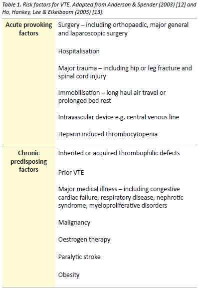
v6_i1_a6a

Thrombophilia

Thrombophilia refers to a predisposition to thrombosis, which may be inherited or acquired. [14] The prevalence of thrombophilia at first presentation  of  VTE  is  approximately  50%,  with  the highest prevalence  found  in  younger  patients  and  those  with unprovoked VTE. [15] Inherited thrombophilias are common in the Caucasian Australian population. The birth prevalence of factor V Leiden heterozygosity and homozygosity, which confers resistance to activated protein  C,  is  9.5%  and  0.7%  respectively. Heterozygosity and homozygosity for the prothrombin gene mutation (G20210A) is another common inherited thrombophilia, with a prevalence of 4.1% and 0.2% respectively. [16] Other significant thrombophilias include antithrombin deficiency, protein C deficiency, protein S deficiency and causes of hyperhomocystinaemia. [16,17] Antiphospholipid syndrome is an acquired disorder characterised by antiphospholipid antibodies and arterial or venous thrombosis or obstetric related morbidity, including recurrent spontaneous abortion. Antiphospholipid syndrome represents an important cause of VTE and may occur as a primary disorder or secondary to autoimmune or rheumatic diseases such as systemic lupus erythematosus. [18]

Testing for hereditary thrombophilia is generally not recommended as it does not affect clinical management of most patients with VTE [19,20] and there is no evidence that such testing alters the risk of recurrent VTE. [21] There are few exceptions such as a fertile women with a family history of a thrombophilia where testing positive may lead to the decision to avoid the oral contraceptive pill or institute prophylaxis in the peripartum period. [22]

Natural history

Most DVT originate in the deep veins of the calf. Thrombi originating in the calf are often asymptomatic and confer a low risk of clinically significant PE. Approximately 25% of untreated calf DVT will extend into the proximal veins of the leg and 80% of patients with symptomatic DVT have involvement of the proximal veins. [9] Symptomatic PE occurs in a significant proportion of patients with untreated proximal DVT; however the exact risk of proximal embolisation is difficult to estimate. [9,23]

Pulmonary vascular remodeling may occur following PE and may result in CTEPH. [24] CTEPH is thought to be caused by unresolved pulmonary emboli and is associated with significant morbidity and mortality. CTEPH develops in approximately 1-4% of patients with treated PE. [25,26]

Post-thrombotic syndrome is an important potential long-term consequence of DVT, which is characterised by leg pain, oedema, venous ectasia and venous ulceration. Within 2 years of symptomatic DVT, post-thrombotic syndrome develops in 23-60% of patients [27] and is associated with poorer quality of life and significant economic burden. [28]

Diagnosis

Signs and symptoms of VTE are often non-specific and may mimic many other common clinical conditions (Table 2). In the primary care setting, less than 30% of patients with signs and symptoms suggestive of DVT have a sonographically proven thrombus. [29] Some of the clinical features of superficial thrombophlebitis overlap with those of DVT. Superficial thrombophlebitis carries a small risk of DVT or PE and contiguous extension of the thrombus. Treatment may be recommended with low-dose anticoagulant therapy or NSAIDs. [30]
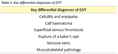
v6_i1_a6b

Deep vein thrombosis

Clinical features

Symptoms of DVT include pain, cramping and heaviness in the lower extremity, swelling and a cyanotic or blue-red discolouration of the limb.  [31]  Signs  may  include  superficial vein  dilation, warmth and unilateral oedema. [31,32] Pain in the calf on forceful dorsiflexion of the foot was described as a sign of DVT by the American surgeon John Homans in 1944. [33] Homans’ sign is non-specific and is an unreliable sign of DVT. [34]

Investigations

Several scoring tools have been evaluated for assessing the pre-test probability of DVT. One such commonly used validated tool is the Modified Wells score, presented in Table 3. [35] The Modified Wells score categorises patients as either likely or unlikely to have a DVT.
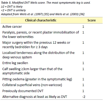
v6_i1_a6c

D-dimer is the recommended investigation in patients considered unlikely to have a DVT, as a negative D-dimer effectively rules out DVT in this patient group. [36] D-dimer measurements have several important limitations with most studies of its use in DVT being performed in outpatients and non-pregnant patients. As D-dimer represents a fibrin degradation product, it is likely to be raised in any inflammatory response. This limits its use in post-operative patients and many hospitalised patients.

Venous compression ultrasound with Doppler flow is indicated as the initial investigation in patients who are considered likely to have DVT (Modified Wells ≥ 2) or in patients with a positive D-dimer. Compression ultrasonography is the most widely used imaging modality due to its high sensitivity and specificity, non-invasive nature and low cost. Limitations include operator-dependent accuracy and reduction of sensitivity and specificity in DVT of pelvic veins, small calf veins or in obese patients. [32]

Pulmonary embolism

Clinical features

10%  of  symptomatic  PE  are  fatal  within  1  hour  of  the  onset of symptoms [9] and delay of diagnosis remains common due to non-specific  presentation.  [37]   Clinical  presentation  will  depend  on several factors including size of the embolus, rapidity of obstruction of the pulmonary vascular bed and patient’s haemodynamic reserve. Symptoms may include sudden or gradual onset dyspnoea, chest pain, cough, haemoptysis, palpitations and syncope. Signs may include tachycardia, tachypnea, fever, cyanosis and the clinical features of DVT. Signs of pulmonary infarction may develop later and include a pleural friction rub and reduced breath sounds. [31] Patients may also present with systemic arterial hypotension with or without clinical features of obstructive shock. [5,38]

Investigations

The first step in the diagnosis of suspected PE is the calculation of the clinical  pre-test  probability  using  a  validated  tool  such  as  the Wells or Geneva score. Clinician gestalt may be used in place of a validated scoring tool; however it may be associated with a lower specificity and therefore increased unnecessary pulmonary imaging. [39] Neither clinician gestalt nor a clinical decision rule can accurately exclude  PE  on its  own.  An  electrocardiogram  (ECG)  will  often  be performed early in the presentation of a patient with suspected PE. A variety of electrocardiographic changes associated with acute PE have been described. Changes consistent with right heart strain and atrial enlargement reflect mechanical pulmonary artery outflow tract obstruction. [40]  Other  ECG  changes  include  sinus  tachycardia, ST segment or T wave abnormalities, QRS axis alteration (left or right), right bundle branch block and a number of others. [40] The S1Q3T3 abnormality, described as a prominent S wave in lead I with a Q wave and inverted T wave in lead III, is a sign of acute corpulmonale. It is not pathognomonic for PE and occurs in less than 25% of patients with acute PE. [40]

In patients with a low pre-test probability, a negative quantitative D-dimer effectively excludes PE. [39] The conventional D-Dimer cut- off value (500 µg/L) are associated with reduced specificity in older patients leading to false positive results. [41] A recent meta-analysis has found that the use of an age specific D-dimer cut off value (age x  10µg/L)  increases  the specificity  of  the  D-dimer  test  with  little effect on sensitivity. [42] The pulmonary embolism rule-out criteria (PERC), as outlined in Table 4, may be applied to patients with a low pre-test  probability  to  reduce  the number  of  patients  undergoing D-dimer testing. [43] A recent meta-analysis demonstrated that in the emergency department, the combination of low pre-test probability and  a  negative  PERC  rule results  in  a  likelihood  of  PE  that  is  so unlikely that the risk-benefit ratio of further investigation for PE is not favourable. [44]
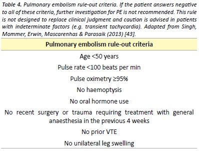
v6_i1_a6d

Patients with a high pre-test probability or with a positive D-dimer test should undergo pulmonary imaging. Multidetector Computed Tomography  Pulmonary  Angiography  (CTPA)  is  largely  considered the imaging modality of choice for PE given its high sensitivity and
specificity and its ability to identify alternative diagnoses. [45,46] CTPA
must be used only with a clear indication due to significant radiation
exposure, risk of allergic reactions and contrast-induced nephropathy.
[47] Concerns have also been raised about over diagnosis of PE with
detection of small subsegmental emboli. [48]


Ventilation-perfusion (V/Q) lung scintigraphy is an alternative pulmonary imaging modality to CTPA. A normal V/Q scan excludes PE; however, a significant proportion of patients will have a ‘non- diagnostic’ result thus requiring further imaging. [49] Non-diagnostic scans are more common in patients with pre-existing respiratory disease or an abnormal chest radiograph and are less likely in younger and pregnant patients. [48,50] Compared with CTPA, V/Q scanning is associated with fewer adverse effects and less radiation exposure and is often employed when a contraindication to CTPA exists. [49,50]

Bedside echocardiography is a useful investigation if CT is not immediately available or if the patient is too unstable for transfer to radiology. [51,52] Echocardiography may reveal right ventricular dysfunction which guides prognosis and the potential for thrombolytic therapy in massive and sub-massive PE. [51]

The diagnosis of PE during pregnancy is an area of controversy. [54] The diagnostic value of D-dimer during pregnancy using the conventional threshold is limited. With both V/Q scans and CTPA, foetal radiation dose is minimal but higher in the former. CTPA is associated with a much higher dose of radiation to maternal breast tissue thus increased risk of breast cancer. [53,54] In light of these risks, some experts advocate for bilateral compression Doppler ultrasound for suspected PE in pregnancy. [54] However, if this is negative and a high clinical suspicion remains, pulmonary imaging is still required.

Prophylaxis

Multiple guidelines exist to direct clinicians on the use of thromboprophylaxis in both medical and surgical patients. [3,55-58] Implementation of thromboprophylaxis involves assessment of the patient’s risk of VTE, risk of adverse effects of thromboprophylaxis, including bleeding and identification of any contraindications.

Patients at high risk include those undergoing any surgical procedure, especially abdominal, pelvic or orthopaedic surgery. Medical patients at high risk include those with myocardial infarction, malignancy, heart failure, ischaemic stroke and inflammatory bowel disease.[3]

Mechanical options for thromboprophylaxis include encouragement of  mobility,  graduated  compression  stockings,  intermittent pneumatic compression devices and venous foot pumps. Mechanical prophylactic measures are often combined with pharmacological thromboprophylaxis. The strength of evidence for each of the anticoagulant varies depending on the surgical procedure or medical condition in  question;  however,  unfractionated heparin  (UFH)  and low-molecular weight heparin (LMWH) remain the mainstay of VTE prophylaxis. [3] The NOACs, also referred to as direct oral anticoagulants, notably  rivaroxaban,  apixaban  and  dabigatran,  have  been  studied most amongst the hip and knee arthroplasty patient groups, where they have been shown to be both efficacious and safe. [59-61] The use of aspirin for the prevention of VTE following orthopaedic surgery remains controversial, despite receiving a recommendation by recent guidelines. [62] It is recommended that pharmacological prophylaxis should be continued until the patient is fully mobile. In certain circumstances such as following total hip or knee arthroplasty and hip fracture surgery, extended duration prophylaxis for up to 35 days post- operatively is recommended. [3,62]

Management

The aim of treatment is to relieve current symptoms, prevent
progression of the disease, reduce the potential for sequelae and
prevent recurrence. Anticoagulation remains the cornerstone of
management of VTE.

Patients with PE and haemodynamic instability (hypotension, persistent
bradycardia, pulselessness), so called ‘massive PE’may require urgent
treatment with thrombolytic therapy. Thrombolysis reduces mortality
in haemodynamically unstable patients, however it is associated with a
risk of major bleeding. [63] Surgical thrombectomy and catheter-based
interventions represent an alternative to thrombolysis in patients withThe aim of treatment is to relieve current symptoms, prevent progression of the  disease,  reduce the potential for sequelae and massive PE where contraindications exist. [64] The use of thrombolytic therapy in patients with evidence of right ventricular dysfunction and myocardial injury without hypotension and haemodynamic instability remains controversial. A recent study revealed that fibrinolysis in this intermediate risk group reduces rates of haemodynamic compromise while significantly increasing the risk of intracranial and other major bleeding. [65]

For the majority of patients with VTE anticoagulation is the mainstay of treatment. Acute treatment involves UFH, LMWH or fondaparinux. [50]

UFH binds to antithrombin III, increasing its ability to inactivate thrombin, factor Xa and other coagulation factors. [66] UFH is usually given as an intravenous bolus initially, followed by a continuous infusion. UFH therapy requires monitoring of the activated partial thromboplastin time (aPTT) and is associated with a risk of heparin- induced thrombocytopenia. [66] The therapeutic aPTT range and dosing regimen vary between institutions. The use of UFH is usually preferred if there is severe renal impairment, in cases where there may be a requirement to rapidly reverse anticoagulation therapy and in obstructive shock where thrombolysis is being considered. [50]

LMWH is administered subcutaneously in a weight adjusted dosing regimen once or twice daily. [51] When compared with UFH, LMWH has a more predictable anticoagulant response and does not usually require monitoring. [67] In obese patients and those with significant renal dysfunction, LMWH may require dose adjustment or monitoring of factor Xa activity. [67]

Therapy with a vitamin K antagonist, most commonly warfarin, should be commenced at the same time as parenteral anticoagulation. Therapy with the parenteral anticoagulant should be discontinued when the international normalised ratio (INR) has reached at least 2.0 on two consecutive measurements and there has been an overlap of treatment with a parenteral anticoagulant for at least five days. [68] This overlap is required as the use of warfarin alone may be associated with an initial transient prothrombotic state due to warfarin mediated rapid depletion of the natural anticoagulant protein C, whilst depletion of coagulation factors II and X takes several days. [69]

The NOACs represent an attractive alternative to traditional anticoagulants for the prevention and management of VTE.

Rivaroxaban is a direct oral anticoagulant that directly inhibits factor Xa.[66] Rivaroxaban has been shown to be as efficacious as standard therapy (parenteral anticoagulation and warfarin) for the treatment of proximal DVT and symptomatic PE. [70,71] When compared with conventional  therapy,  rivaroxaban  may  be  associated  with  lower risks of major bleeding. [70] Rivaroxaban represents an attractive alternative to the standard therapy mentioned above as it does not require parenteral administration, is given as a fixed daily dose, does not require laboratory monitoring and has few drug-drug and food interactions. [70,71]

Dabigatran etexilate is an orally administered direct thrombin inhibitor. Dabigatran is non-inferior to warfarin for the treatment of PE and proximal DVT after a period of parenteral anticoagulation. [72] The safety profile is similar; however dabigatran requires no laboratory
monitoring. [72]

The lack of a requirement for monitoring is a significant benefit over
warfarin for the NOACs. The role of monitoring the anticoagulant
activity of these agents and the clinical relevance of monitoring is a
subject of ongoing research and debate. [73] Anti-factor Xa based
assays may be used to determine the concentration of the anti-factor
Xa inhibitors in specific clinical circumstances. [74,75] The relative
intensity of anticoagulant due to dabigatran can be estimated by
the aPTT and rivaroxaban by the PT or aPTT [76]. There is however,
significant variation in the results based on the reagent the laboratory
uses. Routine monitoring for the NOACs is not currently recommended.

The major studies evaluating the NOACs carried exclusion criteria that included those at high risk of bleeding, with a creatinine clearance of  <30  mL/min,  pregnancy  and  those  with  liver  disease  [70-72], thus caution must be applied with their use in these patient groups. The  NOACs  are  renally  metabolised  to  variable  degrees.  Warfarin or dose adjusted LMWH are preferred for those with reduced renal function (creatinine clearance <30mL/min) who require long-term anticoagulation.

Concern exists regarding a lack of a specific reversal agent for the NOACs. [77,78] Consultation with haematology is recommended if significant bleeding  occurs  during  therapy  with  a  NOAC.  Evidence for the use of agents such as tranexamic acid, recombinant factor VIIa and prothrombin complex concentrate is very limited. [77,78] Haemodialysis may significantly reduce plasma levels of dabigatran, as the drug displays relatively low protein binding. [77,78]

Inferior vena cava (IVC) filters may be placed in patients with VTE and a contraindication to anticoagulation. IVC filters prevent PE however they may increase the risk of DVT and vena cava thrombosis. The use of IVC filters remains controversial due to a lack of evidence. [79]

Recurrence

The risk of recurrence differs significantly depending on whether the initial VTE event was unprovoked or associated with a transient risk factor. [9] Patients with idiopathic VTE have a significantly higher risk of recurrence than those with transient risk factors. Isolated calf DVT carry a lower risk of recurrence than that of proximal DVT or PE. The risk of recurrence after the cessation of anticoagulant therapy is as high as 10% per year in some patients groups. [9]

Duration of anticoagulation therapy should be based on patient preference and a risk-benefit analysis of the risk of recurrence versus the risk of complications from therapy. Generally, anticoagulation should be continued for a minimum of three months and the decision to  continue  anticoagulation  should  be  re-assessed  on  a  regular basis. Recommendations for duration of anticoagulation therapy are presented in Table 5. The currently published guidelines recommend extended anticoagulation therapy with a vitamin K antagonist such as warfarin. Evaluation of the new direct oral anticoagulants for therapy and prevention of recurrence is ongoing. Recent evidence supports the use of dabigatran and rivaroxaban for the secondary prevention of venous thromboembolism with similar efficacy to standard therapy and reduced rates of major bleeding. [70,71,80]
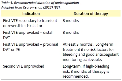
v6_i1_a6e

Aspirin has been shown to be effective in reducing the recurrence VTE in patients with previous unprovoked VTE. After up to 18 months of therapy, aspirin reduces the rate of VTE recurrence by 40%, as compared with placebo. [81]

Acknowledgements

I would like to thank Marianne Turner for her help with editing of the manuscript.

Conflict of interest

None declared.

Correspondence

R Pow: richardeamonpow@gmail.com

Conclusion

VTE  is  a  commonly  encountered  problem  and  is  associated  with significant short and long term morbidity. A sound understanding of the pathogenesis of VTE guides clinical assessment, diagnosis and management. The prevention and management of VTE continues to evolve with the ongoing evaluation of the NOACs. Anticoagulation remains the mainstay of therapy for VTE, with additional measures including thrombolysis used in select cases.  This article has provided medical students with an evidence based review of the current diagnostic and management strategies for venous thromboembolic disease.

References

[1] Oger E. Incidence of venous thromboembolism: a community-based study in Western France. Thromb Haemost. 2000;83(5):657-60.

[2] Cushman M, Tsai AW, White RH, Heckbert SR, Rosamond WD, Enright P, et al. Deep vein thrombosis and pulmonary embolism in two cohorts: the longitudinal investigation of thromboembolism etiology. Am J Med. 2004;117(1):19-25.

[3]  National Health  and  Medical  Research  Council.  Clinical  Practice Guideline  for  the Prevention of Venous Thromboembolism in Patients Admitted to Australian Hospitals. Melbourne: National Health and Medical Research Council; 2009. 157 p.

[4]  National  Institute  of  Clinical  Studies.  Evidence-Practice  Gaps  Report  Volume  1. Melbourne:National Institute of Clinical Studies; 2003. 38 p.

[5] Goldhaber SZ, Visani L, De Rosa M. Acute pulmonary embolism: clinical outcomes in   the   International   Cooperative   Pulmonary   Embolism   Registry   (ICOPER).   Lancet. 1999;353(9162):1386-9.

[6] Huang CM, Lin YC, Lin YJ, Chang SL, Lo LW, Hu YF, et al. Risk stratification and clinical outcomes in patients with acute pulmonary embolism. Clin Biochem. 2011;44(13):1110-5.

[7]  Access  Economics  Pty  Ltd.  The  burden  of  venous  thromboembolism  in  Australia. Canberra: Access Economics Pty Ltd; 2008. 50 p.

[8] Lang IM, Klepetko W. Chronic thromboembolic pulmonary hypertension: an updated review. Curr Opin Cardiol. 2008;23(6):555-9.

[9] Kearon C. Natural history of venous thromboembolism. Circulation. 2003;107(23 Suppl 1):22-30.

[10] Dickinson BC. Venous thrombosis: On the History of Virchow’s Triad. Univ Toronto Med J. 2004;81(3):6.

[11] Heit JA, O’Fallon WM, Petterson TM, Lohse CM, Silverstein MD, Mohr DN, et al. Relative impact of risk factors for deep vein thrombosis and pulmonary embolism: a population-based study. Arch Intern Med. 2002;162(11):1245-8.

[12] Anderson FA, Jr., Spencer FA. Risk factors for venous thromboembolism. Circulation. 2003;107(23 Suppl 1):9-16.

[13] Ho WK, Hankey GJ, Lee CH, Eikelboom JW. Venous thromboembolism: diagnosis and management of deep venous thrombosis. Med J Aust. 2005;182(9):476-81.

[14]   Heit   JA.   Thrombophilia:   common   questions   on   laboratory   assessment   and management. Hematology Am Soc Hematol Educ Program. 2007;1:127-35.

[15] Weingarz L, Schwonberg J, Schindewolf M, Hecking C, Wolf Z, Erbe M, et al. Prevalence of thrombophilia according to age at the first manifestation of venous thromboembolism: results from the MAISTHRO registry. Br J Haematol. 2013;163(5):655-65.

[16] Gibson CS, MacLennan AH, Rudzki Z, Hague WM, Haan EA, Sharpe P, et al. The prevalence of inherited thrombophilias in a Caucasian Australian population. Pathology. 2005;37(2):160-3.

[17]  Genetics  Education  in  Medicine  Consortium.  Genetics  in  Family  Medicine:  The Australian  Handbook  for  General  Practitioners. Canberra:  The  Australian  Government Agency Biotechnology Australia; 2007. 349 p.

[18] Giannakopoulos B, Krilis SA. The pathogenesis of the antiphospholipid syndrome. N Engl J Med. 2013;368(11):1033-44.

[19] Ho WK, Hankey GJ, Eikelboom JW. Should adult patients be routinely tested for heritable  thrombophilia  after  an  episode  of  venous thromboembolism?  Med  J  Aust. 2011;195(3):139-42.

[20]  Middeldorp   S.  Evidence-based  approach  to  thrombophilia  testing.  J  Thromb Thrombolysis. 2011;31(3):275-81.

[21] Cohn DM, Vansenne F, de Borgie CA, Middeldorp S. Thrombophilia testing for prevention of recurrent venous thromboembolism. Cochrane Database Syst Rev[Internet] 2012 [Cited 2014 Sep 10]. Available from: http://www.ncbi.nlm.nih.gov/pubmed/23235639

[22] Middeldorp S, van Hylckama Vlieg A. Does thrombophilia testing help in the clinical management of patients? Br J Haematol. 2008;143(3):321-35.

[23] Markel A. Origin and natural history of deep vein thrombosis of the legs. Semin Vasc Med. 2005;5(1):65-74.

[24] Hoeper MM, Mayer E, Simonneau G, Rubin LJ. Chronic thromboembolic pulmonary hypertension. Circulation. 2006;113(16):2011-20.

[25] Pengo V, Lensing AW, Prins MH, Marchiori A, Davidson BL, Tiozzo F, et al. Incidence of chronic thromboembolic pulmonary hypertension after pulmonary embolism. N Engl J Med. 2004;350(22):2257-64.

[26]  Poli  D,  Grifoni  E,  Antonucci  E,  Arcangeli  C,  Prisco  D,  Abbate  R,  et  al.  Incidence of  recurrent  venous  thromboembolism  and  of  chronic  thromboembolic  pulmonary hypertension  in  patients  after  a  first  episode  of  pulmonary  embolism.  J  Thromb Thrombolysis. 2010;30(3):294-9.

[27] Ashrani AA, Heit JA. Incidence and cost burden of post-thrombotic syndrome. J Thromb Thrombolysis. 2009;28(4):465-76.

[28] Kachroo S, Boyd D, Bookhart BK, LaMori J, Schein JR, Rosenberg DJ, et al. Quality of life and economic costs associated with postthrombotic syndrome. Am J Health Syst Pharm. 2012;69(7):567-72.

[29]  Oudega  R,  Moons  KG,  Hoes  AW.  Limited  value  of  patient history  and  physical examination in diagnosing deep vein thrombosis in primary care. Fam Pract. 2005;22(1):86-91.

[30] Di Nisio M, Wichers IM, Middeldorp S. Treatment for superficial thrombophlebitis of the leg. Cochrane Database Syst Rev [Internet] 2013 [Cited 2014 Sep 10]. Available from: http://www.ncbi.nlm.nih.gov/pubmed/23633322

[31]  Bauersachs  RM.  Clinical  presentation  of  deep  vein  thrombosis  and  pulmonary embolism. Best Pract Res Clin Haematol. 2012;25(3):243-51.

[32] Ramzi DW, Leeper KV. DVT and pulmonary embolism: Part I. Diagnosis. Am Fam physician. 2004;69(12):2829-36.

[33] Homans J. Diseases of the veins. N Engl J Med. 1946;235(5):163-7.

[34] Cranley JJ, Canos AJ, Sull WJ. The diagnosis of deep venous thrombosis. Fallibility of clinical symptoms and signs. Arch Surg. 1976;111(1):34-6.

[35]  Wells  PS,  Anderson  DR,  Bormanis  J,  Guy  F,  Mitchell  M,  Gray  L,  et  al.  Value  of assessment of pretest probability of deep-vein thrombosis in clinical management. Lancet. 1997;350(9094):1795-8.

[36] Wells PS, Anderson DR, Rodger M, Forgie M, Kearon C, Dreyer J, et al. Evaluation of  D-dimer  in  the  diagnosis  of  suspected  deep-vein thrombosis.  N  Engl  J  Med. 2003;349(13):1227-35.

[37] Torres-Macho J, Mancebo-Plaza AB, Crespo-Gimenez A, Sanz de Barros MR, Bibiano-Guillen C, Fallos-Marti R, et al. Clinical features of patients inappropriately undiagnosed of pulmonary embolism. Am J Emerg Med. 2013;31(12):1646-50.

[38] Grifoni S, Olivotto I, Cecchini P, Pieralli F, Camaiti A, Santoro G, et al. Short-term clinical outcome of patients with acute pulmonary embolism, normal blood pressure, and echocardiographic right ventricular dysfunction. Circulation. 2000;101(24):2817-22.

[39] Lucassen W, Geersing GJ, Erkens PM, Reitsma JB, Moons KG, Buller H, et al. Clinical decision  rules  for  excluding  pulmonary  embolism:  a meta-analysis.  Ann  Intern  Med. 2011;155(7):448-60.

[40] Ullman E, Brady WJ, Perron AD, Chan T, Mattu A. Electrocardiographic manifestations of pulmonary embolism. Am J Emerg Med. 2001;19(6):514-9.

[41] Douma RA, Tan M, Schutgens RE, Bates SM, Perrier A, Legnani C, et al. Using an age-dependent D-dimer cut-off value increases the number of older patients in whom deep vein thrombosis can be safely excluded. Haematologica. 2012;97(10):1507-13.

[42] Schouten HJ, Geersing GJ, Koek HL, Zuithoff NP, Janssen KJ, Douma RA, et al. Diagnostic accuracy of conventional or age adjusted D-dimer cut-off values in older patients with suspected   venous   thromboembolism:   systematic   review   and   meta-analysis.   BMJ. 2013;346(2492): 1-13.

[43]  Kline  JA,  Mitchell  AM,  Kabrhel  C,  Richman  PB,  Courtney  DM.  Clinical  criteria  to prevent unnecessary diagnostic testing in emergency department patients with suspected pulmonary embolism. J Thromb Haemost. 2004;2(8):1247-55.

[44] Singh B, Mommer SK, Erwin PJ, Mascarenhas SS, Parsaik AK. Pulmonary embolism rule-out criteria (PERC) in pulmonary embolism–revisited: a systematic review and meta-analysis. Emerg Med J. 2013;30(9):701-6.

[45] den Exter PL, Klok FA, Huisman MV. Diagnosis of pulmonary embolism: Advances and pitfalls. Best Pract Res Clin Haematol. 2012;25(3):295-302.

[46] Stein PD, Fowler SE, Goodman LR, Gottschalk A, Hales CA, Hull RD, et al. Multidetector computed tomography for acute pulmonary embolism. N Engl J Med. 2006;354(22):2317-27.

[47] Mitchell AM, Jones AE, Tumlin JA, Kline JA. Prospective study of the incidence of contrast-induced  nephropathy  among  patients  evaluated  for  pulmonary  embolism  by contrast-enhanced computed tomography. Acad Emerg Med. 2012;19(6):618-25.

[48]   Huisman   MV,   Klok  FA.  How  I  diagnose  acute  pulmonary embolism.  Blood. 2013;121(22):4443-8.

[49] Anderson DR, Kahn SR, Rodger MA, Kovacs MJ, Morris T, Hirsch A, et al. Computed tomographic pulmonary angiography vs ventilation-perfusion lung scanning in patients with suspected pulmonary embolism: a randomized controlled trial. J Am Med Assoc. 2007;298(23):2743-53.

[50] Takach Lapner S, Kearon C. Diagnosis and management of pulmonary embolism. BMJ. 2013 Feb 20;346(757):1-9.

[51] Agnelli G, Becattini C. Acute pulmonary embolism. N Engl J Med. 2010;363(3):266-74.

[52]  Konstantinides  S.  Clinical  practice.  Acute  pulmonary  embolism.  N  Engl  J  Med. 2008;359(26):2804-13.

[53] Duran-Mendicuti A, Sodickson A. Imaging evaluation of the pregnant patient with suspected pulmonary embolism. Int J Obstet Anesth. 2011;20(1):51-9.

[54]  Arya  R.  How  I  manage  venous  thromboembolism  in  pregnancy. Br  J  Haematol. 2011;153(6):698-708.

[55] National Health and Medical Research Council. Prevention of Venous Thromboembolism in Patients Admitted to Australian Hospitals: Guideline Summary. Melbourne: National Health and Medical Research Council; 2009. 2 p.

[56] Australian Orthopaedic Association. Guidelines for VTE Prophylaxis for Hip and Knee Arthroplasty. Sydney: Australian Orthopaedic Association; 2010. 8 p.

[57] The Australia & New Zealand Working Party on the Management and Prevention of  Venous  Thromboembolism.  Prevention of  Venous Thrombormbolism:  Best  practice guidelines for Australia & New Zealand, 4th edition. South Austrlaia: Health Education and Management Innovations;2007. 11 p.

[58] Geerts WH, Bergqvist D, Pineo GF, Heit JA, Samama CM, Lassen MR, et al. Prevention of venous thromboembolism: American College of Chest Physicians Evidence-Based Clinical Practice Guidelines (8th Edition). Chest. 2008;133(6 Suppl):381-453.

[59] Eriksson BI, Borris LC, Friedman RJ, Haas S, Huisman MV, Kakkar AK, et al. Rivaroxaban versus   enoxaparin   for   thromboprophylaxis after  hip   arthroplasty.   N   Engl   J   Med. 2008;358(26):2765-75.

[60]  Lassen  MR,  Ageno  W,  Borris  LC,  Lieberman  JR,  Rosencher  N, Bandel  TJ,  et  al. Rivaroxaban versus enoxaparin for thromboprophylaxis after total knee arthroplasty. N Engl J Med. 2008;358(26):2776-86.

[61]  Eriksson  BI,  Dahl  OE,  Rosencher  N,  Kurth  AA,  van  Dijk  CN, Frostick  SP,  et  al. Dabigatran  etexilate  versus  enoxaparin  for prevention  of  venous  thromboembolism after  total  hip  replacement:  a  randomised,  double-blind,  non-inferiority  trial.  Lancet. 2007;370(9591):949-56.

[62] Falck-Ytter Y, Francis CW, Johanson NA, Curley C, Dahl OE, Schulman S, et al. Prevention of  VTE  in  orthopedic  surgery  patients: Antithrombotic  Therapy  and  Prevention  of Thrombosis, 9th ed: American College of Chest Physicians Evidence-Based Clinical Practice Guidelines. Chest. 2012;141(2 Suppl):278-325.

[63] Wan S, Quinlan DJ, Agnelli G, Eikelboom JW. Thrombolysis compared with heparin for the initial treatment of pulmonary embolism: a meta-analysis of the randomized controlled trials. Circulation. 2004;110(6):744-9.

[64] Jaff MR, McMurtry MS, Archer SL, Cushman M, Goldenberg N, Goldhaber SZ, et al. Management  of  massive and submassive pulmonary embolism, iliofemoral deep vein thrombosis, and chronic thromboembolic pulmonary hypertension: a scientific statement from the American Heart Association. Circulation. 2011;123(16):1788-830.

[65]  Meyer  G,  Vicaut  E,  Danays  T,  Agnelli  G,  Becattini C,  Beyer-Westendorf  J,  et  al. Fibrinolysis  for  patients  with  intermediate-risk  pulmonary  embolism.  N  Engl  J  Med. 2014;370(15):1402-11.

[66] Mavrakanas T, Bounameaux H. The potential role of new oral anticoagulants in the prevention and treatment of thromboembolism. Pharmacol. Ther. 2011 Dec 24;130(1):46-58.

[67] Bounameaux H, de Moerloose P. Is laboratory monitoring of low-molecular-weight heparin therapy necessary? No. J Thromb Haemost. 2004 Apr;2(4):551-4.

[68]  Goldhaber  SZ,  Bounameaux  H.  Pulmonary  embolism  and  deep vein  thrombosis. Lancet. 2012 Apr 10;379(9828):1835-46.

[69] Choueiri T, Deitcher SR. Why shouldn’t we use warfarin alone to treat acute venous thrombosis? Cleve Clin J Med. 2002;69(7):546-8.

[70]  Buller  HR,  Prins  MH,  Lensin  AW,  Decousus  H,  Jacobson  BF, Minar  E,  et al.  Oral rivaroxaban  for  the  treatment  of  symptomatic  pulmonary  embolism.  N  Engl  J  Med. 2012;366(14):1287-97.

[71] Landman GW, Gans RO. Oral rivaroxaban for symptomatic venous thromboembolism. N Engl J Med. 2011;364(12):1178.

[72] Schulman S, Kearon C, Kakkar AK, Mismetti P, Schellong S, Eriksson H, et al. Dabigatran versus  warfarin  in  the  treatment  of  acute  venous thromboembolism.  N  Engl  J  Med. 2009;361(24):2342-52

[73] Schmitz EM, Boonen K, van den Heuvel DJ, van Dongen JL, Schellings MW, Emmen JM, et al. Determination of dabigatran, rivaroxaban and apixaban by UPLC-MS/MS and coagulation assays for therapy monitoring of novel direct oral anticoagulants. J Thromb Haemost. Forthcoming 2014 Aug 20.

[74] Lippi G, Ardissino D, Quintavalla R, Cervellin G. Urgent monitoring of direct oral anticoagulants in patients with atrial fibrillation: a tentative approach based on routine laboratory tests. J Thromb Thrombolysis. 2014;38(2):269-74.

[75] Samama MM, Contant G, Spiro TE, Perzborn E, Le Flem L, Guinet C, et al. Laboratory assessment of rivaroxaban: a review. Thromb J. 2013;11(1):11.

[76] Baglin T, Keeling D, Kitchen S. Effects on routine coagulation screens and assessment of anticoagulant intensity in patients taking oral dabigatran or rivaroxaban: guidance from the British Committee for Standards in Haematology. Br J Haematol. 2012;159(4):427-9.

[77] Jackson LR, 2nd, Becker RC. Novel oral anticoagulants: pharmacology, coagulation measures, and considerations for reversal. J Thromb Thrombolysis. 2014;37(3):380-91

[78] Wood P. New oral anticoagulants: an emergency department overview. Emerg MedAustralas. 2013;25(6):503-14.

[79]   Rajasekhar   A,   Streiff   MB.   Vena   cava   filters   for   management of  venous thromboembolism: a clinical review. Blood rev. 2013;27(5):225-41.

[80] Schulman S, Kearon C, Kakkar AK, Schellong S, Eriksson H, Baanstra D, et al. Extended use  of  dabigatran,  warfarin,  or  placebo  in  venous  thromboembolism.  N  Engl  J  Med. 2013;368(8):709-18.

[81] Becattini C, Agnelli G, Schenone A, Eichinger S, Bucherini E, Silingardi M, et al. Aspirin for preventing the recurrence of venous thromboembolism. N Engl J Med. 2012;366(21):1959-67.

[82] Kearon C, Akl EA, Comerota AJ, Prandoni P, Bounameaux H, Goldhaber SZ, et al. Antithrombotic  therapy  for  VTE  disease: Antithrombotic  Therapy  and  Prevention  of Thrombosis, 9th ed: American College of Chest Physicians Evidence-Based Clinical Practice Guidelines. Chest. 2012;141(2 Suppl):419s-94s.