Categories
Feature Articles

Stopping the silent epidemic: my summer internship with the WHO

Viral hepatitis has been historically under-recognised and under-addressed in the global health community. This is despite causing a serious disease burden worldwide due to chronic liver disease, cirrhosis, and liver cancer. The World Health Organization (WHO) has been involved on a global and regional level in the response to the epidemics of hepatitis B and C. A summer internship with the WHO Regional Office in Manila provided insights into modelling techniques to estimate the disease burden and treatment strategies to combat this silent epidemic. With effective antiviral treatments to cure hepatitis C and suppress hepatitis B, the prevalence, complications, and high costs of these diseases can be dramatically reduced.

Introduction

Health is a state of complete physical, mental and social well-being, and not merely the absence of disease or infirmity [1].”  No prizes to guess whose definition of health this is (pun intended). You’ve probably recited it word-for-word at some point in your medical studies. We look to the World Health Organization (WHO) for leadership in global health. The WHO provides technical guidelines, responds to health and humanitarian emergencies, and advocates and coordinates engagement across health, policymakers, and civil society [2].  What goes on behind the public face we see – the guidelines, press releases, and targets? How does the organisation operate on a global, regional, and country-level? How does the WHO respond to communicable threats such as polio, HIV, and viral hepatitis? What does it take in the quest to achieve the highest attainable quality of health for all?

Curious to understand and experience the well-oiled bureaucratic machine that is the WHO, and specifically to contribute in a small way to its response to viral hepatitis, I undertook a summer internship with the WHO at the Regional Office for the Western Pacific (WPRO). This Regional Office bridges Headquarters in Geneva and the 37 Member States under its care; a diverse belt of countries spanning down East and South-East Asia, the Pacific Islands, Australia, and New Zealand. For 8 weeks, I experienced life as an intern in the HIV, Hepatitis and Sexually Transmitted Infections Unit, within the Division of Communicable Diseases.

I also had a glimpse into life in Manila, the bustling metropolitan capital of the Philippines, where colonial powers and local revolutionaries have crossed for control of the island nation, and where for me, crossing the road proved to be a regular, life-threatening exercise. My first impression of Manila was stepping out of the airport into its balmy, honking embrace. From thereinafter I was accompanied in every waking moment by the cacophony of taxis, jeepneys, and motorcycles. There was not a moment of silence. Which, as it happens, is the exact opposite of the description of viral hepatitis.

The silent epidemic

Viral hepatitis is a silent epidemic in every sense of the word. The majority of people living with chronic infection, mostly hepatitis B and C, don’t know they have the disease. Left untreated, years of asymptomatic infection can lead to the deadly sequelae of liver cancer or cirrhosis. The dearth of testing services available also contributes to the “silence” around chronic hepatitis, especially in resource-limited areas where the highest burden lies. About half of the 218,000 people in Australia with hepatitis B aren’t aware they are infected [3]. This is even higher globally, 70-80% in the United States and more than 90% in many Asian countries (except Japan) [4]. In Europe, only 10-40% of people with hepatitis C are diagnosed [5]. That is to say, there are those who are living with chronic hepatitis, those who know their infection status, and those who are receiving treatment. In between, there are broad gaps, disparities in testing and treatment across both higher and lower income countries.

Whilst viral hepatitis has been unrecognised in the past, its burden is substantial. Hepatitis B and C, the biggest contributors to mortality and morbidity from the group of diseases caused by hepatitis viruses, are endemic in parts of South East Asia, Sub Saharan Africa, and South America. The Western Pacific Region, which includes Australia, bears over a third of global mortality from hepatitis – this translates to 1500 deaths every day [6]. Viral hepatitis is the seventh leading cause of mortality worldwide, according to the Global Burden of Disease Study in 2010 [7]. The WHO estimates that 2 billion people – 1 in 3 people globally – have been exposed to hepatitis B during their lifetime [6]. With 1.3 million deaths each year [6], the mortality from viral hepatitis is on par with those of better-known infectious diseases associated with poverty such as HIV, tuberculosis, and malaria.

Yet, in comparison to these conditions, hepatitis has received little to no public health and political attention. Of the 240 million people worldwide living with chronic hepatitis B [8], and the 80 million living with chronic hepatitis C [9], only a small proportion have access to life-saving antiviral treatment. Highly effective antiviral regimens are now available but high costs, local clinical trial requirements, import licensing, and a lack of drug procurement systems and treatment programs keep them out of reach for millions. Diagnostics and laboratory capacity are also challenging: hepatitis serology, DNA viral load testing, and liver function biochemistry are costly and not widely available.

How has WHO responded to combat this silent epidemic? In 2010, the World Health Assembly made a statement to the world in the form of a resolution on viral hepatitis (WHA63.18). This resolution urged member states to act through prevention, control, and management [10]. This was followed by another resolution (WHA67.6) four years later calling for more rigorous technical guidance and strategies linked to measurable targets [11]. In the Western Pacific Region, there has been significant progress over the last decade, especially in vaccination. Supporting immunisation programmes for the birth dose and triple dose, as well as catch-up vaccination, has helped to reduce childhood prevalence of hepatitis B. Global hepatitis B vaccination coverage reached 82% in 2014, and over 90% in the WHO Region of the Americas and the Western Pacific Region [12]. Indeed, as of January 2016, 13 of the 20 countries in the Western Pacific Region had achieved the target of less than 1% hepatitis B childhood prevalence by 2017. This has substantial public health benefits: from 2011–2020, hepatitis immunisation efforts in 73 countries are estimated to avert a total of 4.9 million deaths [12].

Whilst prevention through immunisation has laid important groundwork, the next challenge and opportunities lie in upscaling treatment. With the advent of new, highly effective antiviral medications that can cure hepatitis C and control hepatitis B, we are reaching a new tipping point in the response to viral hepatitis. The transition to inclusion of treatment in public health programmes is both logical and necessary.

More recently, the Sustainable Development Goals (SDGs), adopted by the United Nations in 2015, calls for the elimination of hepatitis in goal 3.3: “By 2030, end the epidemics of AIDS, tuberculosis, malaria and neglected tropical diseases and combat hepatitis, water-borne diseases and other communicable diseases [13].” Whilst only committing to ‘combat’ and not to end the scourge of viral hepatitis, this is still in progress: hepatitis did not receive even a mention in the Millennium Development Goals (MDGs) [14]. Part of the reason why hepatitis was not included in the MDGs was the misconception that its global burden was not as significant. Contrary, whilst the burden was high, there was limited research demonstrating prevalence and incidence, and also the link between viral hepatitis and liver cancer [15]. This was compounded by inclusion of only acute hepatitis, which accounts for only a minor proportion of mortality from viral hepatitis, and not including deaths from liver cancer and cirrhosis that were related to hepatitis B or C [16]. The SDGs present a new opportunity for public health, clinicians, policymakers, and civil society to galvanise behind the cause towards the goal of eliminating viral hepatitis. It is a promising step in the right direction, but much more is needed to tackle the epidemic.

What if? Modelling future disease burden of hepatitis

Understanding the enemy is one important facet of the response. Analysing the current burden of hepatitis, predicting future trends, and implementing prevention and treatment strategies to reduce morbidity and mortality are part of the solutions. In public health terms, this may typically start with gathering surveillance data. However, the underreported and underserviced nature of viral hepatitis has resulted in limited national data in many severely affected countries. Whilst addressing these issues in surveillance is a priority, alternate methods must be used in the interim to build the epidemiological picture of hepatitis in each country.

Disease burden modelling is successfully being used to estimate disease burden of hepatitis B and C and propose public health strategies for countries across the Western Pacific Region. The methodology uses a mathematical model created and operated by the Center for Disease Analysis (CDA) in the United States, responsible for hepatitis disease burden analysis in over 65 countries worldwide [17]. The model simulates a country’s populations (using baseline demographic statistics) and develops a baseline picture of the burden of hepatitis (using local epidemiological information), and also the costs of diagnostics and treatment. It then predicts the future trends in prevalence, complications, and mortality using the natural history of hepatitis through stages of fibrosis to cirrhosis, liver cancer, and death/liver transplantation. The information is drawn from a variety of sources including national surveys and surveillance data (where available), hospital data, unpublished studies, and local clinician consensus. As such, the process is always a collaborative effort between the CDA, local health authorities, researchers, and clinicians, as well as the WHO.

Once a baseline picture of prevalence and future disease burden is established, the model simulates various treatment strategies. It is important to emphasise that the the focus is on upscaling treatment, as many countries are on track with immunisation, however, older populations with high rates of asymptomatic carriers will be at future risk of disease and death if left untreated. The strategies may involve starting treatment programmes where none have previously existed, or replacing older antivirals with new highly effective regimens. The treatment ‘scenarios’ can be also be goal-driven, for example by increasing treatment to reduce mortality, or to achieve elimination of hepatitis by 2030. By simulating these treatment strategies, the model can predict how many lives can be saved by diagnosing and treating more people for their disease.

The disease burden analysis, together with economic analysis, transforms the case into an advocacy tool to present to policymakers. Whilst the economic analysis involves various processes, one noteworthy financing strategy (not unique to hepatitis treatment programmes) is the use of public-private co-payment. This is where the costs of treatment are shared between the public health budget, health insurance (for example, in the social security budget), and the individual patient. This is modelled at a rate that ensures that patients are able to afford care and treatment without being sent into impoverishment due to catastrophic health expenditure. The cost-effectiveness – even cost-saving – outcomes of these treatment strategies makes a strong case for investment. The reality is that treating complications of cirrhosis and cancer is very expensive. In addition to medical costs is the loss of productivity of a substantial amount of the working population, due to years lived with chronic, disabling illness. Treating earlier not only saves lives, but minimises the population progressing to those end-stage complications. The message has been clear: treating now saves lives.

Reflections

Before my internship with the WHO, I was somewhat apprehensive and sceptical about the practical impact of “epidemiology” and “policy” in public health, and specifically, global health contexts. On some level I knew they were important, but their tangible effect was a mystery. Coming away from this experience, I realise that epidemiology and disease burden modelling have a specific and powerful role in bridging the divide between public health challenges and ways for policy-makers to enact change. Presented in the right way, data becomes a case for investment, which if solidified as policy and action, can transform millions of lives through treatment and other public health measures.

Whilst at times I felt swallowed up within the intense WHO environment, I learnt a lot about the structure of the organisation and gained an appreciation for the amount of ongoing work required to keep it going. One can also feel disillusioned by the constant stream of meetings and seemingly menial details in editing and chasing publications, a sentiment shared by many an intern. Whilst my contribution during the internship was minor in the grand scheme of things, it was incredibly humbling to have a small but tangible role to play in the emerging field of hepatitis with the WPRO. I was able to meet clinicians and public health professionals dedicated to reducing the burden of hepatitis in the Philippines, the Western Pacific, and globally. This was both from day-to-day work at the WPRO as well as during a technical advisory meeting in Ha Noi, Viet Nam where experts met to discuss current challenges and strategies affecting the region. The experience showed me in ways I had never known about the impact of hepatitis on people’s lives, as well as on entire populations. I left without a doubt as to the absolute necessity of the world to listen and act. We are starting to break the silence around viral hepatitis, but it can’t stop there. Much more needs to be done if we are ever to end the epidemic and ensure that no one need suffer from this preventable disease.

The WHO offers internships from 6 – 12 weeks in duration in a variety of locations worldwide. See the website for more details http://www.who.int/employment/internship/interns/en/.

Acknowledgements

The author wishes to acknowledge the support and guidance of WPRO HIV, Hepatitis, and STI Unit, particularly Dr Nick Walsh who supervised and mentored her through the internship.

Conflicts of interest

None declared.

References

[1] World Health Organization. Constitution of WHO: principles [Internet]. 2016 [cited 2016 Mar 27]. Available from: http://www.who.int/about/mission/en/

[2] WHO Western Pacific Region. WHO in the Western Pacific [Internet]. 2016 [cited 2016 Mar 27]. Available from: http://www.wpro.who.int/about/en/

[3] MacLachlan J, Allard N, Towell V, Cowie B. The burden of chronic hepatitis B virus infection in Australia, 2011. Aust NZ J Publ Heal. 2013;37(5):416-422.

[4] Liaw Y. Antiviral therapy of chronic hepatitis B: Opportunities and challenges in Asia. J Hepatol. 2009;51(2):403-410.

[5] Harris M, Ward E, Gore C. Finding the undiagnosed: a qualitative exploration of hepatitis C diagnosis delay in the United Kingdom. J Viral Hepatitis. 2016;23(6):479-486.

[6] WHO Western Pacific Region. Hepatitis [Internet]. 2016 [cited 2016 Mar 27]. Available from: http://www.wpro.who.int/hepatitis/en/

[7] Cowie BC, Carville KS, MacLachlan JH. Mortality due to viral hepatitis in the Global Burden of Disease Study 2010: new evidence of an urgent global public health priority demanding action. Antivir ther. 2013;18(8):953-4.

[8] Schweitzer A, Horn J, Mikolajczyk RT, Krause G, Ott JJ. Estimations of worldwide prevalence of chronic hepatitis B virus infection: a systematic review of data published between 1965 and 2013. Lancet. 2015;386(10003):1546-55.

[9] Gower E, Estes C, Blach S, Razavi-Shearer K, Razavi H. Global epidemiology and genotype distribution of the hepatitis C virus infection. J Hepatol. 2014;61(S1):S45-57.

[10] World Health Organisation. Viral hepatitis [Internet]. Geneva: World Health Organisation; 2010 [cited 2016 Mar 27]. 4 p. WHA63.18: Available from http://www.paho.org/hq/index.php?option=com_docman&task=doc_view&gid=14027&Itemid=

[11] World Health Organization. Hepatitis [Internet]. Geneva: World Health Organisation; 2014 [cited 2016 Mar 27]. 6 p. WHA67.6: Available from http://apps.who.int/gb/ebwha/pdf_files/WHA67/A67_R6-en.pdf?ua=1

[12] World Health Organization. Accelerating progress on HIV, tuberculosis, malaria, hepatitis and neglected tropical diseases. A new agenda for 2016 – 2030. [Internet]. Geneva: World Health Organization; 2015 p. 13. Available from: http://apps.who.int/iris/bitstream/10665/204419/1/9789241510134_eng.pdf

[13] United Nations. Health – United Nations Sustainable Development [Internet]. 2016 [cited 2016 Mar 27]. Available from: http://www.un.org/sustainabledevelopment/health/

[14] On Health. Hepatitis and the Sustainable Development Goals: time for an end run – On Health [Internet]. 2015 [cited 2016 Mar 27]. Available from: http://blogs.biomedcentral.com/on-health/2015/09/28/hepatitis-sustainable-development-goals-time-end-run/

[15] Adriano J. The case for keeping viral hepatitis in the SDGs [Internet]. SciDev.Net. 2016 [cited 2016 May 8]. Available from: http://www.scidev.net/global/disease/editorials/health-viral-hepatitis-sdgs.html

[16] Babbington G. Viral hepatitis more deadly than HIV, but unfunded [Internet]. SciDev.Net South-East Asia & Pacific. 2016 [cited 2016 May 8]. Available from: http://www.scidev.net/asia-pacific/mdgs/feature/viral-hepatitis-more-deadly-than-hiv-but-unfunded.html

[17] Razavi H, Waked I, Sarrazin C, Myers R, Idilman R, Calinas F, et al. The present and future disease burden of hepatitis C virus (HCV) infection with today’s treatment paradigm. J Viral Hepatitis. 2014;21:34-59.

[18] World Health Organisation. WHO employment: WHO internships. [Internet]. 2016 [cited 2016 Mar 27]. Available from: http://www.who.int/employment/internship/interns/en/

Categories
Feature Articles

Opening up the gate on suicide prevention for young Victorians through gatekeeper training

VicHealth released its Bright Futures report in December 2015 identifying youth suicide as a problem facing young Victorians over the next 20 years. Youth suicide rates have fallen in Australia since the National Youth Suicide Prevention Strategy, however, data shows that there is still room for improvement. Prevention strategies to date have been understudied for a variety of reasons, including ethical limitations and being studied as part of a broader suicide policy. The author proposes that gatekeeper training should be piloted across Victoria to determine whether it will independently reduce youth suicide rates. It has potential benefits upon integration in schools, general practices, and rural Victorian settings given that it focuses on training people who are associated with those directly at risk of suicide.

Introduction

For young Australians aged 15 to 24 years, suicide is the leading cause of death [1]. Between 1997 and 2012, suicide rates among this age group have fallen by 47% [1]. This reduction may be attributable to the introduction of the National Youth Suicide Prevention Strategy (NYSPS) in 1997, which reduced access to lethal methods of suicide, changed prescription patterns of antidepressants, and saw the introduction of catalytic converters in new cars [2]. Worryingly, however, 76% of deaths are still “considered potentially avoidable” [1], illustrating the need for further intervention nearly 20 years on.

In 2000, the Australian government established the National Suicide Prevention Strategy (NSPS), which integrated and expanded on the NYSPS [3]. Victoria has integrated this national strategy into the Victorian Mental Health Reform Strategy 2009-2019 [4]. In December 2015, the Victorian Health Promotion Foundation published the ‘Bright Futures report and highlighted suicide as part of a “megatrend” that may threaten the stability of young Victorians over the next 20 years [5]. It was hoped that the report would provide a foundation to guide future policies and improve community understanding of mental health, as well as explore the changing nature of service delivery models. Since this report, new data has been published by the Australian Bureau of Statistics (ABS) flagging the 2014 suicide rate as the highest in 10 years, at 12.0 deaths per 100,000 people in Australia [6]. Unfortunately, intentional self-harm continues to remain the leading cause of death in those between the ages of 15 and 44 [7].

This new data is particularly alarming as the ABS was unable to attribute the 2014 rise to administrative system changes in the coding of these deaths [8], which highlights the need to act on reducing these potentially avoidable deaths. Hence, gatekeeper training should be evaluated in a pilot study aimed at young Victorians. Gatekeeper training aims to equip individuals with the skills to “identify at-risk individuals and direct them to appropriate assessment and treatment” [9]. Gatekeepers are people who have primary contact with at risk groups and are able to identify them through recognition of suicide risk factors [10].

Summary of the Bright Futures report

VicHealth state that the future health and prosperity of Victoria is dependent upon the mental wellbeing of the youth, that is,  those aged between 12 and 25 years [5]. The report highlights that in contrast to the falling rates of suicide across other Organization for Economic Cooperation and Development (OECD) countries, there has been a rise in Australia from 6.2 deaths by suicide per 100,000 people in 2004 to 10.1 per 100,000 people in 2013 across all age groups. Vulnerable groups identified include Indigenous Australians, males, and people living in rural and remote areas, with suicide rates being 66% higher in country areas compared to metropolitan areas. Of particular concern is the alarming rate of suicide among young people aged 19-24, where suicide is the leading cause of death [5]. Finally, the report states that to reduce these risks, efforts need to be made to develop preventative programs that target mental wellbeing [5].

Suicide in Australia and around the world

In 2012, the global age-standardised suicide rate was 11.4 per 100,000 people and accounted for 1.4% of deaths worldwide. More specifically, the World Health Organisation (WHO) estimated that in 2012 the suicide rate for high-income countries was closer to 12.7 per 100,000 people [11]. For young people aged between 15 and 29 it is the second leading cause of death internationally, accounting for 8.5% of deaths in this age group [11].

According to the ABS, there were 2,864 deaths from intentional self-harm in 2014 [7]. This made suicide the 13th leading cause of death in Australia [7]. For males, it was the tenth-leading cause of death, as approximately three-quarters of those who died by suicide were male. The overall suicide rate was 12.0 per 100,000 people in 2014, up from 10.9 in 2013 [7,12].

Suicide only accounts for a small proportion (1.9%) of deaths in Australia overall, however, it is in the context of specific age groups that the results become alarming. For example, suicide was the cause of death for “over a third” of males between the ages of 15 and 19 in 2014 [7].

Finally, the median age of death from suicide was 44.2 years, compared to a median age of 81.8 for deaths via all other causes in 2014 [7]. These figures illustrate that there are many years of life lost to suicide, particularly amongst the youth.

Suicide prevention strategies

Suicide prevention models differ around the world. The WHO divide suicide interventions into three categories that focus on: population level policies, selective preventative strategies to target vulnerable groups, and prevention strategies that target specific vulnerable individuals in a population [11]. Gatekeeper training is an example of a selective preventative strategy that targets vulnerable groups, and is the focus of this article.

Pros and cons of gatekeeper training

Gatekeeper training has been shown to be a promising initiative for suicide prevention [9,10], particularly for vulnerable groups that may be at increased risk of suicide [11]. In a 2016 article, Krysinska et al. state that gatekeeper training, as part of an overall preventative strategy, can lead to reductions in suicide [13]. Mellanby et al. demonstrated that amongst veterinary undergraduates, who are at increased risk of suicide, a suicide workshop provided confidence in identifying the signs of suicide and asking about suicide [14].

The strengths of gatekeeper training include:

  • The ability to equip gatekeepers with targeted skills to work with a specific vulnerable group [10], such as Indigenous youth [15,16]
  • Increasing an individual’s knowledge about suicide [17]
  • Training people who are already within a population and are familiar with the environment, as opposed to “outsiders” [10]
  • Addressing and reducing stigma surrounding suicide through training [9]

There are some limitations surrounding gatekeeper training. A Cochrane Collaboration states that there is an inability to demonstrate longer-term effects, and questions whether suicide prevention programs are effective in post-secondary educational institutions [18]. This is further supported by the WHO, who state that there is “no conclusive link … with reduced rates of suicide or suicide attempts”, but that gatekeeper training is “best practice” [11]. However, a study published by Isaac et al. argues that the limited evidence is attributable to the fact that gatekeeper training exists within a broader suicide prevention strategy, which makes it difficult to isolate the effect of gatekeeper training alone [10,19]. Moreover, a randomised controlled trial published subsequent to the Cochrane Collaboration, demonstrated that Applied Suicide Intervention Skills Training (ASIST), a form of gatekeeper training, was able to improve feelings of hope and reduce pro-suicidal feelings in a Lifeline call centre [20]. This is important, as feelings of hopelessness are closely related to suicidal ideation [21], and this may therefore provide some evidence to illustrate changes in suicide behaviour.

Why Victoria should trial a gatekeeper training model

Gatekeeper training, like many suicide preventative strategies, needs further evaluation [9,16]. Given Victoria’s commitment to the NSPS and their identification of suicide as part of a megatrend, there is a need to explore and develop evidence for prevention strategies.

Firstly, suicide rates are twice as high in rural areas as metropolitan areas, which is precisely why gatekeeper training in particular needs to be introduced [5]. Gatekeeper training can be deployed at the ground level and provide individuals within a regional or remote town, who are already familiar with the local environment, the skills to identify those at risk of suicide.

Moreover, gatekeepers can be introduced within the institutions that young Victorians frequent (i.e. schools and universities). This could be achieved through equipping teachers with the skills to act as gatekeepers, which has been shown to lead to an “increase in recognition” of “suicidal students who manifest explicit warning signs” [22,23].  This is especially pertinent now, as VicHealth has flagged the future rise of artificial intelligence and emerging economies as potential challenges for young Victorians to remain competitive in the job market. Such competition is likely to require young people to seek further education and remain in education institutions for longer [5].

Gatekeeper training may also be applicable to primary healthcare, and act as another avenue to target young Victorians. There is evidence to suggest that training general practitioners can substantially reduce deaths by suicide [13,19]. This is supported by Mann et al., who state that “many suicide [victims] have had contact with primary care physicians within a month of death” [9]. Unfortunately, there is some evidence to suggest that primary care physicians do not routinely screen for suicide risk amongst adolescents, and may lack sensitivity when discussing this issue due to a lack of training [10,24]. More recent qualitative evidence has shown further support for the provision of suicide risk assessment training to general practitioners [25].  Therefore, providing general practitioners with gatekeeper training may enable them to better detect, and subsequently treat, youth who are contemplating suicide.

The flexibility of this approach, the vast Australian landscape, and access to a broad range of potential trainees through the established network of general practitioners across Australia suggests that the gatekeeper training model is a suitable strategy to target the high suicide rate among young Victorians.

Challenges that will be faced in implementing suicide prevention strategies

The research to date has focused on the efficacy of suicide prevention strategies as part of a broader overarching suicide policy [13], which is pre-existent in Victoria and Australia. This is based on the understanding that suicide is multifactorial and one preventative strategy will not provide a definitive solution, as each individual is a unique and complex case [16]. To exacerbate this difficulty, suicide research in general is limited, and many preventative strategies implemented thus far have not been scientifically tested [16]. Some of the reasons for this include [10,16,26,27]:

  • The complexity of causes of suicide leads to difficulty in examining interventions
  • Using control groups is difficult
  • Suicide is a rare event in contrast to other deaths, which limits the research designs that may be utilised
  • Outcomes measured are often qualitative rather than quantitative as they can measure decreased feelings of hopelessness rather than lower rates of suicide

Given these mixed results, Suicide Prevention Australia has provided a basis for future research of gatekeeper training, with a focus on key measurements and identifiable targets [26]. Some identifiable targets include [10]:

  • Quantity of training required
  • Referral patterns of gatekeepers
  • Retraining requirements for gatekeepers

Consequently, I urge VicHealth to undertake a pilot study that quantitatively measures the implications of gatekeeper training alone across Victoria.

Conclusion

To address suicide prevention for young Victorians, a multifaceted approach needs to be taken that ensures that all vulnerable young populations are nurtured. However, further research should focus on quantitative measurements of gatekeeper training, to determine if such policy actually yields results. Ultimately, youth suicide rates are largely avoidable, and this needs to be urgently addressed to ensure that Victoria’s youth develop resilience in the coming 20 years.

Acknowledgements

None.

Conflicts of interest

None declared.

References

[1] Australian Institute of Health and Welfare. Deaths among young people aged 15-24 [Internet]: Australian Institute of Health and Welfare; 2016 [cited 2016 June 4]. Available from: http://www.aihw.gov.au/deaths/premature-mortality/ages-15-24/.

[2] Large MM, Nielssen OB. Suicide in Australia: meta-analysis of rates and methods of suicide between 1988 and 2007. Med J Aust. 2010;192(8):432-7.

[3] Life Is For Everyone. Research and evidence in suicide prevention. Canberra: Department of Health and Ageing, 2008.

[4] Mental Health and Drugs Division, Department of Human Services. Because mental health matters. Victorian Government, 2009.

[5] VicHealth, CSIRO. Bright futures: megatrends impacting the mental wellbeing of young Victorians over the coming 20 years. Melbourne (AU): Victorian Health Promotion Foundation; 2015 48 p.

[6] Australian Bureau of Statistics. Media release: changing patterns of mortality reflect ageing population [Internet] Canberra: Australian Bureau of Statistics; 7 March 2016 [updated 2016 March 8; cited 2016 April 17]. Available from: http://www.abs.gov.au/ausstats/abs@.nsf/Lookup/by Subject/3303.0~2014~Media Release~Changing patterns of mortality reflect ageing population (Media Release)~10048.

[7] Australian Bureau of Statistics. Causes of death, Australia, 2014 [Internet]: Canberra: Australian Bureau of Statistics; 8 March 2016 [updated 2016 March 10; cited 2016 April 17]. Available from: http://www.abs.gov.au/ausstats/abs@.nsf/mf/3303.0.

[8] Australian Bureau of Statistics. Causes of death, Australia, 2014: explanatory notes [Internet]: Canberra: Australian Bureau of Statistics; 8 March 2016 [updated 2016 March 15; cited 2016 April 17]. Available from: http://www.abs.gov.au/AUSSTATS/abs@.nsf/Lookup/3303.0Explanatory Notes32014?OpenDocument.

[9] Mann JJ, Apter A, Bertolote J, Beautrais A, Currier D, Haas A, et al. Suicide prevention strategies: a systematic review. JAMA. 2005;294:2064-74.

[10] Isaac M, Elias B, Katz LY, Belik S, Deane FP, Enns MW, et al. Gatekeeper training as a preventative intervention for suicide: a systematic review. Can J Psychiatry. 2009;54(4):260-8.

[11] World Health Organization. Preventing suicide: a global perspective. 2014.

[12] Australian Bureau of Statistics. Causes of death, Australia, 2013 [Internet] Canberra: Australian Bureau of Statistics; 31 March 2015 [updated 2016 Mar 7; cited 2016 February 7]. Available from: http://www.abs.gov.au/ausstats/abs@.nsf/Lookup/by Subject/3303.0~2013~Main Features~Suicide by Age~10010.

[13] Krysinska K, Batterham PJ, Tye M, Shand F, Calear AL, Cockayne N, et al. Best strategies for reducing the suicide rate in Australia. Aust N Z J Psychiatry. 2016;50(2):115-8.

[14] Mellanby RJ, Hudson NPH, Allister R, Bell CE, Else RW, Gunn-Moore DA, et al. Evaluation of suicide awareness programs delivered to veterinary undergraduates and academic staff. Veterinary Record. 2010;167:730-4.

[15] Clifford AC, Doran CM, Tsey K. A systematic review of suicide prevention interventions targetting indigenous peoples in Australia, United States, Canada and New Zealand. BMC Public Health. 2013;13:463-74.

[16] Suicide Prevention Australia. Transforming suicide prevention research: A national action plan. Volume 1. Sydney (AU): 2015.

[17] Bennett K, Rhodes AE, Duda S, Cheung AH, Manassis K, Links P, et al. A youth suicide prevention plan for Canada: a systematic review of reviews. Can J Psychiatry. 2015;60(6):245-57.

[18] Harrod CS, Goss CW, Stallones L, DiGuiseppi C. Interventions for primary prevention of suicide in university and other post-secondary educational settings. Cochrane Database Syst Rev. 2014;10:1-92.

[19] van der Feltz-Cornelis CM, Sarchiapone M, Postuvan V, Volker D, Roskar S, Grum AT, et al. Best practice elements of multilevel suicide prevention strategies: a review of systematic reviews. Crisis. 2011;32(6):319-33.

[20] Gould MS, Cross W, Pisani AR, Munfakh JL, Kleinman M. Impact of Applied Suicide Intervention Skills training on the national suicide prevention Lifeline. Suicide Life Threat Behav. 2013;43(6):676-91.

[21] Beck A, Steer R, Beck J, Newman C. Hopelessness, depression, suicidal ideation, and clinical diagnosis of depression. Suicide Life Threat Behav. 1993;23(2):139.

[22] Wyman PA, Brown CH, Inman J, Guo J, Pena JB, Cross W, et al. Randomized trial of a gatekeeper program for suicide prevention: 1-year impact on secondary school staff. J Consult Clin Psychol. 2008;76(1):104-15.

[23] Robinson J, Cox G, Malone A, Williamson M, Baldwin G, Fletcher K, et al. A systematic review of school-based interventions aimed at preventing, treating, and responding to suicide-related behaviour in yound people. Crisis. 2013;34(3):164-82.

[24] Bridge JA, Goldstein TR, Brent DA. Adolescent suicide and suicidal behavior. J Child Psychol Psychiatry. 2006;47(3):372-94.

[25] Michail M, Tait L. Exploring general practitioners’ views and experiences on suicide risk assessment and management of young people in primary care: a qualitative study in the UK. BMJ Open. 2016;6:2016.

[26] Suicide Prevention Australia. Transforming suicide prevention research: A national action plan. Volume 2. Sydney (AU): Suicide Prevention Australia, 2015.

[27] Hawton K, van Heeringen K. Suicide. Lancet. 2009;373:1372-81.

Categories
Feature Articles

The role of general practitioners in the management of, and advocacy for, newly resettled refugees in Australia: an overview

“You have to understand

That no one puts their children in a boat

Unless the water is safer than the land” – Warsan Shire


Across the world, millions of people are fleeing the ravages of war and internal conflict, only to find themselves in unfamiliar territory with little to no support. By the time a refugee or asylum seeker has landed in Australia, he or she has undergone immense physical, psychological, and financial hardship. Much of the onus falls on doctors and other healthcare providers to engage with these individuals and help them piece together what they have lost along the way.

In 2014, the number of globally displaced persons was 59.5 million, a figure that is projected by this year to reach an all-time high [1]. It is estimated that one person out of every 122 in the world is fleeing home for fear of persecution or death [2], an alarming indication of the scale of what can accurately be called a global crisis. A subsequent increase in resettled refugees is expected in Australia [3,4], which will create the necessity for a healthcare system that is prepared to address the needs of such a uniquely vulnerable population. Although adequate healthcare is a crucial component for the restoration of refugees’ lives, access to essential healthcare services and medicines has been particularly restricted in this demographic [5-7]. As general practitioners (GPs) are often the first port of call for refugees with health needs, the strengthening and monitoring of primary healthcare programs have been identified as key areas for improvement by the United Nations High Commissioner for Refugees (UNHCR) [8] and by local health authorities [9]. GPs have a unique platform to bring refugee rights into daily discourse and advocate for their right to equal and affordable healthcare. This article aims to provide a brief introduction to the common health concerns faced by resettled refugees in Australia, barriers in terms of access to essential care and medications, and ways in which GPs can improve their service provision to refugees.

Common health concerns of refugees and the importance of intervention

Physical health

The compromised wellbeing of newly resettled refugees is the result of a combination of factors. These include interrupted or reduced access to healthcare before departure; prolonged exposure to harsh environments; and reduced access to basic needs, such as clean drinking water, food, shelter, education, and safety. These may be exacerbated by trauma and extended periods of deprivation, particularly if in detention [9]. Tuberculosis, hepatitis B, and intestinal parasitic diseases all occur more frequently in refugees than Australian-born residents and may complicate chronic conditions and undernutrition, which are often overlooked [10]. Refugees commonly have poor dental and optical health and are incompletely vaccinated [10,11]. Anaemia and iron deficiency are also extremely common and have been found in 10-30% of refugee children in Victoria, frequently accompanied by low vitamin D, B12, and folate levels [11].

Unfortunately, another common occurrence among refugees is the presence of the physical effects of torture and trauma. It is predicted that approximately 35% of the world’s refugees have had at least one experience of torture [10]. Patients may exhibit signs and symptoms of chronic pain, amputation of body parts, disfigurement, poor mobility, reduced hearing, and urogenital complications [12]. A further 125 million women and girls in the world are estimated to have been subjected to female genital mutilation (FGM), which greatly increases the risk of pelvic infections, menstrual blockage, sexual dysfunction, pain, secondary infertility, and complications of childbirth [13].

Mental health

Psychological trauma amongst refugees may be the result of a number of influences, including, but not limited to: violence and torture; deprivation; upheaval; and separation from, and loss of, loved ones. These commonly result in feelings ranging from guilt and shame to helplessness and anger, which often endure post-resettlement due to experiences of discrimination and hostility in their host communities and knowledge of the continued hardship of friends and family abroad [14]. Uncertainty about the future is another prevailing sentiment that inhibits the rehabilitation of many refugee families, particularly those on temporary protection visas, which expire within 3 years [15]. Refugees undergoing resettlement have an increased risk of poor mental health [14]. Although a 2008 systematic review of refugee mental health in Australia identifies that the time course of symptomatology varies greatly, a dose-response relationship between the severity of mental illness and the level of trauma experienced is consistent [16]. The conditions encountered are both complex and diverse, and include syndromes, such as posttraumatic stress disorder, acute stress disorder, anxiety, depression, somatisation, bereavement disorders, and anger reactions [16,17].

Psychiatric conditions in young people are of particular concern, as children make up more than 40% of newly arrived refugees and are particularly vulnerable to the pressures of upheaval and the stresses that accompany a journey to unfamiliar territory [6,10]. Prolonged periods of detention lead to the arrest or delay of developmental milestones, sometimes creating long-term issues in terms of behaviour, physical wellbeing, and mental health [6,10]. These are complicated by the adverse effects of detainment on parenting and family relationships [18]. Furthermore, adults often underestimate the prevalence and degree of trauma in their children and are frequently suspicious of external help [6].

Entitlements and accessibility

Given the extensive list of health needs that individuals from refugee-like backgrounds are likely to have, access to healthcare services becomes a key issue in disease management. However, several external and intrinsic factors undermine the equity of healthcare provision in this population.

Visas and entitlements

Resettled refugees and asylum seekers have reduced access to health care, in comparison to the majority of Australian residents [4-7]. This is due to a combination of institutional restrictions on entitlements and a number of cultural, economic, and linguistic obstacles experienced by the individuals. Entitlements are established by the type of visa under which the refugee has entered the country. Those who have formally sought and gained international protection are categorised as refugees and therefore, may be granted a Permanent Protection Visa (PPV) under the government-funded Offshore Humanitarian Reclamation Program [19,20]. The PPV, much like permanent residency, permits access to Medicare, a healthcare card, the Higher Education Contribution Scheme (HECS), work rights, and income support, along with services pertinent to their status as refugees, such as an initial health assessment and an early intervention program. Initial health assessments are conducted in primary care settings in most states of Australia, providing invaluable opportunities for the screening and engagement of individuals in the Australian healthcare system [10]. However, patients are only eligible for initial health assessments within the first 12 months of their resettlement [21].

Those who seek international protection but have not been granted the authority to enter another country are called asylum seekers. Asylum seekers who arrive without permission in Australia are placed in detention and may remain there for several years with few rights and little advocacy. A small percentage will be given Temporary Protection Visas (TPVs) lasting three years or less [22]. These come with work rights and Medicare assistance, as well as an initial health assessment; however, they are not eligible for other benefits, such as free settlement services, job assistance, or HECS support. Consequently, non-PPV holders rely heavily on volunteer-run and community-based services and networks. Despite these options, the detrimental psychological effects of uncertain migration status and reduced amenities are well-documented amongst these visa holders and can adversely affect their ability to cope [10,22].

Communication and cultural competence

By far the greatest barrier to satisfactory healthcare is that of communication, which affects access at every level, from first contact with a primary practice through to treatment and follow-up [7,9]. Available interpreting services are under-utilised (particularly by specialists), largely due to the extra time required in a consultation. However, it has been documented that the exclusion of this service leads to reduced patient satisfaction and quality of care [23]. Children or other family members are often used instead, causing ethical concerns that children may incorrectly interpret information that they cannot understand and may even be disturbed by the consultation’s sensitive nature [6]. On the other hand, children may also choose to censor or willingly leave out vital information.

Cultural differences also contribute strongly to difficulties in communication and should be addressed as a potential cause of misapprehension. One Australian study involving 76 GPs revealed that almost 50% of participants were confident in handling refugee cases despite having little or no experience in this area [24]. This is contrary to the pool of data highlighting the difficulty that refugees have in opening up to, and being heard by, health practitioners and other figures of authority in their settlement countries [6,25]. It is therefore important to recognise that deficiencies in cultural competence do exist and can affect patient outcomes despite any obvious signs of this.

Education and health literacy

Limited knowledge of health and healthcare systems is another factor restricting the full utilisation of services by refugees [26]. This may be due to the existence of a very different healthcare system in their countries of origin, or the interruption of health education due to oppression and upheaval [7]. Health literacy and the understanding of the need for the prevention and treatment of asymptomatic conditions are particularly important points to consider when educating a patient, and should be assessed to ensure that self-management is carried out appropriately [7].

Cost

Yet another consideration when dealing with refugee families is the often debilitating cost of relocation and unemployment, and the implications for access to some health services [6,10]. Refugees are often unwilling to use non-Medicare covered dental, allied health, and specialist services, and may face several added restrictions in the form of a lack of transport and unfavourable waiting times [6,25].

The role of primary care and available strategies

Consultation and health screening

General practitioners should make full use of the first contact they have with refugees, as there is no guarantee that the patients will be seen again in the same practice. GPs should keep in mind that initial health assessments are only available to refugees who arrived in Australia less than 12 months ago, and so should encourage eligible patients to undertake them as early as possible [21].

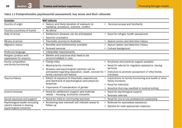
Mental illness makes a large contribution to the disease burden experienced by refugees. It is therefore of utmost importance to adequately screen for sources of psychosocial distress during the consultation. Table 1 is a comprehensive psychological assessment created by the Victorian Foundation for Survivors of Torture Inc [10] and is useful for this purpose.

Table 1: Comprehensive psychosocial assessment: key areas and their rationale. From Promoting Refugee Health: a guide for doctors, nurses and other health care providers caring for people from refugee backgrounds. Victorian Foundation for Survivors of Torture Inc [10]. Reproduced with permission.

v7_i2_f05_f1

Murray et al. [14] encourage a social model of treatment that involves families and the community in the management plan of individuals with psychosocial issues. This method has been found to be highly effective and may play a part in reducing stigma, increasing mental health service utilisation, and adding cultural significance to a patient’s journey through the healthcare system. Communication between community networks and local general practices should therefore be nurtured and maintained. The use of bilingual written material and multimedia have also proved to be helpful and encourage patients to approach their healthcare providers with further questions and concerns [25].

Communication

GPs should also be aware of the free Translating and Interpreting Service (TIS National), as well as the poorer outcomes observed from its omission from consultations [23]. Care should be taken to ensure that the interpreter is politically, culturally, and socially acceptable to the patient and his or her family [6]. The provision of gender-sensitive consultations has produced further improvements in patient outcomes [25]. As with any consultation, doctors should get a complete idea of each individual’s understanding and expectations of their symptoms, illness, and treatment [7]. A thorough history will also determine the barriers to access of treatment for different patients. Referral to refugee support networks can connect patients with less costly or pro bono health and social services, which are particularly helpful for temporary visa holders, who are not eligible for government-funded health programs [25].

Education and training

In 17 different studies, cultural sensitivity training resulted in an increase in patient satisfaction, reporting of symptoms, referrals, health, and access [25]. Bridging the cultural gap can thus prevent valuable missed opportunities for appropriate care and health promotion. However, if unsure about any aspect of the interaction, Bellamy et al. [7] encourage doctors to ask patients how this aspect of the consultation is usually performed in their home countries. This not only displays respect but also allows the patient to feel accepted, and in return may inspire the acceptance of Western practices.

Referral and follow-up

It is always ideal to maintain a relationship of continuing care with one’s patients; however, refugees in their first few years of resettlement will likely relocate several times, making follow-up unfeasible. To facilitate a smoother transition for these patients, GPs should provide them with hardcopies of all their medical records. When making referrals to services such as pathology, radiology, and allied health, GPs are encouraged to call beforehand to ensure that the practices are open to using interpreting services and to prepare them for patients with special needs [10].

Advocacy

Those who have direct contact with refugees have the unique privilege of seeing these groups at their most vulnerable, and often do not face the same time lag that is experienced by research and the media. This gives them the knowledge and credibility to take early action in the support of refugees in their local communities. Health professionals may consider writing to Members of Parliament and Parliamentary Committee Chairs about these issues. The Refugee Council of Australia (RCOA) is the umbrella refugee organisation in Australia and provides some useful information on methods of contact and communication with policy makers [27,28]. Those who choose to voice their concerns directly with the media must maintain patient privacy and informed consent, as many refugees may be put in grave danger if their identities and locations are revealed [27]. Several local organisations also regularly campaign for refugee rights; membership of these committees may open doors for further advocacy and activism.

Table 2 provides a brief summary of recommendations and resources for GPs who find themselves managing a patient from a refugee-like background.

Issue Recommendations Resources
Communication and cultural competence
  • Use of translating services and patient education
  • Atkin N, 2008 [23]
  • Australian Government Translating and Interpreting Service [29]
  • Cultural sensitivity training and further education
  • Gender-sensitive consultations
  • ACEM E-learning courses [30]
  • RACGP E-Learning course 22305 – Refugee Health [31]
  • Victorian Foundation for Survivors of Torture Inc. courses and education [32]
  • Family Planning Victoria, 2012 [12]
Medical problems
  • Initial health assessment
  • Adequate history and examination
  • Australian Family Physician, 2016 [21]
  • Victorian Refugee Health Network [33]
Psychosocial problems
  • Adequate psychosocial assessment
  • The Victorian Foundation for Survivors of Torture Inc., 2012 [10]
Education and health literacy
  • Use of bilingual reading material
  • Patient education and counselling
  • Victorian Refugee Health Network [34]
Follow-up and referral
  • Provide copies of medical records
  • Use of appropriate referral services
  • Call beforehand
  • National Directory of Asylum Seeker and Refugee Service Providers [35]
  • Victorian Refugee Health Network [33]
Advocacy
  • Contact with policy makers and media
  • Activism
  • The Refugee Council of Australia [28]
  • Refugee Action Coalition Sydney [36]

Conclusion

It is evident that providing appropriate and accessible refugee healthcare is a multifaceted task that requires the co-operation of a diverse team of skilled and dedicated professionals. The health of refugees is intrinsically linked to their migration status, education, culture, family situation, and the kaleidoscope of experiences that have landed them in their current situation. General practitioners have a central role in engaging these individuals in health care and the wider community. They must strive to use their position in society to advocate for this underprivileged group and educate resettled refugees on their rights and privileges in their new homes, thus upholding the very pillars of beneficence, autonomy, and justice that encompass the practice of medicine.

Acknowledgements

None.

Conflicts of interest

None declared.

References

[1] Kotschy G. UNHCR Mid-Year Trends 2015. United Nations High Commissioner for Refugees; 2015:3-13.

[2] Gaynor T. 2015 likely to break records for forced displacement – study [Internet]. United Nations High Commissioner for Refugees; 2015. Available from: http://www.unhcr.org/5672c2576.html

[3] World Report 2015: Australia [Internet]. Human Rights Watch; 2015. Available from: https://www.hrw.org/world-report/2015/country-chapters/australia

[4] Wright B. Asylum seekers and Australian politics, 1996-2007 [PhD thesis]. University of Adelaide, School of History and Politics; 2014.

[5] Murray S, Skull S. Hurdles to health: Immigrant and refugee health care in Australia. Aust Health Rev. 2015;29(1):25-9.

[6] Davidson N, Skull S, Burgner D, Kelly P, Raman S, Silove D, et al. An issue of access: Delivering equitable health care for newly arrived refugee children in Australia. J Paediatr Child Health. 2004 Sep 1;40(9‐10):569-75.

[7] Bellamy K, Ostini R, Martini N, Kairuz T. Access to medication and pharmacy services for resettled refugees: A systematic review. Aust J Prim Health. 2015;21(3):273-8.

[8] Bhattarai S. The UNHCR Global Appeal 2014-2015: Providing for Essential Needs. United Nations High Commissioner for Refugees; 2014.

[9] The Victorian refugee and asylum seeker health action plan 2014-2018. Department of Health; 2014.

[10] Promoting refugee health: a guide for doctors, nurses and other health care providers caring for people from refugee backgrounds. 3rd ed. Brunswick: Foundation House – The Victorian Foundation for Survivors of Torture Inc.; 2012. Table 3.3 Comprehensive psychosocial assessment: key areas and their rationale, p.58.

[11] Immigrant Health Service: Overview of health issues in immigrant children [Internet]. The Royal Children’s Hospital Melbourne; 2015. Available from: http://www.rch.org.au/immigranthealth/clinical/Overview_of_health_issues_in_immigrant_children/

[12] Improving the health care of women and girls affected by female genital mutilation/cutting: a service coordination guide. Family Planning Victoria; 2012.

[13] Female genital mutilation/cutting: a statistical overview and exploration of the dynamics of change. UNICEF; 2013.

[14] Murray K, Davidson G, Schweitzer R. Review of refugee mental health interventions following resettlement: Best practices and recommendations. Am J Orthopsychiatr. 2010;80(4):576-85.

[15] Harris M, Zwar N. Refugee health. Aust Fam Physician. 2005 Oct;34(10):825-9.

[16] Murray K, Davidson G, Schweitzer R. Psychological wellbeing of refugees resettling in Australia: A literature review prepared for the Australian Psychological Society. Australian Psychological Society; 2008.

[17] Srinivasa Murthy R. Mass violence and mental health – Recent epidemiological findings. Int Rev Psychiatr. 2007 Jan 1;19(3):183-92.

[18] Silove D, Austin P, Steel Z. No refuge from terror: The impact of detention on the mental health of trauma-affected refugees seeking asylum in Australia. Transcult Psychiatry. 2007 Sep;44(3):359–93.

[19] Australia’s Offshore Humanitarian Programme: 2013-2014. Department of Immigration and Border Protection; 2014.

[20] Guide to social security law: 9.1.2 Visas issued by DIBP [Internet]. Australian Government; [updated 2014 November 10; cited 2016 Jan 4]. Available from: http://guides.dss.gov.au/guide-social-security-law/9/1/2

[21] Benson J, Smith MM. Early health assessment of refugees. Aust Fam Physician. 2007 Jan;36(1-2):41-3.

[22] Policy Brief: Temporary Protection Visas. Refugee Council of Australia; 2013 Sep 24. Available from: http://www.refugeecouncil.org.au/r/pb/THC_140206.pdf

[23] Atkin, N. Getting the message across: Professional interpreters in general practice. Aust Fam Physician. 2008 Mar;37(3):174-6.

[21] Duncan G, Harding C, Gilmour A, Seal A. GP and registrar involvement in refugee health: A needs assessment. Aust Fam Physician. 2013 Jun;42(6):405-8.

[25] Joshi C, Russell G, Cheng IH, Kay M, Pottie K, Alston M, et al. A narrative synthesis of the impact of primary health care delivery models for refugees in resettlement countries on access, quality and coordination. Int J Equity Health. 2013 Nov 7;12(88):1-4.

[26] Cheng I, Wahidi S, Vasi S, Samuel S. Importance of community engagement in primary healthcare: The case of Afghan refugees. Aust J Prim Health. 2015;21(3):262-7.

[27] Cugati A, Lurie C, Piper M. The Advocates’ Help Kit. Refugee Council of Australia; 2002 Dec:10-31. Available from: https://www.refugeecouncil.org.au/docs/resources/VIC_ADVOCACY%20KIT.pdf

[28] Contacting Politicians: Letter Guide [Internet]. Refugee Council of Australia. Available from: http://www.refugeecouncil.org.au/take-action/contacting-politicians/

[29] The Translating and Interpreting Service Website [Internet]. Australian Government Department of Immigration and Border Protection. Available from: https://www.tisnational.gov.au/

[30] Useful Emergency Medicine Resources [Internet]. Australasian College for Emergency Medicine. Available from: http://elearning.acem.org.au/course/index.php?categoryid=29

[31] 22305 – Refugee Health: GP Learning [Internet]. Royal Australian College of General Practitioners. Available from: http://www.racgp.org.au/education/courses/activitylist/activity/?id=17726&q=keywords%3drefugee%2bhealth

[32] Courses and Education [Internet]. The Victorian Foundation for Survivors of Torture Inc. Available from: http://www.foundationhouse.org.au/webinars/

[33] Caring for refugee patients in general practice: a desktop guide. 4th ed. Victorian Refugee Health Network; 2012 Mar.

[34] Translated resources: Translated health information [Internet]. Victorian Refugee Health Network. Available from: http://refugeehealthnetwork.org.au/translated-resources/

[35] National Directory of Asylum Seeker and Refugee Service Providers. Asylum Seeker Resource Centre; 2013 Aug. Available from: http://www.asrc.org.au/wp-content/uploads/2013/07/National-Directory-of-Asylum-Seeker-and-Refugee-Service-Providers-August-2013.pdf

[36] Refugee Action Coalition Website [Internet]. Available from: http://www.refugeeaction.org.au/


Categories
Feature Articles

Music as analgesia in the perioperative setting

Pain is a complex and predictable component of the perioperative experience. Music, as a non-pharmacological pain management modality, is cheap and easy to implement as an analgesic adjunct to reduce patient pain, stress, and anxiety. It has particular benefit in healthcare settings where first-line pharmaceutical pain management options are less available. This review finds an increasing body of evidence supports the use of music to reduce pain in the perioperative setting.  Certain musical elements such as a constant tempo, gentle timbre, and smooth melody combined with patient musical preference have been shown to have the greatest analgesic effect. The mechanism by which music alleviates pain is not clearly defined, but may involve distraction, or regulation of the autonomic nervous system. The perioperative utilisation of music in the perioperative remains low. Standardised guidelines are required to instruct and assist with its application. Music therapy has demonstrated benefit with minimal side effects and should be used liberally in the management of perioperative pain.

Clinical scenario

Late last year, I exited a jumbo jet and took my first steps in the Kingdom of Tonga. This marked the start of a three-week adventure that I now count as one of the best experiences of my medical degree. As part of a four-member medical volunteer delegation, I travelled to the picturesque island of Vava’u, in Tonga’s far north. There, my time was divided between undertaking free community health checks and working with the doctors and staff of the Prince Wellington Ngu Hospital. The hospital was an old sprawling collection of buildings on top of a hillside overlooking the local village. The hospital was operating at capacity and despite the resourcefulness and efficiency of the staff, the hospital suffered from a marked lack of resources. It was under these circumstances, in a sparse medical ward that I first met 63- year-old, Mrs Place (pseudonym to protect patient confidentiality).

Mrs Place had presented to the hospital three weeks prior with a large, infected foot ulcer. The ulcer had first developed after she had innocuously cut her left foot four months earlier. The severity of the wound was compounded by her poorly controlled diabetes and peripheral vascular disease. After the failure of appropriate conservative and medical management, a below-knee amputation of her left leg was conducted.  When I saw Mrs Place two days after her operation, she was grimacing and moaning in pain. She reported her pain score to be 9/10 despite receiving a routine regimen of paracetamol and codeine. Her treating doctor explained that stronger opioid medication would help Mrs Place but was unavailable due to resource limitations. Instead, a novel form of pain management was used in these situations. To my amazement the nurses, doctors, and other patients began to sing local church hymns, in a contained, harmonious, and pleasant manner. No one seemed surprised and everyone was familiar with the songs. The singing continued over the course of the morning, and while doctors and nurses moved on with their daily activities and the hospital continued to operate, a constant melody echoed out of Mrs Place’s ward. Sometimes the music was loud, propagated by family members, and other times it was soft, with a lone voice maintaining the euphony.  Three hours later, Mrs Place was visibly more comfortable. She described her pain score to be 6/10 and credited the lessening of her suffering to the singing for which she was very grateful.  While it is unclear to what extent the patient’s pain improved directly as a result of the music, the apparent effectiveness of music as a pain management modality created an information gap in my medical knowledge that I was determined to explore. A literature review was performed using the electronic databases, Pubmed, Embase, and Medline. The search terms used were “music therapy”, “pain”, “postoperative”, “pain measurement” from 2000 until present.

Discussion

Pain has been described as an unpleasant sensory and emotional experience associated with actual or potential tissue damage [1]. It is a complex problem that is compounded by its subjective nature and the wide spectrum of pain tolerance that exists within the community. The majority of people, at some stage of their life, will undergo a surgical procedure and although pain is a predictable component of the postoperative experience, pain management is often insufficient. The resultant clinical and psychological harm may affect patient morbidity [2]. Negative clinical outcomes associated with inadequate postoperative pain management include deep vein thrombosis, pulmonary embolism, coronary ischaemia, myocardial infarction, pneumonia, poor wound healing, and insomnia [3]. Effective pain management contributes to avoiding the human and economic costs of these sequelae. Pharmacological agents form the current standard of care for the management of postoperative pain [4]. These medications are best applied according to the World Health Organization’s “pain ladder” that advocates for the controlled escalation of pain relief medication [5]. Music, as a non-pharmacological pain management modality, is not widely utilised in the current healthcare environment, whether this relates to a paucity of information within the medical fraternity or an inherent scepticism towards complimentary analgesic methods is uncertain [6]. However, the paradigm is now changing due to a landmark systematic review and meta-analysis conducted by Hole et al. After synthesising the results of 73 randomised control trials, they concluded that music played in the perioperative setting can reduce postoperative pain, anxiety, analgesia requirements, and increase patient satisfaction [7]. This pivotal study adds to an existing body of research that confirms music to be a beneficial compliment to pharmacotherapy in managing perioperative pain [4,8,9].

The first use of music as a therapeutic intervention was recorded in the cuneiform writings of the early Mesopotamia, circa 4000 BC [10]. Since then the use of music in the clinical setting has been commonly employed throughout history. Notably, Florence Nightingale was a strong advocate for its use during the Crimean War, specifically citing the effectiveness of singing and flute melodies to alleviate pain [11]. Today, music may be used in the healthcare setting to reduce emotional distress and the patient’s perception of pain [12]. Given the wide spectrum of music genres and the singular nature of personal preference, determining the most effective music for alleviating pain and anxiety requires careful consideration. It is suggested that there are definitive musical elements that promote analgesia; these include a consistent tempo, gentle timbre, smooth melody, and a limited percussion component [13,14]. In addition Mitchel et al. concluded that music self-selected by the individual provides greater pain control [15]. This improved analgesic experience was postulated to stem from a greater emotional valence with music. For the greatest effect, music used to relieve pain in the perioperative setting should include the known and accepted musical components combined with patient preference [16].

The mechanism by which music alleviates pain is not clearly defined. Modern theories of pain are based on the gate control theory [17] and the subsequent neuromatrix theory [18]. Music is thought to be able to act as a distraction and dampen the patient’s experience of pain. Physiologically, pain impulses travel from the site of injury to the brain via the spinal cord.  Within the spinal cord there are neural gates that can be opened or closed to different degrees that allows for the regulation of pain impulses communicated to the brain. When these gates become obstructed, the patient’s perception of pain may be reduced. The use of music therapy may block these pain gates by causing descending signals to be sent from the brain down the efferent pathways in the spinal cord [19,20]. This means that fewer ascending pain impulses will reach the patient’s awareness. Using magnetic resonance imaging, Valet et al. showed that distraction caused an increase in the activation of the cingulofrontal cortex, the periaqueductal gray, and the posterior thalamus. Through this neural network, distraction may exert a top-down influence in order to reduce the unpleasantness or intensity of a person’s pain [21].

Music may also help reduce pain by acting on the autonomic nervous system activity within the body. Emotional distress adversely affects how an individual experiences pain; anxiety in particular has been identified as a risk factor for developing the chronic sequela of pain [22,23]. Anxiety occurs commonly in patients who are undergoing medical procedures and may decrease a patient’s pain threshold and tolerance causing them to experience more pain [24]. When a person is anxious, their pituitary-adrenal axis and sympathetic nervous system are stimulated, resulting in the release of hormones that are responsible for the patient’s experience of anxiety [25]. The physical expression of anxiety includes increases in heart rate, respiratory rate, and blood pressure [26]. Ozer et al. conducted a case control study that was designed to investigate the impact of music therapy on a number of postoperative physiologic parameters and the patient’s perception of pain. They found that patients who received music therapy had a statistically significant increase in oxygen saturation and a decrease in their reported pain score [4].  In this study, the patient’s oxygen saturation was used as a surrogate marker for their relaxation level and the effectiveness of music therapy as an anxiolytic. Other physiological parameters such as blood pressure, heart rate, and respiratory rate were not shown to have any significant association with music therapy, which corroborates the findings of other studies [27-29].

The role for music in the operating room is less certain. A small percentage of patients undergoing general anaesthetic retain unwanted intraoperative awareness throughout their procedure, which increases their likelihood developing post-traumatic stress disorder [30]. The use of music intraoperatively may benefit these patients by providing distraction. What effect music has on the medical professionals treating the patient is unclear. Allen et al. found that surgeon selected music reduced autonomic reactivity and improved surgical performance, however all of the study participants were biased towards the beneficial effect of music [31].  A more recent literature review suggests that music in the operating theatre is harmful, interrupting communication, hindering work, and risking patient safety [32]. Individual surgical teams can therefore guide the use of intraoperative music, with the option of personal electronic music players on a patient-by-patient basis.

The effectiveness of music therapy in reducing a patient’s pain has been well demonstrated. However, due to the wide range and varying complexity of pain assessment tools that have been employed, the level of analgesic impact is difficult to quantify. A rudimentary assessment has shown that music therapy may improve a patient’s pain by 10% to 40% [9,12,27,33-35]. However, despite the apparent benefit, clinical interest in the use of music remains in its infancy. As such, there are no guidelines available to instruct and assist with its application. This lack of standardisation may damage the future endorsement of this modality due to the potential for uneven application, poor compliance, and unrealistic expectations regarding outcomes. While there are no evident physical side effects from music therapy, exposure to unappealing music may irritate patients, impacting on their mental health and overall wellbeing. Legal issues involving copyright and intellectual property need to also be considered. Further research is required to clearly demarcate the biological pathways that mediate the beneficial effects of music. Without a proper understanding of the physiological basis for the observed positive effect, the most appropriate application cannot be determined and completely explored. For example, does the proposed benefit extend to all forms of audio-visual stimulus or is there something unique about music?  Active distraction through the use of electronic gaming has been shown to increase a patient’s pain tolerance as well as the amount of pain that they reported. Interestingly, this form of active distraction was shown to be superior to passive distraction (watching television), suggesting that other passive distractions, such as listening to music, might be less effective [36]. Future research should evaluate the effects of tailored music that is designed for this specific setting or for individual patients or patient groups. For example, a three-pronged comparative study could be conducted in Tonga to explore the effectiveness of spiritual music given its strong cultural background, in addition to the broader questions regarding efficacy, protocol, and utility.  Likewise, the differences in the effect of music interventions related to patients’ gender, age, and ethnicity should be further evaluated.

Conclusion

Music therapy should not be seen as a primary treatment option for the management of postoperative pain. In most clinical situations it will not completely alleviate the patient’s pain and should instead be used to facilitate patient functionality, improve quality of life, and reduce consumption of pharmacological analgesics. With no known side effects, wide applicability and the ability to be utilised in conjunction with other pain management therapies, the potential for music therapy is significant. In settings where first-line pain management options are not available, the benefits of this non-pharmacological management are magnified. Music therapy has demonstrated benefit and should be used liberally in the management of postoperative pain.

Acknowledgements

None.

Conflicts of interest

None declared.

References

[1] IASP. Part III: Pain Terms, A current list with definitions and notes on usage (pp 209-214). Secondary Part III: Pain Terms, A current list with definitions and notes on usage (pp 209-214) 1994. Available from: http://www.iasp-pain.org.

[2] Tse M, Chan MF, Benzie I. The effect of music therapy on postoperative pain, heart rate, systolic blood pressure and analgesic use following nasal surgery. J Pain Palliat Care Pharmacother. 2005;19(3):21-9.

[3] Apfelbaum JL, Chen C, Mehta SS, Gan aTJ. Postoperative pain experience: results from a national survey suggest postoperative pain continues to be undermanaged. Anesth. Analg. 2003;97(2):534-40.

[4] Ozer N, Karaman Ozlu Z, Arslan S, Gunes N. Effect of music on postoperative pain and physiologic parameters of patients after open heart surgery. Pain Manag Nurs. 2013;14(1):20-8.

[5] World Health Organization. WHO’s Pain Relief Ladder. 2009. Available from: http://www.who.int/cancer/palliative/painladder/en/.

[6] Hupkens S. Implementation of complementary interventions in the Netherlands: experiences of pioneers. Patient Educ Couns. 2012;89(3):411-6.

[7] Hole J, Hirsch M, Ball E, Meads C. Music as an aid for postoperative recovery in adults: a systematic review and meta-analysis. The Lancet. 2015;386(10004):1659-71.

[8] Roy M, Peretz I, Rainville P. Emotional valence contributes to music-induced analgesia. Pain. 2008;134(1-2):140-7.

[9] Bernatzky G, Presch M, Anderson M, Panksepp J. Emotional foundations of music as a non-pharmacological pain management tool in modern medicine. Neurosci Biobehav Rev. 2011;35(9):1989-99.

[10] Conrad C, Niess H, Jauch K-W, Bruns CJ, Hartl WH, Welker L. Overture for growth hormone: Requiem for interleukin-6?*. Crit. Care Med. 2007;35(12):2709-13.

[11] Nightingale F. Notes on nursing: what it is, and what it is not. London: Harrison; 1859.

[12] Zengin S, Kabul S, Al B, Sarcan E, Dogan M, Yildirim C. Effects of music therapy on pain and anxiety in patients undergoing port catheter placement procedure. Complement Ther Med. 2013;21(6):689-96.

[13] Gooding L, Swezey S, Zwischenberger JB. Using music interventions in perioperative care. South Med J. 2012;105(9):486-90.

[14] Nilsson U. The anxiety- and pain-reducing effects of music interventions: a systematic review. AORN J. 2008;87(4):780-807.

[15] Mitchell LA, MacDonald RA, Brodie EE. A comparison of the effects of preferred music, arithmetic and humour on cold pressor pain. Eur J Pain. 2006;10(4):343-51.

[16] Zhang ZS, Wang XL, Xu CL, Zhang C, Cao Z, Xu WD, et al. Music reduces panic: an initial study of listening to preferred music improves male patient discomfort and anxiety during flexible cystoscopy. J Endourol. 2014;28(6):739-44.

[17] Melzack R, Wall PD. Pain mechanisms: a new theory. Science. 1965;150(3699):971-9.

[18] Melzack R. From the gate to the neuromatrix. Pain. 1999;Suppl 6:S121-6.

[19] Roy M, Lebuis A, Hugueville L, Peretz I, Rainville P. Spinal modulation of nociception by music. Eur J Pain. 2012;16(6):870-7.

[20] Li XM, Yan H, Zhou KN, Dang SN, Wang DL, Zhang YP. Effects of music therapy on pain among female breast cancer patients after radical mastectomy: results from a randomized controlled trial. Breast Cancer Res Treat. 2011;128(2):411-9.

[21] Valet M, Sprenger T, Boecker H, Willoch F, Rummeny E, Conrad B, et al. Distraction modulates connectivity of the cingulo-frontal cortex and the midbrain during pain–an fMRI analysis. Pain. 2004;109(3):399-408.

[22] Raichle KA, Osborne TL, Jensen MP, Ehde DM, Smith DG, Robinson LR. Preoperative state anxiety, acute postoperative pain, and analgesic use in persons undergoing lower limb amputation. Clin J Pain. 2015;31(8):699-706.

[23] Theunissen M, Peters ML, Bruce J, Gramke HF, Marcus MA. Preoperative anxiety and catastrophizing: a systematic review and meta-analysis of the association with chronic postsurgical pain. Clin J Pain. 2012;28(9):819-41.

[24] Tan X, Yowler CJ, Super DM, Fratianne RB. The efficacy of music therapy protocols for decreasing pain, anxiety, and muscle tension levels during burn dressing changes: a prospective randomized crossover trial. J Burn Care Res. 2010;31(4):590-7.

[25] Pittman S, Kridli S. Music intervention and preoperative anxiety: an integrative review. Int Nurs Rev. 2011;58(2):157-63.

[26] Nilsson U, Unosson M, Rawal N. Stress reduction and analgesia in patients exposed to calming music postoperatively: a randomized controlled trial. Eur J Anaesthesiol. 2005;22(2):96-102.

[27] Nilsson U. Soothing music can increase oxytocin levels during bed rest after open-heart surgery: a randomised control trial. J Clin Nurs. 2009;18(15):2153-61.

[28] Kshettry VR, Carole LF, Henly SJ, Sendelbach S, Kummer B. Complementary alternative medical therapies for heart surgery patients: feasibility, safety, and impact. Ann Thorac Surg. 2006;81(1):201-5.

[29] Sendelbach SE, Halm MA, Doran KA, Miller EH, Gaillard P. Effects of music therapy on physiological and psychological outcomes for patients undergoing cardiac surgery. J Cardiovasc Nurs. 2006;21(3):194-200.

[30] Leslie K, Chan MT, Myles PS, Forbes A, McCulloch TJ. Posttraumatic stress disorder in aware patients from the B-aware trial. Anesth Analg. 2010;110(3):823-8.

[31] Allen K, Blascovich J. Effects of music on cardiovascular reactivity among surgeons. JAMA. 1994;272(11):882-4.

[32] Shambo L, Umadhay T, Pedoto A. Music in the operating room: is it a safety hazard? AANA J. 2015;83(1):43-8.

[33] Simcock XC, Yoon RS, Chalmers P, Geller JA, Kiernan HA, Macaulay W. Intraoperative music reduces perceived pain after total knee arthroplasty: a blinded, prospective, randomized, placebo-controlled clinical trial. J Knee Surg. 2008;21(4):275-8.

[34] Pothoulaki M, Macdonald RA, Flowers P, Stamataki E, Filiopoulos V, Stamatiadis D, et al. An investigation of the effects of music on anxiety and pain perception in patients undergoing haemodialysis treatment. J Health Psychol. 2008;13(7):912-20.

[35] Salimpoor VN, Benovoy M, Larcher K, Dagher A, Zatorre RJ. Anatomically distinct dopamine release during anticipation and experience of peak emotion to music. Nat Neurosci. 2011;14(2):257-62.

[36] Jameson E, Trevena J, Swain N. Electronic gaming as pain distraction. Pain Res Manag. 2011;16(1):27-32.

Categories
Feature Articles

Medicine you can depend on: opioids and benzodiazepines

Opioids and benzodiazepines are frequently prescribed medications that carry a high risk of dependency and are commonly misused. Long-term use of these agents is associated with significant harm to the individual such as exacerbating the patient’s original symptoms, increasing mortality, and negatively impacting the community by contributing to road trauma. This paper will begin by outlining the regulation of drugs of dependence in Australia and defining misuse, dependency, and associated harms. The evidence for use of these drug classes will then be presented, with reference to best practice guidelines. Finally, the discrepancy between guidelines and prescribing behaviours will be highlighted with reference to specific challenges associated with drugs of dependency. It will be emphasised that doctors, especially the newer generations, have a responsibility to own the problems of drug misuse, and to impact individuals and the community through advocacy and evidence-based clinical governance. It will be shown that whilst opioids and benzodiazepines are effective short-term medicines, there is little support for long-term prescribing. If we are to turn the tide on drug dependency and misuse, change is needed in modern prescribing behaviours to achieve the best long-term outcomes for patients.

The problem

The most commonly misused prescription drugs in Australia are opioids and benzodiazepines, including z-drugs (zolpidem, zopiclone, and zalephon), which are non-benzodiazepines that act on the same receptors [1-3]. Access to these drugs is restricted by the Australian Therapeutic Goods Association Drug Schedule, which determines availability for public consumption [4]. Drugs of dependency include all Schedule 8 drugs (controlled drugs, such as opioids) and some Schedule 4 drugs (prescription only medicines, such as benzodiazepines) [5]. Such restrictions are regularly reviewed and subject to change. For example, alprazolam, a potent benzodiazepine with high toxicity [6], was made Schedule 8 in early 2014 due to a rise in illicit use [7], and there is current debate about the appropriateness of Schedule 4 for codeine, because of increasing codeine-related deaths [8,9].

Drug misuse is an important umbrella term to define in relation to the problems associated with drugs of dependency. Five main categories, that are not mutually exclusive, emerge in the literature:

  1. Overuse: Higher dose or dose frequency than prescribed; often due to tolerance and self-adjusted dosing over long-term use [3].
  2. Abuse: Overuse with the goal of intoxication [3].
  3. Prolonged use: An emerging problem involving continued therapy to achieve a sense of normality. Such individuals have been termed the “hidden population” [2-3]. Similarly, the term “accidental addicts” has emerged in popular media, referring to individuals who become dependent on pharmaceuticals with continued use. Prescribers must be hypervigilant for these patients as they are often of high socioeconomic status and highly functional, in contrast to traditional drug-seeking stereotypes [10].
  4. Substance use disorder: This is a formal diagnosis in the Diagnostic and Statistical Manual of Mental Disorders, fifth edition (DSM-5) based on eleven symptomatology criteria across four categories (impaired control, social impairment, risky use and pharmacology) [11]. For diagnosis, use must cause clinically significant distress or impairment and occur within a twelve-month period [11].
  5. Dependency: A syndrome in which an individual has: (i) a strong drive to use a substance; (ii) difficulty regulating their use; (iii) tolerance; and (iv) withdrawal symptoms on cessation [12].

Opioids and benzodiazepines are included on the World Health Organization list of essential medicines [13], indicated primarily in pain or palliative care (opioids), and anxiety disorders, seizures, acute insomnia, and alcohol withdrawal (benzodiazepines) [3].  Availability of opioids and benzodiazepines has been steadily increasing in recent decades. The number of opioid-dispensing episodes increased from 500,000 to 7.5 million from 1992 to 2012 [14]. Harrison and colleagues evaluated 4666 encounters with general practitioners (GPs) between 2010 and 2011 in which opioids were prescribed or supplied [15]. They found that 3.5% of opioids were prescribed for cancer pain, 43.9% were prescribed for chronic non-cancer pain, with the remainder being prescribed for indications classified as non-chronic [15]. For benzodiazepines, approximately 7 million scripts are written annually [16]. The 2013 National Drug Strategy Household Survey found that 3.3% of people aged 14 or over had used analgesics and 1.6% had used tranquillisers or sleeping pills over the past year for non-medical purposes, with both increasing over each three-yearly iteration of the survey since 2001 [17]. With problematic usage and availability of these drugs in the community (through either personal prescription or diversion), the harm associated with misuse has become increasingly apparent. There is a great degree of harm caused by opioid and benzodiazepine misuse to the individual and community (Table 1).

Table 1: Harms to the individual and community associated with opioid and benzodiazepine misuse

Harms Opioids Benzodiazepines
 

Individual

High risk of dependence [18] and misuse [2] Extremely habit forming: tolerance and dependence can occur in 2-4 weeks [31-33]
Physiological effects

Constipation [19]

Biliary dyskinesia [19]

Cognitive impairment [19]

Increased risk of death [20]/overdose (including inadvertent childhood ingestion) [2]

Sleep apnoea [21]

Sexual and endocrine dysfunction [22]

Immunosuppression [23]

Opioid-induced hyperalgesia [24]

Osteoporosis/increased fracture risk [2]

Dental caries and necrosis secondary to xerostomia [2]

Impaired cognition [2]

Physiological effects

Insomnia [7]

Anxiety [7]

Depression [34]

Poor memory and concentration [35]

Excessive / daytime sedation [36]

Risk of ataxia and falls, disinhibition and amnestic effects [36]

Cognitive impairment [37]/decline and dementia [38]

Increased mortality with long term use [39]


Codeine: gastric bleeding [25], severe metabolic imbalances [26-28], and risk of death [8] Z-drugs: potentially dangerous complex sleep behaviours [16]


Harms with intravenous abuse:  blood clots, amputation, and serious soft tissue injuries [29,30] Harms with intravenous abuse:  blood clots, amputation, and serious soft tissue injuries [29,30]
Community Diversion [40]

Low levels of organised crime [41]

Driving impairment and motor vehicle accidents [42,43]

Doctor shopping affecting GPs [3]


Diversion [7]

Low levels of organised crime [41]

Driving impairment and motor vehicle accidents [42,43]

Doctor shopping impacting GPs [3]

Criminal activity through precipitating aggression and disinhibition [41]

Opioids and benzodiazepines are highly habit-forming and potentially destructive to the individual and society. Physiological side effects of long-term use of both drug classes significantly reduce quality of life and contribute to personal suffering through symptoms such as constipation and balance derangement contributing to falls. Further, opioids and benzodiazepines are both associated with increased mortality. This is through means such as deliberate overdose, accidental overdose, and motor vehicle accidents secondary to impaired driving ability [2,42,43]. In Victoria, overdose deaths involving prescription drugs outnumber deaths from road trauma [44], and alcohol and drug helplines receive nearly threefold more calls regarding prescription opioids than heroin [45]. Importantly, an Australian study investigating 320 oxycodone related deaths reported that 52% were due to unintentional overdose, 20% were deliberate self-harm, and the remainder either awaiting coronial enquiry or with indeterminable intent [46]. Victorian coronial data indicate that benzodiazepines are at least contributory in 48.8% of all drug-related deaths [47]. As such, long-term opioid and benzodiazepine use is associated with a very real increase in mortality, not only in people with the intent to self-harm. Overall, opioids and benzodiazepines carry a high risk of dependence, cause a wide range of physiological harms including increased mortality, and they negatively affect the community through diversion and contributing to motor vehicle accidents.

The evidence

Current evidence supports short-term use of both opioids and benzodiazepines for appropriate indications (for example, less than 90 days and one month, respectively). These drugs are effective when used properly and should be available for people who need them. Long-term use, however, is not supported by evidence for the indications for short-term use. Both classes of medications are known to be highly addictive and associated with poor long-term outcomes if use is not time limited. Notably, recent studies suggest that psychobehavioural interventions are at least equivalent to, or more effective than, pharmacotherapy for pain, anxiety, and insomnia [48-50].

Opioids have convincing evidence supporting their use in acute pain [51], cancer pain [52], palliative care, [53] and addiction [54]. Most of the controversy in opioid prescribing relates to use in chronic non-cancer pain, with reviews suggesting little short-term efficacy [55], and tolerance and opioid-induced hyperalgesia occurring with prolonged use [56]. Furthermore, a Cochrane Database review showed no clear gains in pain and functional outcomes when opioids were used long-term [57], and another study found no significant correlations between opioid dose adjustment and clinical pain scores over time [58]. In line with the biopsychosocial conceptualisation of chronic non-cancer pain, multimodal approaches using non-pharmacological strategies such as exercise and psychological interventions have been shown to be superior to monomodal regimens in terms of long-term impact [59]. Cognitive behavioural therapy (CBT) is recommended as first line psychosocial treatment for chronic, non-cancer pain [60]. A Cochrane review in 2012 found that CBT conferred significant improvement in pain, disability, mood, and catastrophising in comparison to wait-list controls [61]. Effect sizes were small to moderate and the only sustained impacts were seen on mood.

Short acting benzodiazepines and z-drugs are effective at managing acute insomnia [62], generalised anxiety disorder, social anxiety, and panic disorder [63]. Dependency can be rapid (for example, within 2 to 4 weeks) [64] and increased mortality has been associated with long-term use [39]. Unfortunately, randomised controlled trials for long-term use are of inadequate duration (approximately 3 months), and lack representative clinical samples [16]. Reassuringly, however, it has been shown that the majority of patients on benzodiazepine therapy for sleep and anxiety do not increase their doses over time [65]. Moreover, a recent three year follow-up study of users of benzodiazepines and z-drugs found less than 1% of patients developed problems with excessive use, which predominated in patients with a significant drug and alcohol history and increased with duration of therapy [66]. This suggests that careful evaluation of patients to identify those at risk of excessive use is important, and that monitoring and cessation plans at first prescription may be protective for this population. Of note, this paper did not assess experience of adverse effects or harms associated with long-term use of benzodiazepines. Instead, it focussed on excessive use only, defined as more than two daily doses over a three month period.

Insomnia treatment is generally recommended to proceed in a stepwise fashion, with initial management emphasising basic sleep hygiene education and stimulus control, with the latter supported by randomised controlled trial evidence for long-term efficacy [67]. If insomnia persists, more involved behavioural interventions, either with or without medications, should be offered. CBT combines sleep hygiene education, stimulus control, and progressive muscle relaxation into a cognitive therapy framework to address insomnia. A recent meta-analysis of randomised controlled trials has shown lasting benefits for establishing sleep, wake times, and sleep efficiency [68]. However, total sleep time was not significantly increased and the patient samples excluded those with medical and psychological comorbidities. As such, the magnitude of the effects needs to be interpreted with caution. CBT is recommended as first line treatment for insomnia by the Royal Australian College of General Practitioners (RACGP) and the British Association for Pharmacology [69] on the basis of previous systematic reviews, which have included randomised controlled trials and patients with medical and psychiatric comorbidities and shown reliable and sustained improvements on sleep parameters [70]. Similarly, in the treatment of anxiety disorders, CBT is supported by meta-analysis of randomised controlled trials [71] and is recommended as first line therapy, either as monotherapy or in combination with antidepressant medication [72].

Current guidelines are in line with the evidence base for long-term prescribing of opioids and benzodiazepines in Australia. The RACGP published two comprehensive documents in 2015 about prescribing drugs of dependency (with particular reference to benzodiazepines) that summarise best evidence, provide concrete guidance and examples of difficult consultations, and ways in which they can be safely and effectively negotiated [5,16]. For opioid prescribing in chronic, non-cancer pain, the Hunter Integrated Pain Service provides additional evidence-based guidelines [19]. An overview of the key prescribing guidelines can be extracted from these documents (Table 2).

Table 2: Summary of guidance for opioid and benzodiazepine prescribing from Australian evidence-based guidelines

Guideline Authors Summary of recommendations
 

Prescribing drugs of dependence in general practice, part A – clinical governance framework* [5]


RACGP

  • Maintain and develop skills in chronic pain, mental health, drugs of dependency (DOD) and optimise non-pharmacological interventions
  • Inform patients DOD should be prescribed from one practice, one GP and dispensed from one pharmacy
  • In complex cases or if situations deteriorate, be prepared to utilise specialist support
  • Prescribing for drug dependent patients must be based on comprehensive assessment and an authority must be sought when prescribing a Schedule 8 drug
  • Patients have the right to respectful care that promotes their dignity, privacy, and safety
  • Provide patients with information about the purpose, realistic expectations, options, and benefits and risks
  • Develop respectful, non-judgemental, and clear responses to inappropriate requests for DOD
Prescribing drugs of dependence, part B – benzodiazepines[16] RACGP
  • Prescription of benzodiazepines (BZDs) should be with the lowest dose and shortest possible timeframe, continual monitoring, and careful consideration of risks and benefits
  • Discuss with patients the potential for dependence, withdrawal, misuse, and known harmful effects such as falls, cognitive decline, and motor vehicle accidents
  • Avoid prescribing to patients with comorbid alcohol or substance use disorders (these patients are more vulnerable to major harms)
  • Use beyond four weeks should be uncommon, if no alternatives are available, long-term use must be supervised with regular attempts at reduction
  • For insomnia, first line should be non-pharmacological (e.g. CBT) and BZDs/Z-drugs only given on an individual basis, with short-term, intermittent dosing
  • For anxiety disorders, first line therapy should include CBT; SSRI/SNRI are suitable first line pharmacological treatments
  • BZD use in anxiety disorders is mostly limited to severe or treatment resistant cases
  • Short term BZD use as occasional adjunctive therapy may be effective at reducing symptoms in the first few days to weeks of SSRI/SNRI therapy
  • BZD discontinuation can be achieved with minimal interventions (such as GP advice/advisory letters), strong therapeutic alliance and psychological therapies
Reconsidering opioid therapy: a Hunter New England perspective [19] Hunter Integrated Pain Service
  • Opioid therapy is indicated for acute pain; cancer pain, palliative care; opioid dependency/addiction – NOT chronic, non-cancer pain
  • Multidimensional pain assessment is recommended for all types of pain
  • A drug and alcohol history, use of the risk assessment tools to gauge the risk of opioid misuse and contact with the Australian Prescription Shopping Information service is recommended

Opioid therapy for acute pain:

  • Should be time limited and coordinated between hospital and primary care
  • Can usually be ceased within one week of surgery or injury if possible, and weaned and ceased within 90 days in complex cases
  • A daily oral morphine equivalent dose of 100 mg should not be exceeded in primary care
  • A treatment agreement explaining potential benefits, adverse effects, and duration of therapy should be used, with pain and functional outcomes measured
  • Review therapy regularly with the “four A’s”: Analgesia, Activity, Adverse effects, and Aberrant behaviour
  • Should be prescribed with ongoing non-pharmacological supportive care and a focus on evidence-based self-management strategies
  • Involvement of a pain medicine specialist and multidisciplinary pain management team may be helpful
  • Opioid naïve patients prescribed opioids and patients on long term opioids and BZDs should not drive

*These guidelines also provide recommendations for clinical governance and general practice systems of care not summarised here.

The need for evidence-based prescribing behaviours

Evidence does not support long-term therapy with opioids and benzodiazepines. This has been known for some years, yet the problem has only grown. Future generations of practitioners need to engender practice in accordance with guidelines, enhance utilisation of alternative, non-pharmacological interventions, and strive for better patient outcomes.

Consultations that involve the consideration of drugs of dependence present significant challenges to the GP, and usually involve complex and vulnerable patients. GPs report complicating factors such as time pressure, concerns about patient autonomy, patient-centred care and preserving the doctor-patient relationship [73-75]. Studies have shown that GPs have some ambivalence towards prescribing benzodiazepines and may lack consistency in their approach [73]. There is also referral to the notion of the “deserving patient” in the literature, whereby doctors ask themselves whether the patient deserves a prescription rather than whether they should write one [76]. Indeed, it has been reported that some GPs will provide benzodiazepines even when they do not believe it will help, giving reasons such as: having no realistic alternatives; assumptions about the patients’ expectations; problems in saying no; and succumbing to pressure [16]. Importantly, research indicates that many doctors assume that patients want prescriptions or would resist attempts at withdrawal whilst, in fact, patients report wanting explanations and discussion [73,77].

Inevitably, there is often a discrepancy between clinical guidelines and practice. There is some disagreement in the literature regarding the usefulness and clinical impact of best practice guidelines, with some findings demonstrating improved and more consistent management [78,79], whilst others show minimal impacts [80]. Recent studies are suggesting that GPs perceive clinical practice guidelines with a positive attitude [81], but that there are inconsistencies regarding which components are followed [82]. Research into the appropriate prescribing of antibiotics suggests that inappropriate prescribing is more likely to occur in high-volume practices and by those who have been in practice longer [83]. As such, to improve concordance with guidelines, it will be important for junior doctors to emerge into the workplace as proponents of evidence-based prescribing. They should take advantage of their training period, where case loads are lower, to ingrain safe prescribing habits and good clinical governance into their practice. Obviously, there is more to appropriate prescribing than simply following guidelines. There is an art to handling challenging consultations, conveying evidence in a meaningful way, inspiring trust in your patient, and establishing a collaborative management plan, which may or may not meet the patient’s original expectations. In light of this, there are some additional consultation techniques that may facilitate safe prescribing for junior doctors (Table 3).

Table 3: Practical advice for managing patients and drugs of dependency

Areas of Development Strategies
 

Professional Knowledge

Engage in continuing medical education for drug dependence and safe prescribing

Develop skills in cognitive behavioural strategies and structured problem solving [84,85]

Familiarise with typical patient requests and responses from examples [5,16]


Consultation Skills


Time spent in early consultations will save time long-term


Encourage non-pharmacological measures

Assess risk/benefits of any drug prescribed

Make collaborative decision making for prescribing and non-prescribing [5]

Prescribe only for supported indications

Prescribe with plan for reduction [5]

Be honest and explore issues [5]

Involve the patient’s supports [5]

Monitor use and look for signs of dependency (review ‘five As’) [5]

Identify problem use and refer to specialist services [5]

Provide written management plan and fact sheets [5]

Seek advice if unsure

Resources Withdrawal scales for alcohol [86], opioids [87], and benzodiazepines [88]

Therapeutic Guidelines for withdrawal management

Opioid risk tool [89]

Specialist drug and alcohol services

NPS MedicineWise Key Points for managing chronic pain [90]

The patient’s pharmacist [91]

Local support groups

Medicare Australia prescription shopping information service (ph. 1800 631 181) [2]

Junior doctors and emerging medical professionals have the power to be agents of change in the evidence-based management of conditions such as chronic, non-cancer pain; anxiety; and insomnia. Doctors in training are often the most up-to-date on evidence-based guidelines in these areas, with greater emphasis on non-pharmacological management and multidisciplinary collaboration in undergraduate and postgraduate programs than in generations past. In addition, medical school curriculums have been modified to promote highly developed communication skills in graduates that are crucial to the ability to inspire motivational change in patients. As highlighted in Table 3, doctors can up-skill by developing their professional knowledge and consulting skills, and engaging with the wealth of resources available to them. Continuing medical education is paramount and skills in cognitive behavioural strategies such as goal setting and structured problem solving, mindfulness strategies, and motivational interviewing are vital to being an effective clinician. In addition, the RACGP provides examples of consultations and scripted responses to common questions such as “I need something to help me sleep” or “I want medication”, which are helpful for the emerging practitioner whilst they are still building clinical experience [16]. In the consulting room, the junior doctor can advocate evidence-based, non-pharmacological approaches; conduct appropriate risk assessment for dependence; and prescribe appropriately with reduction plans already in place. The ‘five As’ of pain medicine (Analgesia, Activity, Adverse effects, Aberrant behaviour, and Affect) provide a useful framework for assessing risk of dependence [5].

It is best practice to seek advice when there is suspicion of problematic use, and link patients in with specialist services. Managing patients who are substance dependent is highly challenging. Leveraging support organisations, specialist alcohol and drug services, community pharmacists, and prescription tracking services provides greater likelihood of positive patient outcomes. Furthermore, it will protect the primary medical professional from burnout.

In order to improve patient outcomes, doctors need to begin raising patient awareness of the likelihood and seriousness of adverse effects of these medications, and enthusiastically advocate and facilitate evidence-based treatments. Moreover, the right treatments need to be provided for the right indications. This is, after all, at the heart of patient-centred care, rather than allowing motivated and vulnerable patients to choose their own treatment.

Conclusion

While current prescribing behaviours are understandable because of patient expectations and time pressure, particularly limiting the opportunity for patient education, this is an area that will need to be addressed in the current generation of training doctors. Guidelines to assist in this have been provided in this paper along with some practical strategies that can be implemented to drive the changes necessary to improve patients’ long-term outcomes. Whilst opioids and benzodiazepines are drugs of dependency, neither the doctor nor the patient should depend on these drugs to treat a long-term problem that can be resolved, or at least managed, with other effective evidence-based treatments.

Acknowledgements

None.

Conflicts of interest

None declared.

References

[1] Olson L. Hypnotic hazards: adverse effects of zolpidem and other z-drugs. Aust Prescr. 2008;31(6):146-9.

[2] Dobbin M. Pharmaceutical drug misuse in Australia. Aust Prescr. 2014;37(3):79-81.

[3] Australian Drug Foundation. Prevention research quarterly: current evidence evaluated pharmaceuticals. Melbourne: Australian Drug Foundation; 2008.

[4] Therapeutic Goods Administration. Scheduling basics [Internet]. 2011 [cited 2016 Apr 16]. Available from: https://www.tga.gov.au/scheduling-basics.

[5] Royal Australian College of General Practitioners. Prescribing drugs of dependence in general practice, part A – clinical governance framework. Melbourne: Royal Australian College of General Practitioners; 2015.

[6] Isbister GK, O’Regan L, Sibbritt D, Whyte IM. Alprazolam is relatively more toxic than other benzodiazepines in overdose. Br J Clin Pharmacol. Jul 2004;58(1):88-95.

[7] Brett J, Murnion B. Management of benzodiazepine misuse and dependence. Aust Prescr. 2015;38(5):152-5.

[8] Roxburgh A, Hall WD, Burns L, Pilgrim J, Saar E, Nielsen S, et al. Trends and characteristics of accidental and intentional codeine overdose deaths in Australia. Med J Aust. 2015;203(7):299.

[9] Smith P, Ozturk S. Codeine rescheduling like ‘stopping a butcher selling meat’’ [Internet]. Australian Doctor. 2015 [cited 2016 Apr 19]. Available from: http://www.australiandoctor.com.au/news/latest-news/codeine-rescheduling-akin-to-stopping-a-butcher-s.

[10] Nielsen S, Bruno R, Lintzeris N, Fischer J, Carruthers S, Stoove M. Pharmaceutical opioid analgesic and heroin dependence: how do treatment-seeking clients differ in Australia? Drug Alcohol Rev. 2011;30(3):291-9.

[11] American Psychiatric Association. Diagnostic and statistical manual of mental health disorders: DSM-5. Washington DC: American Psychiatric Publishing; 2013.

[12] World Health Organization. Lexicon of alcohol and drug terms published by the World Health Organization [Internet]. 2015 [cited 2016 May 1]. Available from: http://www.who.int/substance_abuse/terminology/who_lexicon/en/

[13] World Health Organisation. 19th WHO model list of essential medicines [Internet]. 2015 [cited 2016 May 1]. Available from: http://www.who.int/medicines/publications/essentialmedicines/EML2015_8-May-15.pdf

[14] Blanch B, Pearson S, Haber PS. An overview of the patterns of prescription opioid use, costs and related harms in Australia. Br J Clin Pharmacol. 2014;78(5):1159-66.

[15] Harrison CM, Charles J, Henderson J, Britt H. Opioid prescribing in Australian general practice. Med J Aust. 2012; 196(6): 380-1.

[16] Royal Australian College of General Practitioners. Prescribing drugs of dependence in general practice, part B – benzodiazepines. Melbourne: The Royal Australian College of General Practitioners; 2015.

[17] Australian Institute of Health and Welfare. National drug strategy household survey detailed report: 2013. Canberra: Australian institute of Health and Welfare; 2013.

[18] Juurlink DN, Dhalla IA. Dependence and addiction during chronic opioid therapy. J Med Toxicol. 2012;8(4):393-9.

[19] Hunter Integrated Pain Service. Reconsidering opioid therapy: a Hunter New England perspective. Hunter, NSW: NSW Government Health; 2014.

[20] Dunn KM, Saunders KW, Rutter CM, Banta-Green CJ, Merrill JO, Sullivan MD, et al. Opioid prescriptions for chronic pain and overdose: a cohort study. Ann Intern Med. 2010;152(2):85-92.

[21] Guilleminault C, Cao M, Yue HJ, Chawla P. Obstructive sleep apnea and chronic opioid use. 2010;188(6):459-68.

[22] Katz N, Mazer NA. The impact of opioids on the endocrine system. Clin J Pain. 2009;25(2):170-5.

[23] Sacerdote P. Opioid-induced immunosuppression. Curr Opin Support Palliat Care. 2008;2(1):14-8.

[24] Lee M, Silverman SM, Hansen H, Patel VB, Manchikanti L. A comprehensive review of opioid-induced hyperalgesia. Pain Physician. 2011;14(2):145-61.

[25] Dutch MJ. Nurofen Plus misuse: an emerging cause of perforated gastric ulcer. Med J Aust. 2008;188(1):56-7.

[26] Chetty R, Baoku Y, Mildner R, Banerjee A, Vallance D, Haddon A, et al. Severe hypokalaemia and weakness due to Nurofen misuse. Ann Clin Biochem. 2003;40(4):422-3.

[27] Dyer BT, Martin JL, Mitchell JL, Sauven NC, Gazzard B. Hypokalaemia in ibuprofen and codeine phosphate abuse. Int J Clin Pract. 2004;58(11):1061-2.

[28] Lambert AP, Close C. Life-threatening hypokalaemia from abuse of Nurofen Plus. J R Soc Med. 2005;98(1):21.

[29] Feeney GF, Fairweather P. Groin tissue necrosis requiring skin graft following parenteral abuse of buprenorphine tablets. Drug Alcohol Rev. 2003;22(3):359-61.

[30] Feeney GF, Gibbs HH. Digit loss following misuse of temazepam. Med J Aust. 2002;176(8):380.

[31] Taylor D, Kerwin R, Paton C. The Maudsley 2005-2006 prescribing guidelines. London: Taylor & Francis; 2005.

[32] Denis C, Fatseas M, Lavie E, Auriacombe M. Pharmacological interventions for benzodiazepine mono-dependence management in outpatient settings. Cochrane Database Syst Rev. 2006(3):Cd005194.

[33] Lingford-Hughes AR, Welch S, Nutt DJ. Evidence-based guidelines for the pharmacological management of substance misuse, addiction and comorbidity: recommendations from the British Association for Psychopharmacology. J Psychopharmacol. 2004;18(3):293-335.

[34] Zitman FG, Couvee JE. Chronic benzodiazepine use in general practice patients with depression: an evaluation of controlled treatment and taper-off: report on behalf of the Dutch Chronic Benzodiazepine Working Group. Br J Psychiatry. 2001;178:317-24.

[35] Ashton H. The diagnosis and management of benzodiazepine dependence. Curr Opin Psychiatry. 2005;18(3):249-55.

[36] Tiller J. The management of insomnia: an update. Aust Prescr. 2003;26(4):78-81.

[37] Barker MJ, Greenwood KM, Jackson M, Crowe SF. Cognitive effects of long-term benzodiazepine use: a meta-analysis. CNS drugs. 2004;18(1):37-48.

[38] Billioti de Gage S, Begaud B, Bazin F, Verdoux H, Dartigues JF, Pérès K et al. Benzodiazepine use and risk of dementia: prospective population based study. BMJ. 2012;345:e6231.

[39] Charlson F, Degenhardt L, McLaren J, Hall W, Lynskey M. A systematic review of research examining benzodiazepine-related mortality. Pharmacoepidemiol Drug Saf. 2009;18(2):93-103.

[40] Sehgal N, Manchikanti L, Smith HS. Prescription opioid abuse in chronic pain: a review of opioid abuse predictors and strategies to curb opioid abuse. Pain Physician. 2012;15(3 Suppl):Es67-92.

[41] Fry C SB, Bruno R, O’Keefe B, Miller P. Benzodiazepine and pharmaceutical opioid misuse and their relationship to crime. An examination of illicit prescription drug markets in Melbourne, Hobart and Darwin [Internet]. 2007 [cited 2016 Apr 23]. Available from: http://www.ndlerf.gov.au/publications/monographs/monograph-21

[42] Kelly E, Darke S, Ross J. A review of drug use and driving: epidemiology, impairment, risk factors and risk perceptions. Drug Alcohol Rev. 2004;23(3):319-44.

[43] Dassanayake T, Michie P, Carter G, Jones A. Effects of benzodiazepines, antidepressants and opioids on driving: a systematic review and meta-analysis of epidemiological and experimental evidence. Drug Saf. 2011;34(2):125-56.

[44] Dwyer J. Coroners Court of Victoria. Coroners Prevention Unit. Drug overdose deaths in Inner North West Melbourne. Presentation to Yarra Drug Health Forum on pharmaceutical misuse. 2013.

[45] Cogger S, Dietze P, Lloyd B. Victorian Drug Trends 2012. Findings form the Illicit Drug Reporting System (IDRS). Australian Drug Trends Series No. 94. Sydney: National Drug and Alcohol Research Centre. University of New South Wales. 2013.

[46] Rintoul AC, Dobbin MDH, Drummer OH, Ozanne-Smith J. Increasing deaths involving oxycodone, Victoria, Australia, 2000-09. Inj Prev. 2010;17:254-9.

[47] Coroners Court of Victoria. Coroners prevention unit – pharmaceutical drugs in fatal overdose: a coroner’s perspective. Melbourne: Coroners Court of Victoria. 2015.

[48] Mitchell MD, Gehrman P, Perlis M, Umscheid CA. Comparative effectiveness of cognitive behavioral therapy for insomnia: a systematic review. BMC Fam Pract. 2012;13:40.

[49] Butler AC, Chapman JE, Forman EM, Beck AT. The empirical status of cognitive-behavioral therapy: a review of meta-analyses. Clin Psychol Rev. 2006;26(1):17-31.

[50] Henschke N, Ostelo RW, van Tulder MW, Vlaeyen MW, Morley S, Assendelft WJ, et al. Behavioural treatment for chronic low-back pain. Cochrane Database Syst Rev. 2010(7):Cd002014.

[51] Macintyre PE, Scott DA, Visser EJ, Walker SM. Acute pain management: scientific evidence. 3rd Edition. Melbourne: Austraian and New Zealand College of Anaesthetists and the Faculty of Pain Medicine; 2010.

[52] Colson J, Koyyalagunta D, Falco FJ, Manchikanti L. A systematic review of observational studies on the effectiveness of opioid therapy for cancer pain. Pain Physician. 2011;14(2):E85-102.

[53] National Institute for Health and Care Excellence. NICE guideline – opioids in paliative care: safe and effective prescribing of strong opioids for pain in palliative care of adults. Cardiff: National Collaborating Centre for Cancer (UK); 2012.

[54] Amato L, Davoli M, Perucci CA, Ferri M, Faggiano F, Mattick RP. An overview of systematic reviews of the effectiveness of opiate maintenance therapies: available evidence to inform clinical practice and research. J Subst Abuse Treat. 2005;28(4):321-9.

[55] Manchikanti L, Ailinani H, Koyyalagunta D, Datta S, Singh V, Eriator I, et al. A systematic review of randomized trials of long-term opioid management for chronic non-cancer pain. Pain Physician. 2011;14(2):91-121.

[56] Ballantyne JC, Shin NS. Efficacy of opioids for chronic pain: a review of the evidence. Clin J Pain. 2008;24(6):469-78.

[57] Noble M, Treadwell JR, Tregear SJ, Coates VH, Wiffen PJ, Akafomo C, et al. Long-term opioid management for chronic noncancer pain. Cochrane Database of Syst 2010(1):Cd006605.

[58] Chen L, Vo T, Seefeld L, Malaric C, Houghton M, Ahmed S, et al. Lack of correlation between opioid dose adjustment and pain score change in a group of chronic pain patients. J Pain. 2013;14(4):384-92.

[59] Schascighini L, Toma V, Dober-Spielmann S, Sprott H. Multidisciplinary treatment for chronic pain: a systematic review of interventions and outcomes. 2008;47(5):670-8.

[60] Ehde DM, Dillworth TM, Turner JA. Cognitive-behavioural therapy for individuals with chronic pain. Am Psychol. 2014;69(2):153-66.

[61] Williams AC, Eccleston C, Morley S. Psychological therapies for the management of chronic pain (excluding headache) in adults. Cochrane Database Syst Rev. 2012(11):CD007407.

[62] Wilson SJ, Nutt DJ, Alford C, Argyropoulos SV, Baldwin DS, Bateson AN, et al. British Association for Psychopharmacology consensus statement on evidence-based treatment of insomnia, parasomnias and circadian rhythm disorders. J Psychopharmacol. 2010;24(11):1577-601.

[63] Bandelow B, Sher L, Bunevicius R, Hollander E, Kasper S, Zohar J, et al. Guidelines for the pharmacological treatment of anxiety disorders, obsessive-compulsive disorder and posttraumatic stress disorder in primary care. Int J Psychiatry Clin Pract. 2012;16(2):77-84.

[64] Baldwin DS, Aitchison K, Bateson A, Curran HV, Davies S, Leonard B, et al. Benzodiazepines: risks and benefits. A reconsideration. J Psychopharmacol. 2013;27(11):967-71.

[65] Bond A, Lader M. Anxiolytics and sedatives. In: Verster JC, Brady K, Galanter M, Conrod P, editors. Drug abuse and addiction in medical illness causes, consequences and treatment. New York: Springer; 2012.

[66] Tvete IF, Bjorner T, Aursnes IA, Skomedal T. A 3-year survey quantifying the risk of dose escalation of benzodiazepines and congeners to identify risk factors to aid doctors to more rationale prescribing. BMJ open. 2013;3(10):e003296.

[67] Pallesen S, Nordhus IH, Kvale G, Nielsen GH, Havik OE, Johnsen BH, et al. Behavioral treatment of insomnia in older adults: an open clinical trial comparing two interventions. Behav Res Ther. 2003;41(1):31-48.

[68] Trauer JM, Qian MY, Doyle JS, Rajaratnam SM, Cunnington D. Cognitive behavioural therapy for chronic insomnia: A systematic review and meta-analysis. Ann Intern Med. 2015;163(3):191-204.

[69] Wilson SJ, Nutt DJ, Alford C, Argyropoulos SV, Baldwin DS, Bateson AN, et al. British association for psychopharmacology consensus statement on evidence-based treatment of insomnia, parasomnias and circadian rhythym disorders. J Psychopharmacol. 2010;24(11):1577-600.

[70] Morin CM, Bootzin RR, Buysse DJ, Edinger JD, Espie CA, Lichstein KL. Psychological and behavioural treatment of insomnia: update of the recent evidence (1998-2004). Sleep. 2006;29(11):1398-414.

[71] Hofmann SG, Smits JA. Cognitive-behavioural therapy for adult anxiety disorders: A meta-analysis of randomised placebo-controlled trials. J Clin Psychiatry. 2008;69(4):621-32.

[72] Bystritsky A, Khalsa SS, Cameron ME, Schiffman J. Current diagnosis and treatment of anxiety disorders. Phys Ther. 2013;38(1):30-57.

[73] Sirdifield C, Anthierens S, Creupelandt H, Chipchase SY, Christiaens T, Siriwardena AN. General practitioners’ experiences and perceptions of benzodiazepine prescribing: systematic review and meta-synthesis. BMC Family Pract. 2013;14:191.

[74] Carlsen B, Norheim OF. “Saying no is no easy matter” a qualitative study of competing concerns in rationing decisions in general practice. BMC Health Serv Res. 2005;5:70.

[75] Walter A, Chew-Graham C, Harrison S. Negotiating refusal in primary care consultations: a qualitative study. Fam Pract. 2012;29(4):488-96.

[76] Rogers A, Pilgrim D, Brennan S, Sulaiman I, Watson G, Chew-Graham C. Prescribing benzodiazepines in general practice: a new view of an old problem. 2007;11(2):181-98.

[77] Dyas JV, Apekey TA, Tilling M, Orner R, Middleton H, Siriwardena AN. Patients’ and clinicians’ experiences of consultations in primary care for sleep problems and insomnia: a focus group study. Br J Gen Pract. 2010;60(574):e180-200.

[78] Grimshaw JM, Russell IT. Effect of clinical guidelines on medical practice: a systematic review of rigorous evaluations. 1993;342(8883):1317-22.

[79] Thomas L, Cullum N, McColl E, Rousseau N, Soutter J, Steen N. Guidelines in professions allied to medicine. Cochrane Database Syst Rev. 2000(2):Cd000349.

[80] Armstrong D, Fry J, Armstrong P. General practitioners’ views of clinical guidelines for the management of asthma. Int J Qual Health Care. 1994;6(2):199-202.

[81] Farquhar CM, Kofa EW, Slutsky JR. Clinicians’ attitudes to clinical practice guidelines: a systematic review. Med J Aust. 2002;177(9):502-6.

[82] Lugtenberg M, Burgers JS, Besters CF, Han D, Westert GP. Perceived barriers to guideline adherence: a survey among general practitioners. BMC Fam Pract. 2011;12:98.

[83] Cadieux G, Tamblyn R, Dauphinee D, Libman M. Predictors of inappropriate antibiotic prescribing among primary care physicians. 2007;177(8):877-83.

[84] Blashki G, Richards JC, Ryan P, Pierce D, McCabe MP, Morgan H, et al. Cognitive behavioural strategies for general practice. Aust Fam Physician. 2003;32(11):910-7.

[85] Blashki G, Morgan H, Hickie IB, Sumich H, Davenport TA. Structured problem solving in general practice. 3. Aust Fam Physician. 2003;32(10):836-42.

[86] Sullivan JT, Sykora K, Schneiderman J, Naranjo CA, Sellers EM. Assessment of alcohol withdrawal: the revised clinical institute withdrawal assessment for alcohol scale (CIWA-Ar). Br J Addict. 1989;84(11):1353-7.

[87] Wesson DR, Ling W. The Clinical Opiate Withdrawal Scale (COWS). Psychoactive 2003;35(2):253-9.

[88] Busto UE, Sykora K, Sellers EM. A clinical scale to assess benzodiazepine withdrawal. J Clin Psychopharmacol. 1989;9(6):412-6.

[89] Webster LR, Webster RM. Predicting aberrant behaviors in opioid-treated patients: preliminary validation of the Opioid Risk Tool. Pain Med. 2005;6(6):432-42.

[90] NPS MedicineWise. MedicineWise Conditions – key points for managing chronic pain [Internet]. 2015 Mar [cited 2016 May 4]. Available from: http://www.nps.org.au/conditions/nervous-system-problems/pain/for-individuals/pain-conditions/chronic-pain/for-health-professionals/key-points.

[91] Majumdar SR, Soumerai SB. Why most interventions to improve physician prescribing do not seem to work. Can Med Assoc J. 2003;169(1):30-1.

Categories
Feature Articles

Diluted medicine: the tension between biomedicine and homeopathy

With a concerning number of individuals with serious health conditions favouring homeopathic medicines over conventional treatment, Australia’s National Health and Medical Research Council (NHMRC) recently declared that homeopathy was not efficacious for treating any health condition.  Given homeopathy has existed  since the 18th century as an alternative medical approach to treatment, the declaration naturally sparked tension with leading homeopathic authoritative bodies and practicing homeopaths.  This article aims to review the laws and regulations underpinning homeopathy and its preparations in Australia, and critically appraises the opinions of major organisations for and against the use of homeopathic medicines in treating serious health conditions.  It provides a brief overview of how clinicians can address the issue of using homeopathic remedies as an alternative to mainstream medicine with patients.  Despite the positions of two leading homeopathic authoritative bodies, the Australian Homeopathy Association and Complementary Medicines Australia, against the NHMRC declaration; there is strong evidence to suggest the need for stricter industry regulations of homeopathic practice and preparations in Australia so as to maximise the health and safety of consumers.  There is also strong scientific evidence to suggest that homeopathic remedies are unlikely to have clinical effects beyond placebo.  Given this, it is up to clinicians to educate their patients and provide them with all the relevant information to not only maximise their health outcomes but also facilitate well-informed decision making regarding homeopathy.

Introduction

Homeopathy, a branch of complementary and alternative medicine founded in the 18th century, is commonly used in Australia and worldwide on a regular basis as a form of treatment for a number of health conditions.  It is based on two principles: 1) minute doses of substances that mimic illness or symptoms in healthy individuals can be used to treat the same illnesses or symptoms in those who are unwell; and 2) highly diluted preparations retain “memory” of original substances. In March 2015, the National Health and Medical Research Council (NHMRC), Australia’s peak medical research organisation, declared that homeopathy was not efficacious for treating any health condition [1], sparking tension with practicing homeopaths.  The investigation was prompted after a concerning number of individuals with serious health conditions put their health at risk or delayed evidence-based treatment by favouring homeopathic medicines [2]. The NHMRC undertook a systematic review, overseen by their Homeopathy Working Committee (HWC) in which only controlled studies were considered i.e. those that compared a group of people who received homeopathic treatment with a similar group of people who did not receive homeopathic treatment [2]. Following this major systematic review, the latest NHMRC Strategic Plan 2013-15 has responded by broadening its aim to facilitate informed decision-making by Australians regarding their healthcare and the use of non-evidence based treatment [3]. The declaration by the NHMRC has sparked tension with leading homeopathic authoritative bodies. This article aims to review the laws and regulations underpinning homeopathy and its preparations in Australia. It critically appraises the opinions of major organisations for and against the use of homeopathic medicines for treating serious health conditions through evidence and case studies. Lastly, a brief overview of how clinicians can address the issue of using homeopathic remedies as an alternative to mainstream medicine with patients is provided.

Basic principles of homeopathy

Homeopathy, first articulated by a German physician in the 18th century, Dr Samuel Hahnemann (1755-1843), refers to a unique scientific system of medicine based on the principle of similarity (“like cures like”) [4,5]. Hahnemann describes this principle as a reactive process whereby giving minute doses of a substance thought capable of inducing a series of symptoms in healthy individuals can be used to cure the same symptoms in those suffering (“similia similibus curantur”) [4,5]. Derived from the Greek words hόmoios meaning similar and pathos meaning suffering, disease; the aim of this “simile-based” therapy is to restore health using a “holistic” approach by individually tailoring homeopathic remedies to enhance the individual’s natural healing processes and strengthen their body’s ability to fight against susceptibility of disease [6,7].

The phenomenon underpinning homeopathy as described by Hahnemann was that for homeopathic substances, the higher the dilution, the higher the potency [8]. In Britain, for example, two common preparations are sold over-the-counter: 6C and 30C dilutions. The 30C dilutions are significantly more dilute than the 6C (1 in 10030 vs. 1 in 1006 respectively), with the former considered to be more potent [4]. To explain this phenomenon, Hahnemann described that when the preparation is vigorously shaken between each dilution, the energy or essence of a substance is transferred to a solvent, a process called “potentiation” and “succession”, whereby a “memory” of the initial substance is retained in the solution [2,5]. This process is how the purported therapeutic effect is achieved [8]. The most common dilution factor is 1060, which can vary depending on the constituents and strengths [5]. Homeopathy is thus a form of therapy based on similarity and dilutions that considers the person as a whole so as to enhance the body’s natural healing processes against various health conditions. However, it is important to highlight here that due to the extensive dilution, standard scientific theory would attest that the final homeopathic preparation is statistically unlikely to contain a single molecule of the original active ingredient [9,10].

Laws and regulations underpinning homoeopathy and homeopathic preparations in Australia

Originally, homeopathy was a self-regulated industry in Australia controlled by individual-state based organisations [11]. In 1997, these organisations merged to form what is known today as the Australian Homeopathy Association (AHA), which is the only national association of professional homeopaths in Australia that maintains the Australian Register of Homeopaths (AROH) [11,12].

Currently, to acquire a private health fund rebate for their services, homeopathic practitioners must be registered with AROH [13]. The AROH guidelines for registration necessitate that all registered practitioners have either passed a competency test for those who have not studied a homeopathic course accredited by AROH or have attained a three-year Advanced Diploma Course at a recognised institution; hold a current first aid certificate; maintain annual professional indemnity insurance; and undergo regular continuing professional development requirements [14]. Furthermore, homeopaths must abide by the AHA Code of Conduct as endorsed by the National Council, which requires them to refer patients with serious illnesses back to medical practitioners for evidence-based treatment [15].

The Australian national regulatory framework for homeopathic practice and its preparations is not as strict as some may anticipate. Homeopathic preparations are seldom registered with the Therapeutic Goods Administration (TGA), the national regulatory organisation that ensures the safety and quality of various therapeutic goods [16]. Those that qualify for registration are homeopathic preparations of no more than 1000-fold dilution, or contain apparent quantities of animal or human ingredients [17]. Most, however, either have undetectable amounts of the “active” ingredient or are too diluted.  Such preparations are thus deemed exempt from the TGA good manufacturing practice requirements, circumventing the rigorous testing for safety and efficacy by TGA that other drugs must undergo [17]. This, in turn, raises the question regarding the safety and efficacy of homeopathic treatment.

On the Australian Register of Therapeutic Goods, complementary medicines including homeopathic preparations can either be “registered” or “listed” depending on the level of risk that the medicines carry [17]. As “registered” medicines carry a high level of risk, they must undergo rigorous and detailed assessment and regulation of all relevant randomised controlled trials so as to provide comprehensive quality, safety, and efficacious data [17]. In contrast, “listed” medicines are not evaluated by TGA as they are of low risk, however, the products must have proven safety, quality, and efficacy as per legislative requirements [17]. Currently, no homeopathic preparations are registered and those listed and claiming to be efficacious must be labelled with the disclaimer “contains homeopathic ingredients without approved therapeutic indications” [17].

The AHA website provides a list of homeopathic medicine suppliers, which is publicly available, with most being sold through health food shops and pharmacies [18]. Such preparations are commercially manufactured and made available to homeopathic practitioners by three Australian suppliers: Pharmaceutical Plant Company, Martin & Pleasance homeopathy lab, and Brauer [18]. In addition, the AHA stress that each homeopathic practitioner has their own unique approach to making custom prescriptions [6], however, no centralised body exists to regulate such practice in Australia [5].

Opinions, the evidence, and case studies

Since the NHMRC declared homeopathy to be ineffective in treating any health condition, a number of disputes have been made by major organisations in favour of homeopathy.  Australia’s two peak industry organisations, Complementary Medicines Australia (CMA) and the AHA, both argue in their letters to the NHRMC that the position was prejudiced based on a draft position statement leaked in 2012 stating “it is unethical for health practitioners to treat patients using homeopathy, for the reason that homeopathy (as a medicine or procedure) has been shown not to be efficacious [19,20].” Furthermore, both the CMA and AHA highlight serious concerns regarding the prelude to and instigation of the work of the NHMRC’s HWC as well as the conduct of the review itself to finalise their conclusion on the use of homeopathy. Several grave issues were raised in both letters with five common key flaws cited: (1) no explanation was provided as to why level 1 evidence including randomised control trials were excluded from the review; (2) the database search used was not broad enough to capture complementary medicine and homeopathic specific content, and excluded non-human and non-English studies; (3) no homeopathic expert was appointed in the NHMRC Review Panel; (4) prior to publication, the concerns raised over the methodology and selective use of data by research contractor(s) engaged for the HWC review were abandoned for unknown reasons; and (5) no justification was provided as to why only systematic reviews were used [19,20]. Other serious accusations made by the AHA in their response letter to the NHMRC involved the blatant bias of the NHMRC evident by: the leakage of their draft position statement in April 2011 and early release of the HWC Draft Review regarding homeopathy to the media; no discussion of prophylactic homeopathy i.e. preventative healthcare; and no reference to the cost-effectiveness, safety, and quality of homeopathic medicines [19].

Despite the NHMRC findings being strongly disputed, they are further supported by positions taken by a number of large and respected organisations.  For example, in 2009, the World Health Organization (WHO) advised against the use of homeopathic medicines for various serious diseases following significant concerns being raised by major health authorities, pharmaceutical industries, and consumers regarding its safety and quality [21]. They reported the clinical effects were compatible with placebo effects [21]. Similarly, in Australia, the Australian Medical Association (AMA) further supports the NHMRC findings by stating in their position statement released in 2012 that there is “limited efficacy evidence” regarding most complementary medicines, thereby posing a risk to patient health [22]. More recently, in May 2015, the Royal College of General Practitioner’s (RACGPs) strongly advocated in their position statement against general practitioner’s prescribing homeopathic medicines, and pharmacists against supporting or recommending it, given the lack of evidence regarding its efficacy [23]. This is particularly pertinent to conventional vaccines given the recent case between the Australian Competition and Consumer Commission (ACCC) vs. Homeopathy Plus! Australia Pty Ltd.  The Federal Court found Homeopathy Plus! Australia Pty Ltd guilty of contravening the Australian Consumer Law by engaging in misleading and deceptive conduct through claiming that homeopathic remedies were a proven, safe, and effective alternative to the conventional vaccine against whooping cough [24].

The positions of the NHMRC, WHO, AMA, and the RACGPs regarding homeopathy is further supported by Cochrane reviews, which provide high-quality evidence with minimal bias [25]. Of the twelve homeopathy Cochrane reviews available in the database, only seven address homeopathic remedies directly and were related to the following conditions: irritable bowel syndrome [26], attention deficit/hyperactivity disorder or hyperkinetic disorder [27], chronic asthma [28], dementia [29], induction of labour [30], cancer [31], and influenza [32]. Given most of these reviews were authored by homeopaths, bias against homeopathy is unlikely [26-32]. The overarching conclusions from these reviews fail to reveal compelling evidence regarding the efficacy of homeopathic remedies [26-32]. For example, Mathie, Frye and Fisher show that there is “no significant difference between the effects of homeopathic Oscillococcinum® and placebo in prevention of influenza-like illness: risk ratio (RR) = 0.48, 95% confidence interval (CI) 0.17-1.34, p-value = 0.16 [31].” The key reasons given for this failure to provide compelling evidence relate to low quality or unclear data, and lack of replicability, suggesting homeopathic remedies are unlikely to have clinical effects beyond placebo [26-32].

Sadly, the ACCC vs. Homeopathy Plus! Australia Pty Ltd is not the only case that has made headlines in Australia in recent years.  An article in the Journal of Law and Medicine coincided with the NHMRC report regarding the number of deaths attributable to favouring homeopathy over conventional medical treatment in recent years [33]. One such case was that of Jessica Ainscough, who passed away earlier this year after losing her battle with a rare form of cancer – epithelioid sarcoma – after rejecting conventional treatment in favour of alternative therapies [34]. Although doctors recognise Ms. Ainscough’s right to choose her own cancer treatments and understand why she refused the disfiguring surgery to save her life, they fear her message may influence others to reject conventional treatments that could ultimately save their lives [35]. Another near death case was that of an eight-month-old boy whose mother was charged with “reckless grievous bodily harm and failure to provide for a child causing danger to death” after ceasing conventional medical and dermatological treatment for her son’s eczema as advised by her naturopath (an umbrella term that includes homeopathy) [36]. The “all-liquid treatment plan” left the boy severely malnourished and consequently, he  now suffers from developmental issues [37]. This case is rather similar to that of R vs. Sam in 2009, where the parents of a nine-month-old girl were convicted of manslaughter by criminal negligence after favouring homeopathic treatment over conventional medical treatment for their daughter’s eczema.  The girl died from septicaemia after her eczema became infected [36,37].

How clinicians can make a difference

As the aim of the latest NHMRC Strategic Plain 2013-15 is to assist Australians in making informed decisions regarding their healthcare, the NHMRC published a resource in April 2014 “Talking with patients about Complementary Medicine – a Resource for Clinicians” [38]. This was developed to facilitate discussion between clinicians and their patients regarding their use of complementary medicines so patients can make well-informed decisions about their healthcare options [38]. Available through the NHMRC website, this resource provides background information on complementary medicines and its regulation in Australia as well as suggestions on how clinicians can initiate discussion with patients regarding its evidence, reliability, effectiveness, and potential risks [38]. For example, to initiate discussion, clinicians can ask their patients if “they have tried anything else to help with their problem” [38]. Such discussion is imperative as many patients fail to disclose they are taking complementary medicines, which in itself may lead to adverse outcomes.  The resource also provides further additional information, resources, and links to help clinicians in providing patients with all relevant information so he/she can make a truly autonomous well-informed decision as per the AMA Code of Ethics [38,39].

Conclusion

Significant repercussions have followed the major review and final position statement by the NHMRC HWC regarding the efficacy of homeopathic remedies in treating health conditions.  Despite the defensive position of the AHA and CMA, there is strong evidence to suggest that there is a need for stricter industry regulations for homeopathic practice and preparations.  Furthermore, given that the efficacy of homeopathic remedies for treating any health condition is unlikely, it is now up to clinicians to provide and ensure that their patients have all the relevant information so they can make a well-informed decisions regarding their health.  With a concerning number of individuals with serious health conditions favouring homeopathic medicines over conventional treatment, collaboration between leading regulatory bodies, clinicians and patients is vital in maximising patient safety and health outcomes.

Acknowledgements

None.

Conflicts of interest

None declared.

References

[1] National Health and Medical Research Council. NHMRC Statement: Statement on Homeopathy [Internet]. Canberra ACT: Australian Government NHMRC; 2015. 2 p. NHMRC ref #CAM02A [cited 2015 Oct 02]. Available from: https://www.nhmrc.gov.au/_files_nhmrc/publications/attachments/ cam02_nhmrc_statement_homeopathy.pdf.

[2] National Health and Medical Research Council. NHMRC Information Paper: evidence on the effectiveness of homeopathy for treating health conditions. Canberra ACT: Australian Government NHMRC; 2015. 40 p. NHMRC ref #CAM02A.

[3] National Health and Medical Research Council. Complementary medicines [Internet]. Canberra ACT: Australian Government NHMRC; 2015 [updated 2015 March 04; cited 2015 Oct 02]. Available from: https://www.nhmrc.gov.au/health-topics/complementary-medicines.

[4] British Homeopathic Association. The medicines [Internet]. Place unknown: BHA; 2015 [cited 2015 Oct 02]. Available from: http://www.britishhomeopathic.org/what-is-homeopathy/the-medicines/.

[5] National Centre for Immunisation Research and Surveillance. NCIRS fact sheet homeopathy and vaccination [Internet]. Westmead NSW: NCIRS Kids Research Institute at The Children’s Hospital at Westmead; 2014 [cited 2015 Oct 05].  Available from: http://www.ncirs.edu.au/assets/provider_resources/fact-sheets/homeopathy-vaccination-fact-sheet.pdf.

[6] Australian Homeopathic Association. Homeopathy explained [Internet]. Place unknown: AHA; 2015 [cited 2015 Oct 05]. Available from: http://www.homeopathyoz.org/homoeopathy.html.

[7] Shaw D. Homeopathy and medical ethics. Focus Altern. Complement. Ther. 2011;16(1):17-21.

[8] Teixeira MZ, Guedes CHFF, Barreto PV, Martins MA. The placebo effect and homeopathy. Homeopathy. 2010;99(2):119-29.

[9] Gold PW, Novella S, Roy R, Marcus D, Bell I, Davidovitch N, et al. Homeopathy-quackery or a key to the future of medicine? Homeopathy. 2008;97(1):28-33.

[10] Vallance AK. Can biological activity be maintained at ultra-high dilution? An overview of homeopathy, evidence, and Bayesian philosophy. J Altern Complement Med. 2008; 4(1):49-76.

[11] Australian Homeopathic Association. Introduction to AHA [Internet]. Place unknown: AHA; 2015 [cited 2015 Oct 05]. Available from http://www.homeopathyoz.org/about-aha.html.

[12] Torokfalvy P, Armstrong B. History of homeopathy in Australia [Internet]. Place unknown: AHA; 2015 [cited 2015 Oct 05]. Available from http://www.homeopathyoz.org/homoeopathy-history/timeline.html

[13] Australian Register of Homeopaths Ltd. Recognition by health funds [Internet]. Place unknown: AROH; 2013 [updated 2013 Sep 15; cited 2015 Oct 05]. Available from http://www.aroh.com.au/page-1270715.

[14] Australian Register of Homeopaths Ltd. How to register [Internet]. Place unknown: AROH; 2012 [cited 2015 Oct 05]. Available from http://www.aroh.com.au/page-1186033.

[15] Australian Homeopathic Association. Code of professional conduct [Internet]. Place unknown: AHA; 2001 [cited 2015 Oct 05]. Available from http://www.homeopathyoz.org/images/aha/codeofconduct.pdf.

[16] Therapeutic Goods Administration. Who we are & what we do [Internet]. Canberra ACT: Australian Government Department of Health; 2014 [cited 2015 Oct 05]. Available from https://www.tga.gov.au/who-we-are-what-we-do.

[17] Therapeutic Goods Administration. Australian regulatory guidelines for complimentary medicines [Internet]. Canberra ACT: Australian Government Department of Health; 2015. 143 p. Report No. V5.3 [cited 2015 Oct 05]. Available from: https://www.tga.gov.au/sites/default/files/australian-regulatory-guidelines-complementary-medicines-argcm.pdf.

[18] Australian Homeopathic Association. Suppliers [Internet]. Place unknown: AHA; 2015 [cited 2015 Oct 05]. Available from: http://www.homeopathyoz.org/general-info.html.

[19] Australian Homeopathic Association. AHA media release & NHMRC response [Internet]. Place unknown: AHA; 2015 [updated 2015 March 11; cited 2015 Oct 05]. Available from: http://www.homeopathyoz.org/news-8.html.

[20] Complementary Medicines Australia. CMA disappointed with NHMRC statement on homeopathy [Internet]. Canberra ACT: CMA; 2015. [cited 2015 Oct 05]. Available from: http://www.cmaustralia.org.au/Media-Releases/3246880.

[21] World Health Organisation. Safety issues in the preparation of homeopathic medicines [Internet]. Geneva Switzerland: WHO; 2009. 63 p. Report No. WB 930 [cited 2015 Oct 05]. Available from: http://www.who.int/medicines/areas/traditional/Homeopathy.pdf.

[22] Australian Medical Association. AMA position statement: complementary medicine – 2012 [Internet]. Barton ACT: AMA; 2012. [updated 2012 Aug 28; cited 2015 Oct 05]. Available from: https://ama.com.au/position-statement/complementary-medicine-2012.

[23] The Royal Australian College of General Practitioners. RACGP position statement: homeopathy [Internet]. East Melbourne Victoria: RACGP; 2015. [updated 2015 May; cited 2015 Oct 05]. Available from: http://www.racgp.org.au/download/Documents/Policies/Health%20systems/PPI-PositionStatement-Homeopathy-v1.pdf.

[24] Australian Competition and Consumer Commission. Court finds Homeopathy Plus! vaccine claims misleading [Internet]. Canberra ACT: ACCC; 2014. [updated 2014 Dec 23; cited 2015 Oct 05]. Available from: https://www.accc.gov.au/media-release/court-finds-homeopathy-plus-vaccine-claims-misleading.

[25] Higgins JPT, Green S (editors). Cochrane handbook for systematic reviews of interventions [Internet]. London UK: The Cochrane Collaboration; 2011. Report No. V5.1.0. [cited 2015 Oct 05]. Available from: http://handbook.cochrane.org.

[26] Peckham EJ, Nelson EA, Greenhalgh J, Cooper K, Roberts ER, Agrawal A. Homeopathy for treatment of irritable bowel syndrome. Cochrane Database of Syst Rev. 2013; 11:CD009710.

[27] Heirs M, Dean ME. Homeopathy for attention deficit/hyperactivity disorder or hyperkinetic disorder. Cochrane Database of Syst Rev. 2007; 4:CD005648.

[28] McCarney RW, Linde K, Lasserson TJ. Homeopathy for chronic asthma. Cochrane Database of Syst Rev. 2004; 1:CD000353.

[29] McCarney RW, Warner J, Fisher P, van Haselen R. Hoemopathy for dementia. Cochrane Database of Syst Rev. 2003; 1:CD003803.

[30] Smith CA. Homeopathy for induction of labor. Cochrane Database of Syst Rev. 2003; 4:CD003399.

[31] Kassab S, Cummings M, Berkovitz S, van Haselen R, Fisher P. Homeopathic medicines for adverse effects of cancer treatments. Cochrane Database of Syst Rev. 2009; 2:CD004845.

[32] Mathie RT, Frye J, Fisher P. Homeopathic Oscillococcinum ® for preventing and treating influenza and influenza-like illness. Cochrane Database of Syst Rev. 2015; 1:CD001957.

[33] Freckelton I. Death by homeopathy: issues for civil, criminal and coronial law and for health service policy. J Law Med. 2012;19(3):454-78.

[34] Corderoy A. Cancer death of ‘Wellness Warrier’ Jess Ainscough brings focus onto alternative treatments [Internet]. Sydney NSW: The Sydney Morning Herald; 2015 [cited 2015 Oct 06]. Available from: http://www.smh.com.au/national/health/cancer-death-of-wellness-warrior-jess-ainscough–brings-focus-onto-alternative-treatments-20150304-13vp0z.html.

[35] Carne L, Mullany A. Sydney naturopath arrested after health plan left baby near death [Internet]. Surry Hills NSW: The Daily Telegraph; 2015 [cited 2015 Oct 06]. Available from: http://www.dailytelegraph.com.au/news/nsw/sydney-naturopath-arrested-after-health-plan-left-baby-near-death/news-story/8aab19171013f34caffc55d6a1eb6c92.

[36] Alexander H. Parents guilty of manslaughter over daughter’s eczema death [Internet]. Sydney NSW: The Sydney Morning Herald; 2009 [cited 2015 Oct 06]. Available from: http://www.smh.com.au/national/parents-guilty-of-manslaughter-over-daughters-eczema-death-20090605-bxvx.html.

[37] State Library of New South Wales. Gloria Thomas – R V Thomas Sam – R V Manju Sam – Manslaughter by criminal negligence [Internet]. Sydney NSW: State Library of New South Wales; 2016 [cited 2016 Jun 12]. Available from: http://guides.sl.nsw.gov.au/content.php?pid=242811&sid=2189510

[38] National Health and Medical Research Council. Talking with your patients about complementary medicine – a resource for clinicians [Internet]. Canberra ACT: Australian Government NHMRC; 2014. 10p. Report No. CAM001 [updated 2014 April; cited 2015 Oct 06]. Available from: http://www.nhmrc.gov.au/_files_nhmrc/publications/attachments/cam001_complementary_medicine_resource_clinicians_140409.pdf.

[39] Australian Medical Association. AMA code of ethics – 2004 editorially revised 2006 [Internet]. Barton ACT: AMA; 2006. [updated 2006 Nov 20; cited 2015 Oct 05]. Available from: https://ama.com.au/position-statement/ama-code-ethics-2004-editorially-revised-2006.

Categories
Review Articles

Addressing primary risk factors for strongyloidiasis in rural and remote Australian Indigenous communities through health promotion: A narrative review

Strongyloidiasis is a disease caused by infection with the soil-transmitted helminth Strongyloides stercoralis. The infection can become life threatening if it progresses to complicated strongyloidiasis. S. stercoralis infection is a concern primarily in developing countries with tropical or subtropical climates. In Australia, the disease is significant for Indigenous people in rural and remote communities in these climates. Conditions that facilitate infection with S. stercoralis overlap considerably with the conditions found in rural and remote Australian Indigenous communities.  These conditions include the physical and social context of the people living in these communities. Of particular emphasis in this review are their hygiene beliefs and behaviours. These beliefs and behaviours, which include beliefs surrounding defecation and general hygiene, as well as behaviours such as toilet use versus open-defecation, and use of footwear in areas with fecally contaminated soil, provide a focal point for intervention. Addressing harmful hygiene beliefs and behaviours through health education as a means of primary prevention of strongyloidiasis is considered, and a framework for implementing an educative programme is provided. There are considerable gaps in the research relating to the risk factors for and prevention of strongyloidiasis, as well as the role of the social determinants of health in facilitating infection with S. stercoralis. Furthermore, more research is required to consider the value of educative strategies versus anthelminthic chemotherapy in combatting this disease. The significance of strongyloidiasis in rural and remote Australian Indigenous communities must be recognized, and culturally safe, evidence-based means of preventing it need to be investigated.

Introduction

“We know that this parasite kills; we know that the infection can be lifelong in the absence of effective treatment; we know that the prevalence is probably much higher than previously estimated…we know enough to call for action now [1].”

Strongyloidiasis is a soil-transmitted helminthiasis caused by infection with the intestinal parasite Strongyloides stercoralis, a nematode that is endemic to tropical and subtropical regions [2–4]. Though under-recognised and poorly represented in the literature, strongyloidiasis is an important health issue in Australia.

Estimates have suggested that up to 100 million people are infected worldwide [5]. Poor disease surveillance has made it challenging to determine the prevalence of strongyloidiasis however, and the number of infected people is thought to be much higher [1]. In Australia, the infection is of particular significance for Indigenous people living in rural and remote communities (RRCs) in the northern parts of the country [6] who are by far the most affected population group [7,8]. Recent research indicates that the prevalence of the disease in these communities is as high as 60% [9,10]. With a prevalence rivaling that of the world’s most destitute countries, and an estimated fatality rate of over 80% in complicated cases [11], it is time for strongyloidiasis in Australia to be given due attention. This article addresses the underlying factors that have permitted and perpetuated the success of S. stercoralis in rural and remote Australian Indigenous communities. Additionally, it discusses existing and potential approaches to manage or eliminate these factors.

Clinical presentation of S. stercoralis infection

Uncomplicated cases can present asymptomatically or with non-specific symptoms. The exception is when the patient presents with larva currens.

Infection with S. stercoralis has manifestations of varying severity. It has the potential to become a life threatening illness if it progresses to complicated strongyloidiasis [11,12]. Uncomplicated cases can present asymptomatically or with mild, non-specific gastrointestinal, respiratory, and dermatologic symptoms. The ambiguous and subdued clinical presentation can make it difficult to diagnose the disease. The exception is when the patient presents with larva currens, a unique rash that is pathognomonic to S. stercoralis infection [6,12,13]. Complicated strongyloidiasis, which refers to either hyperinfective syndrome or disseminated strongyloidiasis, manifests less obscurely and more severely [5]. Occurring in 2.5% of cases, most often due to a compromise in the patient’s immune status, complicated strongyloidiasis may present with potentially life-threatening complications. These complications include shock, disseminated intravascular coagulation, meningitis, renal or respiratory failure, and septicemia, all of which contribute to its 50-86% case fatality rate [6,11,12,14].

Risk factors for strongyloidiasis infection

Understanding of the risk factors for developing strongyloidiasis is important for developing effective interventions in rural and remote Australian Indigenous communities (RRAICs). These risk factors stem from the interaction between factors pertaining to the individual (the host), the pathogen (S. stercoralis), and the sociocultural and physical environment. Many of these individual and contextual factors are present in RRAICs, explaining the high prevalence of the disease in this population.

Pathogen factors refer to the characteristics of S. stercoralis that allow it to infect humans. These include its mode of transmission, life cycle, and reservoir of infection. S. stercoralis may be transmitted through penetration of exposed skin (through contact with the faeces of an infected individual) or via the faecal-oral route. The parasite also has a unique ability to propagate itself via an auto-infective lifecycle [14]. Infected humans are the primary reservoir of infection, however the environment may also act as a reservoir in the case of continual habitation and contamination by infected humans [10,15].

Individual determinants of infection risk include the person’s health behaviours and beliefs, while environmental determinants refer to the individual’s social context and physical environment [16]. Individual and environmental risk factors that are particularly relevant to RRAICs include: contact of bare skin with infectious faeces and transport of faeces to the mouth via contaminated hands or food [14]; close living quarters; living in a warm climate [8,17]; and being socioeconomically disadvantaged [15]. Individual conditions seen in RRAICs that facilitate either skin or oral contact with infectious faeces are: limited use of appropriate footwear [18,19], the practice of open-defecation among children, the belief that open-defecation and the faeces of young children pose no health risk, not appreciating the health risks associated with faecally contaminated soil, poor general hygiene, and the unsafe disposal of nappies and faecal material [20]. Further contributing to the risk of contact with faeces is the lack of adequate sanitation hardware, as there is a high level of non-functional sanitation facilities in RRAICs [20–22]. Overcrowding is a significant issue in RRAICs, with 20% of remote and 53% of very remote Indigenous households thought to be overcrowded, exacerbated by the marked socioeconomic disadvantage faced by Indigenous people living in RRCs [23].

Clearly, the risk factors for S. stercoralis infection overlap significantly with the determinants of health of Indigenous Australians in RRCs. It is this overlap that has ensured that the insidious presence of strongyloidiasis in these communities has endured for so long.

Barriers to effective strongyloidiasis management in Australia

A number of barriers to effective management of strongyloidiasis in Australia have been identified. Firstly, there is sparse literature on the topic: among the soil-transmitted helminthiases (STHs), strongyloidiasis is the least well-researched [24]. Secondly, clinicians in RRAICs often lack adequate knowledge about the disease and its presentation, which may result in a delayed or missed diagnosis. Thirdly, there is poor knowledge surrounding appropriate treatment, as well as a lack of follow-up on unconfirmed cases [6]. Fourthly, the cost of screening and treatment has also been an obstacle to control, as has the lack of a coordinated public health strategy [4,7]. To aid in the management of S. stercoralis, effective primary prevention of should be employed to prevent infection.

Primary prevention of strongyloidiasis

The interplay of the risk factors for infection, determinants of health of Indigenous Australians in RRCs, barriers to control, and characteristics of RRAICs is reflected in the variety of strategies suggested. Here strategies are presented in isolation, but it should be emphasised that a homogenous approach would be ineffective in controlling a disease that has such a multifaceted foundation. Furthermore due to the scarcity of research on controlling strongyloidiasis in Australia, evidence must be drawn from studies undertaken in other countries, as well as those relating to hygiene-associated diarrhoeal diseases (HADDs) and STHs other than strongyloidiasis.

Strategies to break the transmission of S. stercoralis have the most published evidence, including reducing skin contact with faecally-contaminated soil and preventing faecal-oral transmission. These include wearing appropriate footwear, possessing and utilising adequate sanitation facilities, and improving hygiene beliefs and behaviours (HBBs). A systematic review found that wearing shoes was found to strongly lower the odds of infection with hookworm (OR 0.29, 95% CI 0.18-0.47), a soil-transmitted helminth that also penetrates exposed skin [24]. The results of a study conducted in Cambodia are also supportive of this intervention, as the authors found a negative association with possession of shoes and S. stercoralis infection (OR 0.4, 95% CI 0.2-0.9, P = 0.031) [25]. Possession and use of adequate sanitation facilities to be negatively linked to S. stercoralis infection (OR 0.3, 95% CI 0.1-0.5, P < 0.001) [25,26]. The possession and use of functional sanitation hardware also reduced the number of cases of soil-transmitted helminthiases (OR 0.46-0.58) [27,28]. Contrary to this however, a study conducted in Zanzibar, found no significant association between new strongyloidiasis cases and having access to a latrine at home (OR 0.97, 95% CI 0.51-1.86, P = 0.987) [29]. It is worth mentioning that, though not very well represented in the literature, there is yet another possible avenue for primary prevention of the disease through environmental control strategies. Unfortunately, limited research on such strategies precludes meaningful evaluation of this approach [15].

Studies evaluating the effect of improving HBBs on the incidence of strongyloidiasis, other STHs, or HADDs have validated the use of HBBs as a target for strongyloidiasis prevention programs. A review evaluating the effectiveness of hygiene interventions for improving general health outcomes in RRAICs found that encouraging hand washing with soap was effective in reducing STHs and HADDs in children [30]. Though the review did not pertain to strongyloidiasis, its supportive findings encourage the utilisation of hygiene interventions as a possible strategy for controlling the disease. One study evaluated in this review specifically addressed STHs, supporting the validity of improving HBBs as a control measure for strongyloidiasis [31].. The usefulness of a strategy targeting HBBs is dependent not only on the degree of protection from infection that it would afford, but also on the practicality of implementation. Previous methods used to promote healthy hygiene behaviours in RRAICs have, unfortunately, been neither comprehensive nor long lasting, confirming the need for alternative, evidence-based approaches [30].

Approaching strongyloidiasis through health promotion

Health promotion to improve HBBs surrounding defecation may represent a viable strategy in the primary prevention of S. stercoralis infection. There are numerous avenues through which HBBs in RRAICs could be improved, though the suggested strategy discussed in this review focuses on improvement through health promotion. Health education is an essential component of any program to control STHs. The World Health Organization (WHO) recommends that health education be the starting point for preventing STHs because it ensures an environment in which the success of other methods of prevention will be facilitated [32,33]. The foundational nature of health education and the absence of current programs in Australia that utilise health promotion bespeak the urgency with which such an approach should be evaluated [4].  Strategies employing chemotherapeutic deworming strategies provide only short term benefit, and cannot prevent re-infection [27]. Addressing the disease through the lens of health promotion provides a long-term strategy to eradicate S. stercoralis infection through behavior change [27].

Here we propose a strategy focused on health education and based on two of the five tenets of the Ottawa Charter for Health Promotion: creating supportive environments and developing personal skills [34].

We propose a strategy focused on health education, based on the Ottawa Charter for Health Promotion
The primary focus of the strategy is to achieve the following objectives through health education. The first objective of this approach is to educate community members about S. stercoralis, and to ensure that they understand how it causes disease. When educating about S. stercoralis, it would be necessary to establish microscopic literacy, so as to create awareness of the microscopic world, and to facilitate an understanding of the concept that microorganisms can cause disease [35]. This would contextualise and give meaning to the remaining objectives [36]. The second objective is to encourage the use of toilets, if available, and to educate about alternative means of hygienic waste management. The third objective is to make community members aware of the health risks associated with faeces, with an emphasis on the importance of ensuring appropriate disposal of the faeces of S. stercoralis-infected individuals. The fourth objective is to educate community members about the importance of discouraging open-defecation by young children, and to enable caretakers to potty-train young children. The final objective is to educate individuals about the rationale behind hand washing, particularly after defecating, disposing of children’s faeces, or contact with soil. The components of this approach have been synthesised based on strategies for preventing strongyloidiasis, STHs, or other HADDs that have been suggested or evaluated in the existing literature. Additionally, the evidence from which this strategy was synthesised was derived primarily from studies in communities with similar socioeconomic contexts and sanitation infrastructure to RRAICs.

A suitable framework would be required in order to implement a program focusing on improving HBBs. The WHO has outlined one such framework that could be modified to increase its relevance to strongyloidiasis in RRAICs [32,33]. The action plan they have developed relates to hygiene behaviours beyond those surrounding defecation, but the principles are still applicable. Briefly, the framework details the need to: ascertain and understand local hygiene beliefs and traditional defecation behaviours, learn about the dynamics and the perceived needs of the target community, involve community members and build on or establish community organisations, direct the hygiene education at children and their caretakers, utilise a variety of participatory learning activities, and capitalise on human resources by involving community elders and other respected members of the community, as well as health professionals. It is emphasised that the educational strategies should be used alongside the provision of functional hand-washing and defecation facilities [32,33]. In order for the hygiene education framework to be applied to RRAICs, the principles of effective Aboriginal health promotion, as outlined in the 2002 Sydney Consensus Statement on Principles for Better Practice in Aboriginal Health Promotion, would need to be considered [37]. These principles include: acknowledging the social, economic, and cultural contexts of the target communities, ensuring continued community participation and evaluation, and maintaining transparency when designing and implementing any interventions [37]. Ensuring that the education program is culturally appropriate and acceptable by involving community members in its design and application is paramount to the efficacy of the intervention [20].

There are a number of limitations and barriers to this strategy. The scarcity of evidence for the effectiveness of health promotion and educative strategies to control strongyloidiasis is striking. Additionally, the cost of implementing a comprehensive behavioural intervention strategy could prove to be prohibitive. Research on the control of hookworm has suggested that the influence of health education would be negligible without first improving the economic status of the people living in these communities [38]. Additionally relevant is the sensitive nature of the topic of hygiene: indicating that there is a need for ‘better’ HBBs implies that the current practices are ‘bad’ or ‘wrong’[30]. This is a damaging sentiment that could hinder the implementation of this strategy. Another limitation of this strategy is the dependence on adequate sanitation infrastructure – a significant limitation when considering the previously discussed low levels of functioning sanitation hardware in RRAICs. Lastly, hygiene interventions for the control of strongyloidiasis have not been unanimously supported in the literature, as at least one study reported that there was no significant link between hand washing and S. stercoralis infection [24].

The strengths of this strategy lie in its capacity to lay the foundation for long-lasting control of the disease. As mentioned previously, health education is needed to set a facilitative stage for further control measures. This strategy also addresses the underlying factors that contribute to the hyperendemicity of strongyloidiasis in RRAICs. Most promising is the potential for long-lasting prevention that can come from educating children and their caretakers or elders about healthy hygiene behaviours. Improving the HBBs of caretakers and elders establishes a home environment in which said HBBs will be taught to children. Without this education and reinforcement in the home environment, it would be unlikely for the children to adopt these new behaviours [35]. Thus, by educating elders, the education of children is ensured. The effective education of children is critical, as children can apply the learnt behaviours in their adult lives and pass them on to the next generation [32,33]. This multigenerational education could ensure that the improved HBBs become ingrained in the norms of the community. Additionally, although the initial cost of implementing such a comprehensive intervention may be significant, there is the potential for a greater cost-effectiveness in the long run. This long-term reduction in cost could arise if the more permanent educative solution negates the need for regular use of screening and anti-helminthic chemotherapy in the future. The strategy’s final strength is its potential to empower target communities. By focusing on educating community members about S. stercoralis, the strategy would enable them to take the initiative to reduce their risk of infection, as opposed to cultivating a paternalistic situation in which they are exclusively reliant on the instructions of external parties.

Regular evaluation of outcomes of the strategy is essential to ensure its effectiveness.

This could be achieved by: regularly screening for infection, frequently discussing the perceived efficacy of the strategy with community members, and continually reassessing the hygiene needs of the community. However, it has been reported that measuring the success of hygiene interventions can be a complex and difficult undertaking [30].

Conclusions

The enduring presence of strongyloidiasis in RRAICs is appalling: a disease that is otherwise almost entirely restricted to impoverished countries thrives in the wealthy nation of Australia. Without addressing the social, cultural, and economic factors that underlie the health disparity seen in RRAICs, effectively controlling this disease will prove to be challenging. Health promotion and health education as a means of controlling strongyloidiasis should be further investigated, as the role of such strategies has, for the most part, only been incidentally discussed to date. There are a considerable number of gaps in the research relating to strongyloidiasis and health promotion strategies for improving hygiene in RRAICs. Areas that require more research include: culturally acceptable, non-confrontational methods of hygiene education, the short- and long-term cost-effectiveness of anthelminthic chemotherapy and screening, with or without health promotion programs, the role of the social determinants of health in strongyloidiasis in RRAICs, the role of overcrowding in the transmission of strongyloidiasis, strategies to reduce overcrowding in RRAICs; the influence of rurality on the prevalence of the disease, the role of the faecal-oral route of transmission and its significance to preventative strategies, and the potential role of the environmental stages of S. stercoralis as a target for control strategies.

The medical community of Australia must recognise the significance of strongyloidiasis in rural and remote Australian Indigenous populations. Furthermore, the need for comprehensive, coordinated control strategies – that prevent not only initial infection but also the potentially fatal complications of existing infection – must be made a priority when the health of Indigenous Australians is discussed.

Glossary of abbreviations

HBBs Hygiene beliefs and behaviours
HADDs Hygiene-associated diarrhoeal diseases
RRAICs Rural and remote Australian Indigenous communities
RRC Rural and remote communities
STHs Soil-transmitted helminthiases
WHO World Health Organization


Acknowledgements

None.

Conflicts of interest

None declared.

References

[1] Murray JM, Prestage G, Grierson J, Middleton M, McDonald A. Increasing HIV diagnoses in Australia among men who have sex with men correlated with the growing number not taking antiretroviral therapy. Sex Health. 2011;8(3):304-10. doi: 10.1071/SH10114.

[2] Kirby Institute. HIV, viral hepatitis and sexually transmissible infections in Australia: Annual surveillance report 2015. University of New South Wales; 2015.

[3] Wilson DP, Hoare A, Regan DG, Law MG. Importance of promoting HIV testing for preventing secondary transmissions: modelling the Australian HIV epidemic among men who have sex with men. Sex Health. 2009;6(1):19-33.

[4] Grant RM, Lama JR, Anderson PL, McMahan V, Liu AY, Vargas L, et al. Preexposure chemoprophylaxis for HIV prevention in men who have sex with men. N Engl J Med. 2010;363(27):2587-99.

[5] McCormack S, Dunn DT, Desai M, Dolling DI, Gafos M, Gilson R, et al. Pre-exposure prophylaxis to prevent the acquisition of HIV-1 infection (PROUD): effectiveness results from the pilot phase of a pragmatic open-label randomised trial. Lancet. 2016;387(10013):53-60.

[6] Baeten JM, Donnell D, Ndase P, Mugo NR, Campbell JD, Wangisi J, et al. Antiretroviral prophylaxis for HIV prevention in heterosexual men and women. N Engl J Med. 2012;367(5):399-410.

[7] Thigpen MC, Kebaabetswe PM, Paxton LA, Smith DK, Rose CE, Segolodi TM, et al. Antiretroviral preexposure prophylaxis for heterosexual HIV transmission in Botswana. N Engl J Med. 2012;367(5):423-34.

[8] Choopanya K, Martin M, Suntharasamai P, Sangkum U, Mock PA, Leethochawalit M, et al. Antiretroviral prophylaxis for HIV infection in injecting drug users in Bangkok, Thailand (the Bangkok Tenofovir Study): a randomised, double-blind, placebo-controlled phase 3 trial. 2013;381(9883):2083-90.

[9] Center for Disease Control. FDA approves first drug for reducing the risk of sexually acquired HIV infection [Internet] 2012, July [cited 2016 June 27] Available from: http://www.fda.gov/NewsEvents/Newsroom/PressAnnouncements/ucm312210.htm.

[10] Van Damme, Corneli A, Ahmed K, Agot K, Lombaard J, Kapiga S, et al. Preexposure prophylaxis for HIV infection among African women. N Engl J Med. 2012;367(5):411-22.

[11] Marrazzo JM, Ramjee G, Richardson BA, Gomez K, Mgodi N, Nair G, et al. Tenofovir-based preexposure prophylaxis for HIV infection among African women. N Engl J Med. 2015;372(6):509-18.

[12] Anderson PL, Glidden DV, Liu A, Buchbinder S, Lama JR, Guanira JV, et al. Emtricitabine-tenofovir exposure and pre-exposure prophylaxis efficacy in men who have sex with men. Sci Transl Med. 2012;4(151):151ra125.

[13] Anderson PL, Kiser JJ, Gardner EM, Rower JE, Meditz A, Grant RM. Pharmacological considerations for tenofovir and emtricitabine to prevent HIV infection. J Antimicrob Chemother. 2011;66(2):240-50.

[14] van der Straten A, Stadler J, Montgomery E, Hartmann M, Magazi B, Mathebula F, et al. Women’s experiences with oral and vaginal pre-exposure prophylaxis: the VOICE-C qualitative study in Johannesburg, South Africa. PLoS One. 2014;9(2):e89118.

[15] Golub SA, Gamarel KE, Rendina HJ, Surace A, Lelutiu-Weinberger CL. From efficacy to effectiveness: facilitators and barriers to PrEP acceptability and motivations for adherence among MSM and transgender women in New York City. AIDS Patient Care STDS. 2013;27(4):248-54.

[16] Brookmeyer R, Boren D, Baral SD, Bekker LG, Phaswana-Mafuya N, Beyrer C, et al. Combination HIV prevention among MSM in South Africa: results from agent-based modeling. PLoS One. 2014;9(11):e112668.

[17] Smith DK, Herbst JH, Rose CE. Estimating HIV protective effects of method adherence with combinations of preexposure prophylaxis and condom use among African American men who have sex with men. Sex Transm Dis. 2015;42(2):88-92.

[18] Pettifor A, Nguyen NL, Celum C, Cowan FM, Go V, Hightow-Weidman L. Tailored combination prevention packages and PrEP for young key populations. J Int AIDS Soc. 2015;18(2, S1):19434.

[19] Punyacharoensin N, Edmunds WJ, De Angelis D, Delpech V, Hart G, Elford J, et al. Effect of pre-exposure prophylaxis and combination HIV prevention for men who have sex with men in the UK: A mathematical modelling study. Lancet HIV. 2016;3(2):e94-e104.

[20] Lea T, Murphy D, Rosengarten M, Kippax S, de Wit J, Schmidt H, et al. Gay men’s attitudes to biomedical HIV prevention: Key findings from the PrEPARE Project 2015. Sydney: Centre for Social Research in Health, UNSW Australia; 2015.

[21] Zablotska IB, Prestage G, de Wit J, Grulich AE, Mao L, Holt M. The informal use of antiretrovirals for preexposure prophylaxis of HIV infection among gay men in Australia. J Acquir Immune Defic Syndr. 2013;62(3):334-8.

[22] Australian commentary: US Public Health Service clinical practice guidelines on prescribing PrEP. Australasian Society for HIV Medicine; 2015.

[23] Schneider K, Gray RT, Wilson DP. A cost-effectiveness analysis of HIV preexposure prophylaxis for men who have sex with men in Australia. Clin Infect Dis. 2014;58(7):1027-34.

[24] Juusola JL, Brandeau ML, Owens DK, Bendavid E. The cost-effectiveness of preexposure prophylaxis for HIV prevention in the United States in men who have sex with men. Ann Intern Med. 2012;156(8):541-50.

[25] Holt M, Lea T, Murphy D, Ellard J, Rosengarten M, Kippax S, et al. Willingness to use HIV pre-exposure prophylaxis has declined among Australian gay and bisexual men: results from repeated national surveys, 2011-2013. J Acquir Immune Defic Syndr. 2014;67(2):222-6.

[26] Poynten IM, Jin F, Prestage GP, Kaldor JM, Imrie J, Grulich AE. Attitudes towards new HIV biomedical prevention technologies among a cohort of HIV-negative gay men in Sydney, Australia. HIV Med. 2010;11(4):282-8.

[27] Karris MY, Beekmann SE, Mehta SR, Anderson CM, Polgreen PM. Are we prepped for preexposure prophylaxis (PrEP)? Provider opinions on the real-world use of PrEP in the United States and Canada. Clin Infect Dis. 2014;58(5):704-12.

[28] Krakower D, Ware N, Mitty JA, Maloney K, Mayer KH. HIV providers’ perceived barriers and facilitators to implementing pre-exposure prophylaxis in care settings: a qualitative study. AIDS Behav. 2014;18(9):1712-21.

[29] Sharma M, Wilton J, Senn H, Fowler S, Tan DH. Preparing for PrEP: perceptions and readiness of Canadian physicians for the implementation of HIV pre-exposure prophylaxis. PLoS One. 2014;9(8):e105283.

[30] Puro V, Palummieri A, De Carli G, Piselli P, Ippolito G. Attitude towards antiretroviral Pre-Exposure Prophylaxis (PrEP) prescription among HIV specialists. BMC Infect Dis. 2013;13:217.

[31] Lehman DA, Baeten JM, McCoy CO, Weis JF, Peterson D, Mbara G, et al. Risk of drug resistance among persons acquiring HIV within a randomized clinical trial of single- or dual-agent preexposure prophylaxis. J Infect Dis. 2015; 211(8): 1211–

[32] Baeten JM, Haberer JE, Liu AY, Sista N. Pre-exposure prophylaxis for HIV prevention: where have we been and where are we going? J Acquir Immune Defic Syndr. 2013;63(02): S122–S129.

[33] Scott HM, Klausner JD. Sexually transmitted infections and pre-exposure prophylaxis: challenges and opportunities among men who have sex with men in the US. AIDS Res Ther. 2016;13:5.

[34] Gilmore HJ, Liu A, Koester KA, Amico KR, McMahan V, Goicochea P, et al. Participant experiences and facilitators and barriers to pill use among men who have sex with men in the iPrEx pre-exposure prophylaxis trial in San Francisco. AIDS Patient Care STDS. 2013;27(10):560-6.


Categories
Review Articles

Appraisal of the significant considerations associated with oral pre-exposure prophylaxis (PrEP) within the Australian context: Existing challenges and future opportunities

This review critically appraises the major considerations associated with oral pre-exposure prophylaxis (PrEP) for men who have sex with men (MSM) within the Australian context, and suggests implications for future research. Daily oral PrEP, tenofovir disoproxil fumarate (TDF)/emtricitabine (FTC) has demonstrated efficacy in preventing human immunodeficiency virus (HIV) transmission in MSM with an estimated risk reduction between 44.0 – 86.7% and even up to 99%, with consistent daily adherence. However, uptake has been slow, driven by high costs, limited availability, poor acceptance, and low concept awareness. Implementation of PrEP will rely heavily on primary care providers, who are at the forefront of health care services, in identifying high-risk patients, providing education, assessing readiness, and prescribing antiretroviral medication. Clinician scepticism and reluctance to prescribe PrEP can significantly impair access to this effective preventative strategy. Future research is essential to inform the best strategies in developing programs to support PrEP uptake, utilisation, and adherence in Australia. This will require collaboration and coordination between community health organisations, the health sector, and the general public. Open label and implementation research modelling real world effects, is urgently needed to respond to this gap in knowledge and is pivotal in driving the introduction of this effective primary prevention modality in Australia.

Background

Despite biotechnical and pharmaceutical advancements in primary, secondary, and tertiary prevention, human immunodeficiency virus (HIV) notifications have been on the rise in Australia since 1999 [1]. In Australia, 27,150 persons were living with HIV at the end of 2014 and 1,081 new cases are diagnosed annually, of which 80% are attributed to men who have sex with men (MSM) [1,2]. Furthermore, undiagnosed infections in MSM account for 31% of newly acquired HIV cases in Australia [3]. This urgently calls for original and unprecedented approaches in prevention and treatment to reduce the unrelenting high rates of infection.

A single pill taken daily has been shown to dramatically reduce HIV acquisition
A promising new strategy, combining both treatment and prevention, is pre-exposure prophylaxis (PrEP). A single pill taken daily, tenofovir disoproxil fumarate (TDF)/emtricitabine (FTC) (200 mg/300 mg) has been shown to dramatically reduce HIV acquisition in uninfected, high-risk individuals [4-8]. TDF and FTC are nucleoside and nucleotide reverse transcriptase inhibitors and synergistically stop viral replication by interfering with viral DNA polymerase [2]. These agents have classically been used to treat HIV infection and are now recommended as pre-exposure prophylaxis. Several trials have indicated that PrEP is safe and effective in preventing HIV amongst MSM [4,5], serodiscordant couples [6], and injecting drug users [8]. This prompted the United States Center for Disease Control and Prevention (CDC) to recommend immediate utilisation in 2012 [9]. Although PrEP was recently approved by the Therapeutic Goods Administration (May 2016), this costly drug is yet to be funded through the Australian Pharmaceutical Benefits Scheme (PBS).

This paper focuses on critically appraising the significant considerations associated with introducing oral PrEP in the Australian MSM population, and suggests implications for future research. This review discusses the efficacy of HIV chemoprophylaxis, evaluates current awareness, accessibility and acceptance of PrEP in Australia, and finally, examines the considerations for future drug implementation within the Australian context.

Establishing efficacy of oral HIV pre-exposure chemoprophylaxis

Clinical trials investigating PrEP efficacy and safety in MSM

Research investigating the efficacy of HIV chemoprophylaxis in MSM commenced in 2005 and evidence of success was first demonstrated in the Gates Foundation-funded Phase III multinational Preexposure Prophylaxis Initiative (iPrEx) study (Table 1) [4]. Daily oral TDF/FTC was evaluated in 2499 MSM participants from the United States, South America, Thailand, and Africa [4]. Subjects were randomised into either a placebo or a daily oral TDF/FTC cohort [4]. Follow up was conducted every 4 weeks and included provision of study medications, compliance counselling, and comprehensive prevention services. In total, adjusting for modified intention-to-treat, there was a higher seroconversion rate in the placebo arm (64/1248 subjects) compared to the TDF/FTC arm (36/1251 subjects), corresponding with an overall relative reduction in HIV risk by 44% (95% confidence interval [CI] 15-63%; p = 0.005) [4]. Adherence across participants was 50% based on pill counting and self-reported data, and estimated at 51% based on plasma detection of tenofovir [4].  Longer follow up did not reveal improvements in antiretroviral efficacy (p = 0.44). Of importance, the independent contribution of access to standard preventative services (HIV/STI testing, provision of condoms, risk reduction counselling, etc.) in the final outcome analysis is unknown. However, it is likely to have contributed positively to efficacy rates.

Table 1: Major studies on oral pre-exposure prophylaxis.

Trial and location Population and number of participants HIV risk reduction with oral PrEP Estimated adherence level based on detectable plasma tenofovir levels
iPrEx [4]

Brazil, Ecuador Peru, South Africa, Thailand, US

2499 HIV negative MSM TDF/FTC 44% TDF/FTC 50%
Partners PrEP [6]

Uganda, Kenya

4747 HIV negative heterosexual men and women with serodiscordant partners TDF/FTC 75%

TDF 67%

TDF/FTC 80.6%

TDF 83.1%

FEM-PrEP [10]

Kenya, South Africa, Tanzania

202 HIV negative women No efficacy observed TDF/FTC 37%
TDF2 [7]

Botswana

1219 HIV negative heterosexual men and women TDF/FTC 62.2% TDF/FTC 80%
VOICE [11]

South Africa, Uganda, Zimbabwe

5029 HIV negative women No efficacy observed TDF/FTC 29%

TDF 30%

PROUD [5]

England

544 HIV negative MSM TDF/FTC 86% Unreported

The open-label trial sponsored by the UK Medical Research Council, Pre-exposure Prophylaxis to Prevent the Acquisition of HIV-1 Infection (PROUD) study, evaluated the efficacy of TDF/FTC (245 mg/ 200 mg) in 544 HIV negative MSM reporting recent high-risk sexual activity (receptive anal intercourse without a condom) [5]. Subjects recruited from 13 British sexual health clinics were randomised into two cohorts – an immediate group, which received daily oral TDF/FTC at the time of enrolment, or into a deferred group, receiving the study medication one year later [5]. High levels of post-exposure prophylaxis (PEP) was utilised in both the deferred (n = 85) and immediate (n = 12) groups, acting as a confounding variable, thus altering final outcomes [5]. Despite this, rates of HIV were lower in the immediate group, 3/243 person-years of follow up (1.2 cases per 100 person-years) (90% CI 0.4-2.9) compared to the deferred group, 20/222 person-years (9.0 per 100 person-years) (90% CI 6.1-12.8; p = 0.0001) producing an 86.7% (90% CI 64–96%; p = 0.0001) relative risk reduction [5]. The demonstrated efficacy of PrEP was higher in this trial, compared to the iPrEx study. PROUD participants knew they were taking PrEP whereas iPrEx subjects were blinded to their treatment groups. This open label design gave participants the knowledge that they were taking PrEP that would prevent HIV, thereby improving motivation to adhere to study medication and generating overall better efficacy rates. Further non-blinded research should be conducted within the context of a real life, implementation based framework. Non-blinded study design can improve access of non-subsidized TDF/FTC to at-risk populations and heighten participant confidence and motivation while taking study medication.

Clinical trials investigating PrEP efficacy and safety in heterosexual couples and women

Two other trials reproduced similar results in serodiscordant heterosexual couples (Table 1). The Partners PrEP study indicated a 75% (95% CI 55–87%; p < 0.001) risk reduction with daily oral TDF/FTC in Kenyan and Ugandan serodiscordant couples [6].  The TDF 2 trial investigated TDF/FTC in 1219 high-risk heterosexual men and women in Botswana and demonstrated an overall 62.2% (95% CI 21.5-83.4%; p = 0.03) reduction in HIV acquisition compared to placebo [7].

In contrast, the FEM PrEP (Pre-exposure Prophylaxis Trial for HIV Prevention among African Women) and VOICE (Vaginal and Oral Interventions to Control the Epidemic) trials did not observe any therapeutic benefit with TDF/FTC in uninfected, high-risk women (Table 1) [10,11]. In addition to investigating oral TDF/FTC, the VOICE study also evaluated oral TDF alone and tenofovir (TFV) as an antiretroviral vaginal gel [11]. Research arms in both studies were prematurely terminated following a rise in HIV infections and due to a lack of evidence to support efficacy of oral [10,11] and vaginal preparations [11]. The vastly differing outcomes between these studies and previous studies are currently the focus of investigation.

Factors influencing PrEP efficacy

Adherence and quality of drug protection

The varying rates of PrEP efficacy across the aforementioned studies can be attributed to suboptimal medication adherence rates [4-11]. Adherence was measured by pill counting and participant self-reporting. Both methods are unreliable indicators, given the potential for misreporting and the assumption that subjects had actually taken the number of pills counted [4,6,7,10,11]. Pharmacokinetic measurements of plasma tenofovir in seronegative subjects are more reliable [12].

In the iPrEx study, overall reduction in HIV risk was 44%, with a higher rate of protection of 92% (95% CI 1.7 -99.3; p < 0.001) in those with detectable plasma tenofovir levels compared to those with undetectable plasma drug levels [4].

A quantitative study analysed the impact of varying doses against antiretroviral efficacy, based on plasma tenofovir data obtained from an iPrEx substudy and a separate trial involving monitored oral TDF dosing [12]. This study found a 99% (95% CI 96 to > 99%; p = 0.016) risk reduction when PrEP was taken 7 days a week [12]. Adherence based on detectable levels of plasma tenofovir was suggested to be a key predictor of TDF/FTC efficacy in HIV prevention [4-7,12]. However, cautious interpretation of plasma drug concentrations as a sole measure of adherence should be maintained. Factors that could alter plasma levels include individual pharmacokinetic variability, pharmacogenetic responses, dosing regimens, and co-administration of other drugs [13].

Perhaps of greater clinical importance is to appreciate the factors associated with low adherence rates. In a qualitative New York study, MSM participants rated concerns about short- and long-term side effects of chronic PrEP exposure as the greatest barrier to PrEP adherence [15]. Across clinical trials, no serious events were documented, however, long-term consequences are unknown [4,5]. Self-limiting and short-term side effects, including mild nausea and headaches, were reported [4,5] along with mild elevations in serum alanine aminotransferase [10] and serum creatinine [10,11]. Future educational programs should directly address these concerns by presenting the available evidence to suitable candidates to support their informed decision to take PrEP.

Comparatively, research participants in the VOICE trial based in South Africa, Uganda, and Zimbabwe emphasised poor cultural and social acceptance as reasons underpinning poor adherence [14]. Specifically, these barriers were the cultural stigmata of being perceived as HIV positive, fear of scrutiny, and lack of partner support [14]. Furthermore, there was a prevailing view among research participants (70%) of being at low risk for HIV acquisition, which could have undermined the motivation to take the study’s drugs [10]. In Africa, cultural, societal, and perceptual influences appeared to be the strongest barriers to adherence, while North American attitudinal studies suggested primary concerns revolved around health consequences and medication safety. Meanwhile, future research should investigate the unique Australian factors that would predispose to poor adherence, as this information is lacking in the literature.

Access to preventative health services

Adherence alone cannot fully explain the substantial variability observed in drug efficacy rates. Experimental design in these trials included comprehensive HIV/STI prevention services [4,6,7]. The exact impact of these services in contributing to medication efficacy in the final analysis is unknown, however, prevention services alongside PrEP are likely to be important for reducing HIV infection.

Although PrEP has been demonstrated to be an effective tool in HIV prevention, use of this drug is complicated by concerns over increased risky sexual behaviour and higher rates of sexually transmitted infections [15]. There is conflicting data outside of clinical trials concerning risk compensation, emphasising a need for open label implementation research to better inform these associations in Australia. Meanwhile, applications of PrEP within the Australian context should involve a synergistic, multimodal approach, which includes expansion of prevention services providing intense and frequent HIV/STI testing, behavioural counselling, and provision of condoms [16-19].

Current status of PrEP in Australia

Outcomes from the aforementioned clinical trials will shape the future landscape of HIV prevention in Australia. These studies establish PrEP as an effective tool in preventing HIV transmission in MSM [4,5]. The current status of PrEP in Australia and the likelihood of successful implementation will be led by discussion of three key factors – awareness, accessibility, and acceptance.

Awareness

Advocacy organisations, HIV foundations, and research groups have recently made strong messages supporting PrEP utilisation in Australia. These messages seem to have garnered considerable awareness of PrEP, evidenced by a 2015 Australian study reporting 76.2% of homosexual and bisexual respondents had previously heard of PrEP [20]. Despite these efforts, few Australian MSM, 38/1251 (3.0%), have ever used PrEP, [20] with utilisation more likely to be associated with high-risk sexual practices (unprotected anal sex with casual partners rather than with regular partners) (adjusted odds ratio [AOR]  2.36, 95% CI 1.24-4.48; p < 0.10) [21]. Public health campaigns targeted at high-risk populations at sexual health clinics, at social venues, and through gay community media should continue raising awareness with the goal of improving PrEP uptake. Despite high levels of awareness, the actual utilisation of PrEP in Australia is complicated by several factors that are subsequently discussed.

There is considerable awareness of PrEP utilisation in Australia; however, funding remains unavailable

Accessibility

Oral TDF/FTC was recently approved for the indication of pre-exposure prophylaxis in Australia.  However, funding to subsidise this medication remains unavailable through the Australian PBS. TDF/FTC can be acquired through participation in clinical trials, from overseas vendors at a cost of AU$1,300/year, as a prescription costing AU$13,500/year or obtained from another person prescribed TDF/FTC for HIV infection [22]. Undoubtedly, improved uptake of PrEP will require financial subsidisation and support through the PBS. From a population viewpoint, PrEP is expensive and extending coverage to all MSM would not be cost effective in the Australian context [23]. However, one study suggested PrEP would be cost effective if one dose cost less than $15/day and had > 75% efficacy rate [24]. Conflicting research demands further investigation to ascertain the specific groups best suited for this drug, taking into consideration the individual biopsychosocial benefits and cost to society.

Acceptance and attitudes

Willingness to take PrEP among uninfected or sero-status unknown Australian MSM fell from 327/1161 (28.2%) in 2011 to 285/1223 (23.3%) in 2013 (AOR = 0.83, 95% CI 0.68-1.00, p = 0.05) [25]. Another Australian study suggested that less than half of MSM (43.2%) were willing to participate in research trials evaluating antiretroviral prophylaxis [26]. These studies demonstrate negative attitudes and lack of acceptance towards PrEP. Encouragingly, however, very high-risk MSM in serodiscordant partnerships were more interested in PrEP (AOR = 3.23, 95% CI 1.48-7.05, p = 0.003), representing a group likely to derive the greatest benefit from this drug [25]. Future Australian research should be conducted to inform of the best strategies aimed at improving understanding and developing greater acceptance of PrEP amongst other MSM groups with hesitations towards antiretroviral prevention.

Future agenda for PrEP in Australia

Clinician aspects

Primary care clinics are often the first point of health care contact for many consumers in Australia, including MSM who would be suitable candidates for PrEP. A survey of 1175 physicians in the US and Canada revealed that only 9% had ever prescribed PrEP and 26% were unsure or would not prescribe PrEP to high-risk persons [27]. Multiple studies analysing physician attitudes towards PrEP describe concerns regarding real world effectiveness, non-adherence, drug resistance, toxicity, and risk compensation as reasons against PrEP [25,28-30].

Drug resistance is rare, however, can develop when PrEP is taken in acute seronegative HIV infection [31]. Management of drug resistance is challenging given that exclusion of acute infection is practically difficult in persons engaging in frequent sexual activity and delaying treatment can increase the risk of HIV infection. Close monitoring and intensive HIV surveillance after initiation of PrEP is vital in minimising the risk of HIV drug resistance [31].

Risk compensation associated with PrEP is another concern amongst clinicians. Behavioural disinhibition and risky sexual practices can increase STI incidence [32,33]. Early detection and management of STIs through frequent and routine screening every three months can reduce transmission and reduce infection incidence [32,33]. Longitudinal and real world data analysing drug resistance and risk compensation are limited. Further inquiry is needed to inform the safest and most effective therapeutic application of PrEP within the Australian context.

The surmounting evidence supporting PrEP in MSM is compelling and with recent approval in Australia, there has been general acceptance of this effective measure across public sexual health centres and general practice clinics. Future frameworks should embody programs supporting physician awareness of PrEP, improving comfort in discussing sexual health topics, assessing HIV risk, prescribing TDF/FTC, supporting frequent screening for STIs, managing adherence, and monitoring for resistance and toxicity (Figure 1). Primary care providers will be instrumental in improving the delivery of PrEP and monitoring for drug safety as PrEP becomes more available in Australia.

Figure 1: Factors influencing successful implementation of PrEP in Australia

v7_i2_r15_f1

Facilitating PrEP in Australia

In order to implement PrEP in Australia, facilitators for uptake and adherence will need to be considered. Respondents in US studies have named affordability of PrEP, availability of free HIV testing centres, accessibility to sexual health services, individualised counselling programs to assist with antiretroviral therapy, and a coherent understanding of what pre-exposure prophylaxis entails, as important factors facilitating uptake [15,34]. Future Australian policy development will need to account for these same issues to maximise PrEP impact.

This can only be achieved with more research to fill a major gap in the current understanding of PrEP in Australia. Future research could run as an Australian nationwide, implementation-based, open label study and assess the impact of TDF/FTC in high-risk MSM with three-monthly consultations. Outcomes would ideally inform on real world effectiveness, adherence, long-term safety considerations, risk compensation and issues surrounding utilisation. Successful PrEP rollout in Australia will require a systematic, multi-sectoral approach involving clinician training, expansion of preventative services, support from community health organisations, and increased community engagement (Figure 1). These partnerships will improve understanding, broaden acceptance, and maximise the positive impacts of PrEP in Australia.

Conclusion

This review aimed to evaluate the factors influencing PrEP implementation in Australia and to suggest an immediate research agenda. Implementation trials would bridge the gap between clinical studies and real world application through examination of PrEP within the Australian environment. Outcome variables would address adherence, awareness, accessibility, acceptance, clinician factors, cost/benefit analysis, patient motivations, and patient experiences with PrEP. Analysis of these findings will produce strategies to improve delivery of services, programs, and policies. In doing so, we optimise the successful impact of PrEP in a new primary prevention model and work towards a stronger and healthier future.

Acknowledgements

None.

Conflicts of Interest

None declared.

References

[1] Murray JM, Prestage G, Grierson J, Middleton M, McDonald A. Increasing HIV diagnoses in Australia among men who have sex with men correlated with the growing number not taking antiretroviral therapy. Sex Health. 2011;8(3):304-10. doi: 10.1071/SH10114.

[2] Kirby Institute. HIV, viral hepatitis and sexually transmissible infections in Australia: Annual surveillance report 2015. University of New South Wales; 2015.

[3] Wilson DP, Hoare A, Regan DG, Law MG. Importance of promoting HIV testing for preventing secondary transmissions: modelling the Australian HIV epidemic among men who have sex with men. Sex Health. 2009;6(1):19-33.

[4] Grant RM, Lama JR, Anderson PL, McMahan V, Liu AY, Vargas L, et al. Preexposure chemoprophylaxis for HIV prevention in men who have sex with men. N Engl J Med. 2010;363(27):2587-99.

[5] McCormack S, Dunn DT, Desai M, Dolling DI, Gafos M, Gilson R, et al. Pre-exposure prophylaxis to prevent the acquisition of HIV-1 infection (PROUD): effectiveness results from the pilot phase of a pragmatic open-label randomised trial. Lancet. 2016;387(10013):53-60.

[6] Baeten JM, Donnell D, Ndase P, Mugo NR, Campbell JD, Wangisi J, et al. Antiretroviral prophylaxis for HIV prevention in heterosexual men and women. N Engl J Med. 2012;367(5):399-410.

[7] Thigpen MC, Kebaabetswe PM, Paxton LA, Smith DK, Rose CE, Segolodi TM, et al. Antiretroviral preexposure prophylaxis for heterosexual HIV transmission in Botswana. N Engl J Med. 2012;367(5):423-34.

[8] Choopanya K, Martin M, Suntharasamai P, Sangkum U, Mock PA, Leethochawalit M, et al. Antiretroviral prophylaxis for HIV infection in injecting drug users in Bangkok, Thailand (the Bangkok Tenofovir Study): a randomised, double-blind, placebo-controlled phase 3 trial. 2013;381(9883):2083-90.

[9] Center for Disease Control. FDA approves first drug for reducing the risk of sexually acquired HIV infection [Internet] 2012, July [cited 2016 June 27] Available from: http://www.fda.gov/NewsEvents/Newsroom/PressAnnouncements/ucm312210.htm.

[10] Van Damme, Corneli A, Ahmed K, Agot K, Lombaard J, Kapiga S, et al. Preexposure prophylaxis for HIV infection among African women. N Engl J Med. 2012;367(5):411-22.

[11] Marrazzo JM, Ramjee G, Richardson BA, Gomez K, Mgodi N, Nair G, et al. Tenofovir-based preexposure prophylaxis for HIV infection among African women. N Engl J Med. 2015;372(6):509-18.

[12] Anderson PL, Glidden DV, Liu A, Buchbinder S, Lama JR, Guanira JV, et al. Emtricitabine-tenofovir exposure and pre-exposure prophylaxis efficacy in men who have sex with men. Sci Transl Med. 2012;4(151):151ra125.

[13] Anderson PL, Kiser JJ, Gardner EM, Rower JE, Meditz A, Grant RM. Pharmacological considerations for tenofovir and emtricitabine to prevent HIV infection. J Antimicrob Chemother. 2011;66(2):240-50.

[14] van der Straten A, Stadler J, Montgomery E, Hartmann M, Magazi B, Mathebula F, et al. Women’s experiences with oral and vaginal pre-exposure prophylaxis: the VOICE-C qualitative study in Johannesburg, South Africa. PLoS One. 2014;9(2):e89118.

[15] Golub SA, Gamarel KE, Rendina HJ, Surace A, Lelutiu-Weinberger CL. From efficacy to effectiveness: facilitators and barriers to PrEP acceptability and motivations for adherence among MSM and transgender women in New York City. AIDS Patient Care STDS. 2013;27(4):248-54.

[16] Brookmeyer R, Boren D, Baral SD, Bekker LG, Phaswana-Mafuya N, Beyrer C, et al. Combination HIV prevention among MSM in South Africa: results from agent-based modeling. PLoS One. 2014;9(11):e112668.

[17] Smith DK, Herbst JH, Rose CE. Estimating HIV protective effects of method adherence with combinations of preexposure prophylaxis and condom use among African American men who have sex with men. Sex Transm Dis. 2015;42(2):88-92.

[18] Pettifor A, Nguyen NL, Celum C, Cowan FM, Go V, Hightow-Weidman L. Tailored combination prevention packages and PrEP for young key populations. J Int AIDS Soc. 2015;18(2, S1):19434.

[19] Punyacharoensin N, Edmunds WJ, De Angelis D, Delpech V, Hart G, Elford J, et al. Effect of pre-exposure prophylaxis and combination HIV prevention for men who have sex with men in the UK: A mathematical modelling study. Lancet HIV. 2016;3(2):e94-e104.

[20] Lea T, Murphy D, Rosengarten M, Kippax S, de Wit J, Schmidt H, et al. Gay men’s attitudes to biomedical HIV prevention: Key findings from the PrEPARE Project 2015. Sydney: Centre for Social Research in Health, UNSW Australia; 2015.

[21] Zablotska IB, Prestage G, de Wit J, Grulich AE, Mao L, Holt M. The informal use of antiretrovirals for preexposure prophylaxis of HIV infection among gay men in Australia. J Acquir Immune Defic Syndr. 2013;62(3):334-8.

[22] Australian commentary: US Public Health Service clinical practice guidelines on prescribing PrEP. Australasian Society for HIV Medicine; 2015.

[23] Schneider K, Gray RT, Wilson DP. A cost-effectiveness analysis of HIV preexposure prophylaxis for men who have sex with men in Australia. Clin Infect Dis. 2014;58(7):1027-34.

[24] Juusola JL, Brandeau ML, Owens DK, Bendavid E. The cost-effectiveness of preexposure prophylaxis for HIV prevention in the United States in men who have sex with men. Ann Intern Med. 2012;156(8):541-50.

[25] Holt M, Lea T, Murphy D, Ellard J, Rosengarten M, Kippax S, et al. Willingness to use HIV pre-exposure prophylaxis has declined among Australian gay and bisexual men: results from repeated national surveys, 2011-2013. J Acquir Immune Defic Syndr. 2014;67(2):222-6.

[26] Poynten IM, Jin F, Prestage GP, Kaldor JM, Imrie J, Grulich AE. Attitudes towards new HIV biomedical prevention technologies among a cohort of HIV-negative gay men in Sydney, Australia. HIV Med. 2010;11(4):282-8.

[27] Karris MY, Beekmann SE, Mehta SR, Anderson CM, Polgreen PM. Are we prepped for preexposure prophylaxis (PrEP)? Provider opinions on the real-world use of PrEP in the United States and Canada. Clin Infect Dis. 2014;58(5):704-12.

[28] Krakower D, Ware N, Mitty JA, Maloney K, Mayer KH. HIV providers’ perceived barriers and facilitators to implementing pre-exposure prophylaxis in care settings: a qualitative study. AIDS Behav. 2014;18(9):1712-21.

[29] Sharma M, Wilton J, Senn H, Fowler S, Tan DH. Preparing for PrEP: perceptions and readiness of Canadian physicians for the implementation of HIV pre-exposure prophylaxis. PLoS One. 2014;9(8):e105283.

[30] Puro V, Palummieri A, De Carli G, Piselli P, Ippolito G. Attitude towards antiretroviral Pre-Exposure Prophylaxis (PrEP) prescription among HIV specialists. BMC Infect Dis. 2013;13:217.

[31] Lehman DA, Baeten JM, McCoy CO, Weis JF, Peterson D, Mbara G, et al. Risk of drug resistance among persons acquiring HIV within a randomized clinical trial of single- or dual-agent preexposure prophylaxis. J Infect Dis. 2015; 211(8): 1211–

[32] Baeten JM, Haberer JE, Liu AY, Sista N. Pre-exposure prophylaxis for HIV prevention: where have we been and where are we going? J Acquir Immune Defic Syndr. 2013;63(02): S122–S129.

[33] Scott HM, Klausner JD. Sexually transmitted infections and pre-exposure prophylaxis: challenges and opportunities among men who have sex with men in the US. AIDS Res Ther. 2016;13:5.

[34] Gilmore HJ, Liu A, Koester KA, Amico KR, McMahan V, Goicochea P, et al. Participant experiences and facilitators and barriers to pill use among men who have sex with men in the iPrEx pre-exposure prophylaxis trial in San Francisco. AIDS Patient Care STDS. 2013;27(10):560-6.

Categories
Review Articles

Evidence-based approach for the management of persistent occiput posterior position in labour: A review of the current literature

Purpose of review: To determine the maternal and neonatal outcomes after operative vaginal delivery of foetuses in the persistent occiput posterior position, and examine the efficacy and outcomes of techniques to rotate the foetus into an occiput anterior position prior to vaginal delivery. Summary of findings: A literature search of the MEDLINE/PubMed databases was conducted to identify all study types examining the management of persistent occiput posterior position during labour and subsequent maternal and neonatal outcomes.

Maternal posturing in the last four weeks of pregnancy or during labour has no benefit in reducing the incidence of occiput posterior position at delivery. Rotational operative vaginal deliveries tend to have a low failure rate, however may be associated with anal sphincter injury, despite the overall risk being low. The most current evidence supports the use of rotational forceps in achieving a successful vaginal delivery. Manual rotation followed by direct traction forceps is a commonly performed method of delivery for the occiput posterior positioned foetus, however has only been directly compared to rotational forceps or vacuum in one study, with no demonstrable statistical difference in maternal or neonatal outcomes. Further evidence from the POP-OUT study currently in progress may further support an increasing role for manual rotation in the management of occiput posterior position at delivery.

Introduction

Occiput posterior (OP) describes the foetal head position where the base of the skull abuts the mother’s sacrum, and the forehead abuts the mother’s symphysis pubis. It is the most common foetal malpresentation and is of clinical important due to its associations with increased maternal and neonatal adverse outcomes. The management of the persistent OP position in labour continues to be an area of debate among obstetricians. Historically there has been conflicting opinion on the morbidity and mortality associated with the OP position. A number of early studies highlighted the increased incidence of operative deliveries and adverse maternal and neonatal outcomes [1, 2]. Interestingly, the basic principles for management of the OP position in labour remain largely unchanged into the modern era of obstetrics – conservative management in the first stage, and conflicting opinions in the second.

Recent studies suggest OP is the most common foetal malpresentation, with reported prevalence of 15-32% at the onset of labour and 5-8% at delivery [3, 4]. Approximately 90% of foetuses in the OP position at the onset of labour rotate into an anterior position without significant prolongation of labour [5]. Therefore the majority of OP positions at delivery arise through failure of rotation to the occiput anterior (OA) position during labour.

Maternal age greater than 35, nulliparity, or gestational age greater than 41 weeks at delivery have a higher incidence of OP position at delivery [4]. Studies have also shown an association between the use of epidural anaesthetic or oxytocin augmentation during labour, and increased rates of OP position at delivery [3]. Although not an established cause, studies have suggested inefficient uterine contractility during labour may account for a proportion of OP positions and may not always be correctible with oxytocin [6].

The OP position is associated with a number of adverse maternal outcomes including increased length of labour, augmentation of labour, chorioamnionitis, anal sphincter injury, and post-partum haemorrhage. Adverse outcomes to the neonate include lower 5-minute Apgar scores, cord blood acidosis, birth trauma, admission to neonatal intensive care units (NICU), and longer duration of stay in hospital [3, 4].

The increased rate of operative deliveries in the persistent OP position is similarly well documented. Studies suggest less than half the number of spontaneous vaginal deliveries occur in OP compared to OA. Furthermore, OP accounts for a disproportionate amount of assisted vaginal deliveries and caesarean sections [6].

Fetal head position has traditionally been determined by digital vaginal examination, by palpating the fontanelles and suture of the foetal skull. However, this has become problematic, as recent evidence suggests that vaginal examination fails to correctly determine the position of the foetal head in 72% of cases in the first stage, and 64% in the second [7]. This finding directly impacts the principles of management, as the exact position of the foetal head should be determined prior to any operative vaginal delivery to ensure safe and correct positioning of the instrument and a successful outcome. Furthermore, this brings into question the reliability of a number of studies in which the diagnosis of OP position was made with digital vaginal examination alone, as there is likely to be a large margin of error. Intrapartum ultrasonography continues to be an operator-dependent method that does not establish the exact position of the foetal head, however a recent study has shown promising results using an algorithm for quantitatively evaluating the position of the foetal head in the maternal pelvis [8]. Advances in ultrasonography techniques will likely continue to improve the utility of this technique, aiding clinicians in deciding whether or not to allow a vaginal delivery. More recent studies, such as the POP-OUT study, are implementing the routine use of transabdominal ultrasound early in the second stage of labour to reliably establish the diagnosis of OP position [9].

A number of large retrospective population based studies have examined the maternal and neonatal outcomes of instrumental vaginal deliveries, however there is limited data comparing the use of forceps and vacuum in the persistent OP position [10]. Furthermore, evidence is lacking for the effectiveness of these techniques in rotating the foetal head to an anterior position to improve outcomes during vaginal delivery.

This review aims to examine the current literature on maternal and neonatal outcomes after instrumental vaginal delivery of foetuses in the persistent OP position, as well as to examine the efficacy and outcomes of techniques to rotate the foetus into the OA position prior to vaginal delivery.

Methods

The criteria outlined below were used for consideration of studies to be included in this review.

Types of studies

All published studies of any type including randomised controlled trials, retrospective and prospective cohort studies, retrospective and prospective case-control studies, case series and systematic reviews, examining the management of persistent OP position during labour and subsequent maternal and neonatal outcomes were included.

Types of interventions

The interventions of interest were maternal posturing, operative vaginal delivery including forceps and vacuum, and manual rotation.

Types of outcomes

The primary maternal outcomes to be assessed were the occurrence of anal sphincter injury and postpartum haemorrhage.

The primary neonatal outcomes to be assessed were the occurrence of birth trauma, shoulder dystocia, 5-minute Apgar scores <7, and NICU admission.

For the purposes of this review, only the above short-term outcomes were assessed. However, it should be noted that there are a number of important longer-term outcomes that can be used to assess maternal morbidity. These include bowel and bladder dysfunction and haemorrhoids, which tend to occur more commonly in operative vaginal deliveries, as well as an increased risk of pelvic organ prolapse [11-13]. Recent evidence in this area suggests vacuum extraction shares a comparable risk of pelvic organ prolapse to spontaneous vaginal deliveries, however further discussion is outside the scope of this review [14]. Long-term sequelae of neonatal injury during operative vaginal deliveries appear to be uncommon [15].

Search strategy

An electronic keyword search of the MEDLINE/PubMed databases was conducted to identify studies according to the criteria above. The keywords labor, labour, and occiput posterior were used initially and combined as appropriate with rotation, rotational, operative, instrumental, forceps, vacuum, vaginal delivery using the Boolean operators AND and OR. The reference lists of review articles and other retrieved articles were also searched for relevant studies. The language was restricted to English. All studies up to the 1st of November 2015 were included in the search strategy.

Selection of studies

The author assessed all potential studies identified as a result of the search strategy for inclusion. Only articles with full electronic text available were included in the analysis. To be included in the analysis studies needed to examine at least one maternal or neonatal outcome for an intervention of interest.

Results

A total of 198 studies were identified in the initial electronic search. 123 studies were available with full electronic text in English. 16 studies were screened for inclusion. An additional 8 studies were identified through reference lists and additional electronic searching. 14 studies were included for the final analysis based on the inclusion criteria above (Table 1). Articles were identified for all interventions to be examined. The majority of the included studies were retrospective cohort or population studies.

Table 1: Articles identified from the search strategy

Author (Year) Study Design Intervention(s) Primary outcomes
Damron & Capeless (2004) ReCS Forceps

Vacuum

Anal sphincter injury
Demissie et al. (2004) ReCS Forceps

Vacuum

Various maternal and neonatal outcomes
Johnson et al. (2004) ReCS Forceps

Vacuum

Various maternal and neonatal outcomes
Kariminia et al. (2004) RCT Maternal posturing Incidence of OP at delivery
Benavides et al. (2005) ReCS Forceps Anal sphincter injury
Wu et al. (2005) ReCS Vacuum Anal sphincter injury
Le Ray et al. (2007) ReCCS Manual rotation Manual rotation failure
Bahl et al. (2013) PCS Manual rotation

Rotational vacuum

Rotational forceps

PPH

Anal sphincter injury

Neonatal trauma

Desbriere et al. (2013) RCT Maternal posturing Incidence of OP at delivery
Stock et al. (2013) ReCS Rotational forceps Various maternal and neonatal outcomes
Tempest et al. (2013) ReCS Rotational forceps

Rotational vacuum

Various maternal and neonatal outcomes
Phipps et al. (2014) SR Prophylactic manual rotation Various maternal and neonatal outcomes
Al Wattar et al. (2015) SR Rotational forceps Various maternal and neonatal outcomes
Phipps et al. (2015) RCT (in progress) Manual rotation Various maternal and neonatal outcomes

RCT – Randomized control trial; ReCS – Retrospective cohort study; PCS – Prospective cohort study; ReCCS – Retrospective case-control study; SR – Systematic review

Discussion

Maternal positioning

A number of studies have examined the effect of maternal posturing and pelvic rocking exercises as an intervention for malposition of the foetus, however substantive evidence has been lacking. One study investigated the effect of ten minutes of hands and knees positioning with slow pelvic rocking beginning in the 37th week of gestation and continuing to the time of labour, as a prophylactic intervention to reduce the incidence of foetal malposition [16]. This large, multi-centre randomised controlled trial showed no difference in the incidence of persistent OP position at delivery. In this study foetal position was determined through ultrasonography at the onset of labour. Persistent OP accounted for 32% of all OP neonates at delivery, with the remaining likely to have come about through malrotation, consistent with the literature. This suggests maternal posturing before the start of labour would be of no benefit.

This was further explored in another randomised controlled trial where the intervention group were randomised at the onset of labour after ultrasonography confirmation of the foetal head in the OP position [17]. This study found that intervention with three different maternal postures corresponding to various stages of descent of the foetal head into the maternal pelvis had no significant effect on the foetal head rotation to the anterior position.

Non-rotational operative vaginal delivery

Several studies were identified examining maternal and neonatal outcomes in operative vaginal deliveries, however in two of these the data was not separated for OP position and will therefore not be discussed in detail. Briefly, Demissie and colleagues [18] found vacuum deliveries resulted in a lower risk of birth injuries, neonatal seizures, and anal sphincter injury. However in this study, rates of shoulder dystocia and postpartum haemorrhage were higher for vacuum deliveries. This study was limited by the data being sourced from birth certificates and administrative data, however the large sample size provided sufficient power to detect important differences in outcomes. In a smaller retrospective cohort study, the investigators showed vacuum deliveries resulted in less episiotomies (81.8% vs. 90.5%, p = 0.01), and a lower incidence of anal sphincter injury (27.9% vs. 44.4%, p < 0.001) [19]. Interestingly, more periurethral tears were seen in the vacuum-assisted group. Similar Apgar scores and NICU admissions were seen in both groups.

This data suggests vacuum-assistance is superior to forceps in operative vaginal deliveries, however neither of these studies were specific to deliveries in the OP position. A retrospective case study by Damron and Capeless [20] obtained data from 364 operative deliveries in the OP position. The authors found vacuum-assistance had a higher risk of primary instrument failure (33.1% vs. 13.6%, p < 0.0001), but a lower risk of anal sphincter injury (33.1% vs. 71.6%, p < 0.0001). The overall increase in the risk of anal sphincter injury with the use of forceps compared to vacuum has been shown in previous studies [21]. Interestingly this study showed the risk of anal sphincter injury was further increased in the OP position compared to OA (OR 3.25 vs OR 5.25). There was no evaluation of neonatal outcomes measured in this study. The authors’ exclusion of operative rotational procedures limited this study for the purposes of comparing the techniques.

Two further retrospective cohort studies examined the risk of anal sphincter injury in the OP vs. OA position with forceps-assisted or vacuum-assisted vaginal deliveries. Benavides and colleagues [22] showed that anal sphincter injury in forceps-assisted vaginal delivery was significantly more common in the OP position (51.5% vs. 32.9%, p = 0.003). In this study the absolute risk was shown to be lower than previously described [20]. The authors also excluded rotational forceps in the initial analysis, however a subsequent analysis showed that in 39 foetuses rotated from the OP to OA position with forceps, 31% resulted in anal sphincter injury, a rate comparable to the absolute risk for the OA position. A concurrent study examining vacuum-assisted vaginal deliveries also showed anal sphincter injury was more common in the OP position (41.7% vs 22.0%, p = 0.003) [23]. In comparison to the absolute risk shown previously [22], vacuum-assisted vaginal deliveries in the OP position appear to be associated with a reduced risk of anal sphincter injury. None of the above-mentioned studies were designed to show superiority, and further comparative studies would aid clinicians in deciding which instrument to choose.

Rotational operative delivery

Survey suggest that rotational forceps (RF) are being much less frequently used when when malposition delays delivery, attributed to a rise in use of rotational vacuum (RV) or caesarean section [24]. In a comparative retrospective study, women were eight times more likely to undergo caesarean section if RV was selected to assist birth rather than RF [25]. The low numbers in the RV group compared to RF in this study (107 vs. 1038) did not allow statistical comparison of maternal outcomes. Of the results obtained however, the absolute incidence of anal sphincter injury with RF remained low (2.4%), and no cases of anal sphincter injury were observed with the use of RV. The incidence of maternal haemorrhage was similar in both groups (1.8% RF vs. 1.9% RV). No significant differences were seen in neonatal outcomes such as lower 5-minute Apgar scores, admission to NICU, or cord blood acidosis, however there was a non-significant increase in shoulder dystocia in RF deliveries (6.2% vs. 3.7%). While this data suggests that RF is associated with the highest chance of achieving a vaginal birth without significant increase in maternal or neonatal morbidity, further studies are required for a definitive conclusion. Furthermore, delivery of the malpositioned foetus was either conducted or directly supervised by experienced obstetrician with at least 6-7 years of specialist training. This may have introduced bias as the skill level of the clinician may have impacted the outcomes of the chosen technique. This is further highlighted by the overall risk of anal sphincter injury being much lower than previous studies [20].

In a similar study, Stock and colleagues [26] also examined the maternal and neonatal outcomes of RF deliveries. This study was primarily designed for descriptive purposes and included a comparison with other types of deliveries in a secondary analysis. Thus, caution should be taken when interpreting this data. The initial analysis of 873 rotational forceps deliveries found a rate of anal sphincter injury of 6.1%. The secondary comparative analysis was limited by the inclusion of only successful RF deliveries in a single year (2008) and the lower number of cases in the RF group (n = 150). No comparison was made to RV, nor did the study include data on patients undergoing manual rotation prior to instrumental delivery. The rate of anal sphincter injury after RF delivery was 9.3% and comparable with non-rotational forceps (8.5%, p = 0.64), but higher than vacuum (1.9%, p = 0.005) or spontaneous delivery (2.9%, p < 0.001). No statistically significant differences in postpartum haemorrhage were observed between RF and non-rotational forceps (5.3% vs. 7.2%, p = 0.49) or ventouse delivery (5.3% vs. 2.5%, p = 0.25), however lower rates were observed for spontaneous delivery (5.3% vs. 2.3%, p = 0.027). It should be noted that the comparison groups are not specific for the OP position. There were no statistical differences in NICU admissions or neonatal encephalopathy between the modes of vaginal delivery. Interestingly, delivery by emergency caesarean showed an increase in NICU admission compared to RF (3.3% vs 11.2%, p = 0.002). Despite the limitations, this study showed RF deliveries overall had a low incidence of neonatal and maternal morbidity.

A recent meta-analysis compared the safety and efficacy of RF and RV [27]. Data was obtained for 5870 rotational vaginal deliveries performed with forceps. Most of this data came from retrospective cohort studies, with only one prospective study included. In direct comparison to RF, RV showed a significantly higher risk of failure in achieving vaginal delivery. There were no significant differences in maternal outcomes such as anal sphincter injury, haemorrhage, or extended tears. Furthermore, there was a significantly lower risk of neonatal trauma with RF, and no significant difference in NICU admission, neonatal jaundice, or shoulder dystocia. This study currently provides the most robust evidence in this area. However, it is important to note that no randomised controlled trials have been performed and the authors state the quality of included studies was generally poor with significant sources of bias such as moderate selection bias and a high risk of comparability and outcome assessment bias.

Manual rotation

Manual rotation (MR) of the foetal head from OP to OA position during the second stage of labour is a relatively simple intervention that may increase the chance of normal vaginal delivery. In one study, investigators showed that MR was a successful intervention in 90.3% of cases, with 69.6% successful on the first attempt [28]. Interestingly, none of the fourth or fifth attempts were successful, suggesting that more than three attempts may have no benefit. Rotation failure was more common in nulliparity and a maternal age >35 years. The authors showed the risk of anal sphincter injury was minimal in both successful and unsuccessful rotations, and that although manual rotation may induce foetal heart rate abnormalities, there is no association between foetal heart rate abnormalities after manual rotation and caesarean delivery.

Overall, this study suggests that MR may be an effective technique for reducing the caesarean delivery rate in patients with an OP position during labour. Although this opinion is shared by 97% of obstetricians in Australia and New Zealand, only a minority regularly perform MR [29]. Furthermore, it has been suggested that more obstetricians would be willing to perform the procedure if more robust evidence showed a reduction in the operative delivery rate [29].

Bahl and colleagues [30] conducted a prospective cohort study of 381 nulliparous women who had rotational operative vaginal deliveries, comparing MR with RV and RF. It is important to note that in this study, direct traction forceps followed MR. In this study MR was the most commonly performed method of rotation accounting for 42.8% of deliveries (followed by RF 38.1% and RV 19.1%). There were no significant differences in anal sphincter injury or postpartum haemorrhage between the three interventions. Additionally, there were no significant differences between the groups when comparing neonatal outcomes including 5-minute Apgar scores <7, cord blood acidosis, birth trauma, NICU admissions or shoulder dystocia. Compared with RV, MR was significantly less likely to result in sequential use of instruments (OR 0.01; 95% CI 0.002-0.09 p<0.05).  This data showed rotational operative vaginal deliveries including MR, RV and RF had a low failure rate (6.8%) and are associated with few adverse maternal and neonatal outcomes. This study was limited by its cohort design and low number of cases. Additionally, the authors presented no significance levels for their findings.

A recent systematic review investigating the efficacy of prophylactic MR to reduce operative delivery found only one small pilot study, which showed no clear difference in the operative delivery rate [31, 32]. Thus, there remains insufficient evidence to determine the efficacy of MR. However the POP-OUT study, a randomised controlled trial currently in progress, will determine the effect of MR at full dilatation in reducing the operative delivery rate, as well as a number of secondary maternal and neonatal outcomes [9]. The results of this trial may provide the robust evidence needed to inform future practice amongst obstetricians.

Recommendations

Based on the current available evidence, this review has demonstrated the following. Maternal posturing in the last four weeks of pregnancy or during labour has no benefit in reducing the incidence of OP position at or before delivery. Therefore, no posture should be imposed on women with OP position during labour. Rotational operative vaginal deliveries tend to have a low failure rate when performed by experienced clinicians, however may be associated with anal sphincter injury, despite the overall risk being low. Despite current trends favouring the use of rotational vacuum, the most current evidence supports the use of rotational forceps in achieving a successful vaginal delivery with no increase in maternal morbidity, and a lower rate of neonatal trauma. Manual rotation followed by direct traction forceps is a commonly performed method of delivery for the OP positioned foetus, however has only been directly compared to rotational forceps or vacuum in one study, with no demonstrable statistical difference in maternal or neonatal outcomes. At the completion of the POP-OUT study, new evidence may support an increasing role for manual rotation in the management of OP position at delivery.

Acknowledgements

The author would like to acknowledge Associate Professor Andrew Bisits from the Royal Hospital for Women for his mentorship and continuing passion for teaching medical students.

Conflicts of Interest

None declared.

References

[1] Kutcipal RA. The Persistent Occiput Posterior Position. A review of 498 cases. Obstet Gynecol. 1959;14(3):296-304.

[2] Phillips RD, Freeman M. The Management of the Persistent Occiput Posterior Position. A Review of 552 Consecutive Cases. Obstet Gynecol. 1974;43(2):171-7.

[3] Ponkey SE, Cohen AP, Heffner LJ, Lieberman E. Persistent Fetal Occiput Posterior Position: Obstetric Outcomes. Obstet Gynecol. 2003;101(5):915-20.

[4] Cheng YW, Shaffer BL, Caughey AB. The association between persistent occiput posterior position and neonatal outcomes. Obstet Gynecol. 2006;107(4):837-44.

[5] Gardberg M, Laakkonen E, Salevaara M. Intrapartum sonography and persistent occiput posterior position: a study of 408 deliveries. Obstet Gynecol. 1998;91(5):746-9.

[6] Fitzpatrick M, McQuillan K, O’Herlihy C. Influence of Persistent Occiput Posterior Position on Delivery Outcome. Obstet Gynecol. 2001;98(6):1027-31.

[7] Malvasi A, Tinelli A, Barbera A, Eggebo TM, Mynbaev OA, Bochicchio M, et al. Occiput posterior position diagnosis: vaginal examination or intrapartum sonography? A clinical review. J Matern Fetal Neonatal Med. 2014;27(5):520-6.

[8] Malvasi A, Bochicchio M, Vaira L, Longo A, Pacella E, Tinelli A. The fetal head evaluation during labor in the occiput posterior position: the ESA (evaluation by simulation algorithm) approach. J Matern Fetal Neonatal Med. 2014;27(11):1151-7.

[9] Phipps H, Hyett JA, Kuah S, Pardey J, Ludlow J, Bisits A, et al. Persistent Occiput Posterior position – OUTcomes following manual rotation (POP-OUT): study protocol for a randomised controlled trial. Trials. 2015;16:96:DOI 10.1186/s13063-015-0603-7.

[10] Broekhuizen FF, Washington JM, Johnson F, Hamilton PR. Vacuum extraction versus forceps delivery: indications and complications, 1979 to 1984. Obstet Gynecol. 1987;69(3):338-42.

[11] Bahl R, Strachan B, Murphy DJ. Pelvic floor morbidity at 3 years after instrumental delivery and cesarean delivery in the second stage of labor and the impact of a subsequent delivery. Am J Obstet Gynecol. 2005;192(3):789-94.

[12] Handa VL, Blomquist JL, Knoepp LR, Hoskey KA, McDermott KC, Munoz A. Pelvic Floor Disorders 5-10 Years After Vaginal or Cesarean Childbirth. Obstet Gynecol. 2011;118(4):777-84.

[13] Johanson RB, Heycock E, Carter J, Sultan AH, Walklate K, Jones PW. Maternal and child health after assisted vaginal delivery: five-year follow up of a randomised controlled study comparing forceps and ventouse. Br J Obstet Gynaecol. 1999;106(6):544-9.

[14] Gyagen M, Bullarbo M, Nielsen TF, Milsom I. Prevalence and risk factors for pelvic organ prolapse 20 years after childbirth: a national cohort study in singleton primiparae after vaginal or caesarean delivery. BJOG. 2013;120(2):152-60.

[15] Ngan HY, Miu P, Ko L, Ma HK. Long-term neurological sequelae following vacuum extractor delivery. Aust N Z J Obstet Gynaecol. 1990;30(2):111-4.

[16] Kariminia A, Chamberlain ME, Keogh J, Shea A. Randomised controlled trial of effect of hands and knees posturing on incidence of occiput posterior position at birth. BMJ. 2004;328(7438):490.

[17] Desbriere R, Blanc J, Le Dû R, Renner J-P, Carcopino X, Loundou A, et al. Is maternal posturing during labor efficient in preventing persistent occiput posterior position? A randomized controlled trial. Am J Obstet Gynecol. 2013;208(1):60.e1-.e8.

[18] Demissie K, Rhoads GG, Smulian JC, Balasubramanian BA, Gandhi K, Joseph KS, et al. Operative vaginal delivery and neonatal and infant adverse outcomes: population based retrospective analysis. BMJ. 2004;329(7456):24.

[19] Johnson JH, Figueroa R, Garry D, Elimian A, Maulik D. Immediate Maternal and Neonatal Effects of Forceps and Vacuum-Assisted Deliveries. Obstet Gynecol. 2004;103(3):513-8.

[20] Damron DP, Capeless EL. Operative vaginal delivery: A comparison of forceps and vacuum for success rate and risk of rectal sphincter injury. Am J Obstet Gynecol. 2004;191(3):907-10.

[21] Wen SW, Liu S, Kramer MS, Marcoux S, Ohlsson A, Sauvé R, et al. Comparison of Maternal and Infant Outcomes between Vacuum Extraction and Forceps Deliveries. Am J Epidemiol. 2001;153(2):103-7.

[22] Benavides L, Wu JM, Hundley AF, Ivester TS, Visco AG. The impact of occiput posterior fetal head position on the risk of anal sphincter injury in forceps-assisted vaginal deliveries. Am J Obstet Gynecol. 2005;192(5):1702-6.

[23] Wu JM, Williams KS, Hundley AF, Connolly A, Visco AG. Occiput posterior fetal head position increases the risk of anal sphincter injury in vacuum-assisted deliveries. Am J Obstet Gynecol. 2005;193(2):525-8.

[24] Patel RR, Murphy DJ. Forceps delivery in modern obstetric practice. BMJ. 2004;328(7451):1302-5.

[25] Tempest N, Hart A, Walkinshaw S, Hapangama DK. A re-evaluation of the role of rotational forceps: retrospective comparison of maternal and perinatal outcomes following different methods of birth for malposition in the second stage of labour. BJOG. 2013;120(10):1277-84.

[26] Stock SJ, Josephs K, Farquharson S, Love C, Cooper SE, Kissack C, et al. Maternal and Neonatal Outcomes of Successful Kielland’s Rotational Forceps Delivery. Obstet Gynecol. 2013;121(5):1032-9.

[27] Al Wattar BH, Al Wattar B, Gallos I, Pirie AM. Rotational vaginal delivery with Kielland’s forceps: a systematic review and meta-analysis of effectiveness and safety outcomes. Curr Opin Obstet Gynecol. 2015;27(6):438-44.

[28] Le Ray C, Serres P, Schmitz T, Cabrol D, Goffinet F. Manual rotation in occiput posterior or transverse positions: risk factors and consequences on the cesarean delivery rate. Obstet Gynecol. 2007;110(4):873-9.

[29] Phipps H, de Vries B, Lee PN, Hyett JA. Management of occiput posterior position in the second stage of labour: A survey of obstetric practice in Australia and New Zealand. Aust N Z J Obstet Gynaecol. 2012;52(5):450-4.

[30] Bahl R, Van de Venne M, Macleod M, Strachan B, Murphy DJ. Maternal and neonatal morbidity in relation to the instrument used for mid-cavity rotational operative vaginal delivery: a prospective cohort study. BJOG. 2013;120(12):1526-33.

[31] Phipps H, de Vries B, Hyett J, Osborn DA. Prophylactic manual rotation for fetal malposition to reduce operative delivery. Cochrane Database Syst Rev. 2014 (12)

[32] Graham K, Phipps H, Hyett JA, Ludlow JP, Mackie A, Marren A, et al. Persistent occiput posterior: OUTcomes following digital rotation: a pilot randomised controlled trial. Aust N Z J Obstet Gynaecol. 2014;54(3):268-74.

Categories
Review Articles

Meniscal repairs: A review of past, current, and future options

Meniscal tears are amongst the most common knee injuries. The purpose of the current article is to identify the most common meniscal tears, current treatment options, and potential future treatment methods. The traditional approach to meniscal tears was total meniscectomy. However, this approach has been largely abandoned due to the emergent relationship between even partial meniscectomy and the early onset and development of osteoarthritis. Complete meniscectomy is indicated only if the meniscus is torn beyond repair, however, preservation of the meniscal rim is always a priority. Tears in the peripheral one-third region are well suited for repair and these have high success rates. When irreparable damage is encountered, removal and replacement of the meniscus with natural or synthetic scaffolds presents a promising option if its efficacy can be definitively demonstrated in future trials.

Introduction

The menisci are a pair of semilunar, wedge-shaped fibrocartilaginous structures sitting between the femur and the tibia within the tibiofemoral joint of each knee. They are involved in load transmission and produce significant load-spreading across the articular surfaces in normal knees [1,2], as well as shock-absorption, which is reduced in painful knees [3]. Other meniscal functions include joint lubrication [4], structural stability [5], and proprioception [6].

Meniscal tears are among the most common knee injuries and can be secondary to trauma or degenerative changes [7]. The former occurs when excessive force is applied to a normal meniscus while the latter occurs from normal forces acting on a degenerative structure [8]. Acute tears are usually caused by a traumatic, twisting motion of the knee, frequently during sports [7]. Sports-related meniscal tears are often associated with anterior cruciate ligament (ACL) rupture [7,9].

In addition to the two aetiological categories mentioned, meniscal tears can be classified according to the pattern of rupture seen at arthroscopy [8]. The most commonly described patterns of meniscal injury are as follows:

  • Horizontal (cleavage) tears: These tears result from shear stresses generated by inferior and superior sections of the meniscus, causing the meniscus to cleave into two layers [7,8]. Horizontal tears are difficult to manage and are not usually amenable to repair.
  • Vertical (longitudinal/circumferential) tears (Figure 1): These are the most common type of meniscal tear to be repaired. They can vary in size between ~1mm to the full length of the meniscus [8]. The Bucket-handle tear (Figure 2) is a type of vertical longitudinal tear in which the inner margin is displaced from the remainder of the meniscus. Depending on the extent of injury, it can be unstable and cause mechanical symptoms such as the classical “lock knee” [8]. Owing to its more secure attachment to the tibial plateau, the medial meniscus is more prone to shear forces and is more commonly affected [8].

v7_i2_r1_f1

Figure 1: Peripheral longitudinal tear. Source: Dr Andrew Stuart

v7_i2_r1_f2

Figure 2: Bucket-handle tear. Source: Dr Andrew Stuart

  • Oblique (flap/parrot-beak) tears (Figure 3): These are tears that proceed towards the periphery at an acute angle to the meniscal margin. Oblique tears can occur at any point along the meniscus but most often between the posterior- and middle-thirds of the meniscus. Propagation of the tear or mechanical symptoms can be elicited by a free flap catching in the articular interface [8]. Along with vertical tears, they make up 81% of all tears [8].

v7_i2_r1_f3

Figure 3: Oblique (flap) tear. Source: Dr Andrew Stuart

  • Radial (transverse) tears: These are tears that occur across the circumferential fibers of the meniscus (Figure 4). They are commonly seen following ACL disruption. Radial tears may extend to the periphery if not treated, at which point they disrupt the natural hoop stresses of the meniscus and interfere with its natural load-spreading and shock-absorptive functions [8].

v7_i2_r1_f4

Figure 4: Types of meniscal tears. Source: Hope Orthopaedics

  • Complex tears: These are degenerative in nature, composed of several different tears in multiple planes resulting from persistent hoop and sheer stresses, and are often associated with degeneration of articular cartilage [10]. Together, they feature in the pathology of degenerative arthritis, which is positively associated with older age [8,11]. The healing potential of chronic degenerative menisci remains poor and conservative treatment is recommended except in refractory cases [10].

Methods

A literature search of publications relating to meniscal repairs was undertaken. The databases PubMed, CINAHL, and Ovid were searched. The search terms “Meniscus OR Meniscal”, “Tear”, “Treatment OR Repair OR Surgery”, “Knee”, “Options” and “Outcome” were used. This search yielded 453 publications. Additional articles were obtained from bibliographic screening. After the search, articles that were not relevant to meniscal tears of the knee and treatment options were excluded. In the final analysis, 37 articles were included in the current review.

Meniscectomy

The traditional approach to meniscal tears, until the 1970s, was total meniscectomy [11]. However, this approach has been largely abandoned due to the emergent relationship between even partial meniscectomy and the early onset and development of osteoarthritis (OA) [7,12-15]. Meniscectomies have been shown to cause joint-space narrowing, lowering the rate of regeneration, leading to an increase in the degenerative changes in the surrounding cartilage, and hence a higher incidence of OA [16-20]. In a normal knee, the menisci transmit 50% of joint-compressive forces in full extension and 85% of the load at 90° of flexion [21]. However, following a total meniscectomy, the tibiofemoral contact area is reduced by approximately 75%, while contact forces increase by 235% [1,2]. Today, repair and preservation of the menisci is the universally-accepted approach to meniscal tears.

Untreated meniscal damage is a potent risk factor for the development of OA [22]. When meniscal repair is not possible, partial meniscal resection is preferred to total meniscectomy, and is associated with less radiographic OA over time [22,23]. A systematic review of clinical outcomes found lateral meniscectomy to be associated with significantly greater frequency of radiographic OA, reduced knee function and future instability than medial meniscectomy [24]. The lateral meniscus has been reported to carry a higher load in the knee compared with the medial meniscus, and its loss may result in increased cartilage contact stress [22]. Furthermore, degenerative meniscal tear and cartilage changes at the time of surgery were associated with radiographic OA more frequently than were longitudinal tear and absence of cartilage changes, respectively [22].

Current treatment methods

Meniscus surgery procedures have rapidly developed from open procedures to arthroscopic surgery over the past two decades [25]. Reparability of the meniscus depends on several factors, such as vascularity, type of tear, chronicity, and size [15,26]. Complete meniscectomy is indicated only if the meniscus is torn beyond repair, however, preservation of the meniscal rim is always a priority [15,26,27].

The indications for meniscus repair include: active patients with tibiofemoral joint line pain, patients younger than 50, or patients between 50 and 60 who are athletically active. Repair may also be undertaken with concurrent knee-ligament reconstruction, a reducible meniscus tear, and good tissue integrity [28].

Tears in the peripheral one-third region (zone 1, also known as the red-red zone due to its high vascularity; Figure 5) are well suited for repair, and these have high success rates. Tears in the middle-third region (zone 2, also known as the red-white zone) are often reparable with reasonable success rates. Longitudinal, radial, and horizontal tears confined to zones 1 and 2 are patterns that are usually amenable to repair [28-30]. The vascularity of the menisci is important, as it is critical to the success of a healing response. Meniscal vascularity has been described by Arnoczky and Warren [31]. The degree of vascular penetration was found to be 10-30% of the width of the medial meniscus, and 10-25% of the width of the lateral meniscus. Therefore, tears in the vascular zone have the highest chance of healing. Cannon and Vittori [32] reported a decline in healing rates as tear location moved from peripheral to central: 90% for tears within 2 mm of the periphery, 74% for tears within 3 mm of the periphery and 50% for tears within 4-5 mm of the periphery. The length of the tear has also been suggested to affect repair outcome. Cannon and Vittori [32] reported 90% and 50% healing rates for tears less than 2 cm and greater than 4 cm, respectively. Bach et al. [33] documented significantly earlier failure in larger tears. However, other data has been unable to demonstrate a significant difference in repair outcomes based on tear length [33].

v7_i2_r1_f5

Figure 5: Types of meniscal tears. Source: Hope Orthopaedics

The identification of meniscal tears suitable for repair is usually made at the time of arthroscopy. The three general meniscal repair techniques are as follows:

  • Inside-out repair technique: This technique uses sutures placed in the menisci from within the joint cavity and tied over the capsule through a limited open approach. This technique is the gold standard with which other repair techniques are compared [34,35]. However, comparable results are reported with other techniques that are less technically demanding [35].
  • Outside-in repair technique: Sutures are introduced into the knee through the lumen of a standard spinal needle, where they are visualised by the arthroscope. The suture is then drawn through a portal and interference knot tied. The knot is pulled back into the knee where it tamps down the torn meniscal fragments. This technique is mostly suited to tears in the anterior and adjacent middle segments of the meniscus [36].
  • All-inside repair technique: This is a more recent technique, favoured by most clinicians due to its lower complication rate and lower morbidity [37,38]. It can be divided into two types: one that uses resorbable, rigid arrows (darts and staples), which provide rigid fixation, and one that employs flexible, suture-based repair devices, which deploy anchors for stability [39]. These arrows and sutures are designed to hold the meniscal fragments together while healing occurs. They are popular, as they do not necessitate an additional skin incision. However, some of the rigid devices have demonstrated inferior mechanical properties compared with sutures [39-41]. Suture-based devices were developed in an attempt to avoid the complications associated with rigid devices and to allow a more flexible fixation of the meniscal fragments [42]. Success rates of 83-88% have been reported for this technique [43,44].

Arthroscopic trephination of vascular channels at the free meniscal edges has also shown improved meniscal healing. Trephination is a safe and easy procedure that involves the creation of a channel through the meniscus from the vascularised zone to the tear. Fox et al. [45] reported good results in 90% of cases and Zhang et al. [46,47] showed trephination led to healing of all tears, either partially or fully [47], with an improved healing rate and lower re-tear rate compared with suturing alone [46].

Not all meniscal tears can or should be repaired, particularly if considerable damage has been sustained. Contraindications include: tears located in the inner-third region (zone 3, also known as the white-white zone, where the tissue is entirely devoid of a vascular supply), and tears with major degeneration. Longitudinal tears measuring less than 10 mm in length and incomplete radial tears that do not extend into the outer one-third of the meniscus should also not be repaired. Older patients (above 60 years of age) and those unwilling to follow post-operative rehabilitation programs would also be unsuitable candidates [28].

Degenerative (complex) tears in older patients are among those tears historically treated by unnecessarily invasive means. Sihvonen et al. [48] conducted a randomised sham-controlled trial in patients with a degenerative meniscal tear and no knee OA. Patient outcomes after arthroscopic surgery were no better than those after a sham operation. A subsequent meta-analysis showed a small, clinically insignificant benefit from surgical intervention that is entirely absent within two years [49]. Current evidence does not support a role for arthroscopic debridement, washout, or partial meniscectomy for middle-aged and older patients with knee pain with or without signs of OA [49,50]. First-line treatment comprises non-operative modalities, such as education; self-management; exercise; weight loss, if overweight or obese; walking aids, if indicated, paracetamol; non-steroidal anti-inflammatory drugs; and intra-articular glucocorticoids [50]. Knee pain refractory to conservative therapy may respond to partial meniscectomy in the absence of OA when combined with a physiotherapy program, however, relief does not persist in the long-term [10].

Meniscus replacement

Potential future treatments have expanded their focus beyond the conventional methods. In some cases, there may be an indication to resect and replace the entire meniscus. Meniscal replacement has a greater capacity to protect the joint surfaces and is a suitable option in cases where patients have suffered degenerative changes due to prior meniscectomy or when an irreparable tear is encountered [16]. Meniscal replacement surgery uses natural or synthetic scaffolds to guide tissue repair or regeneration in three dimensions while providing a temporary construct for mechanical function [51]. Clinical application of both allograft transplantation and synthetic replacement scaffolds show promising results [52], however, their superiority to partial meniscectomy still needs to be demonstrated [53].

Conclusion

Meniscal tears are a common orthopaedic pathology that may have an acute traumatic or chronic degenerative cause. Due to the likely complication of OA, alternatives are now preferred to total meniscectomy. There are several surgical approaches suited to treating different patterns of meniscal tear. Where indicated, meniscal repair is the preferred method of treatment, with evidence supporting very good long-term results. When irreparable damage is encountered, removal and replacement of the meniscus with natural or synthetic scaffolds presents a promising option, provided its efficacy can be definitively demonstrated in future trials.

Acknowledgements

The authors wish to acknowledge Hope Orthopaedics for supplying Figure 4 and Figure 5.

Conflicts of interest

None declared.

Correspondence

K Wegrecki: kamil.wegrecki1@my.nd.edu.au

References

[1] Fukubayashi T, Kurosawa H. The contact area and pressure distribution pattern of the knee. A study of normal and osteoarthrotic knee joints. Acta Orthop Scand. 1980;51(6):871-9.

[2] Baratz ME, Fu FH, Mengato R. Meniscal tears: the effect of meniscectomy and of repair on intraarticular contact areas and stress in the human knee. A preliminary report. Am J Sports Med. 1986;14(4):270-5.

[3] Voloshin AS, Wosk J. Shock absorption of meniscectomized and painful knees: a comparative in vivo study. J Biomed Eng. 1983;5(2):157-61.

[4] Mow V, et al. Structure and function relationships of the menisci of the knee, in Knee Meniscus: Basic and clinical foundations, V. Mow, S. Arnoczky, and D. Jackson, Editors. 1992, Raven: New York:p37-57.

[5] Walker PS, Erkman MJ. The role of the menisci in force transmission across the knee. Clin Orthop Relat Res, 1975(109):184-92.

[6] Wilson AS, Legg P, McNeur JC. Studies on the innervation of the medial meniscus in the human knee joint. Anat Rec. 1969;165(4):485-91.

[7] Maak TG, Fabricant PD, Wickiewicz TL. Indications for meniscus repair. Clin Sports Med. 2012;31(1):1-14. Epub 2011 Oct 11.

[8] Greis PE, et al. Meniscal injury: I. Basic science and evaluation. J Am Acad Orthop Surg. 2002;10(3):168-76.

[9] Xu C, Zhao J. A meta-analysis comparing meniscal repair with meniscectomy in the treatment of meniscal tears: the more meniscus, the better outcome? Knee Surg Sports Traumatol Arthrosc. 2015.;23(1):164-70. Epub 2013 May 14.

[10] Howell R, Kumar NS, Patel N, Tom J. Degenerative meniscus: pathogenesis, diagnosis, and treatment options. World J Orthop. 2014;5(5):597-602.

[11] Boyd KT, Myers PT. Meniscus preservation; rationale, repair techniques and results. Knee. 2003;10(1):1-11.

[12] Englund M, Roos EM, Lohmander LS. Impact of type of meniscal tear on radiographic and symptomatic knee osteoarthritis: a sixteen-year follow-up of meniscectomy with matched controls. Arthritis Rheum. 2003;48(8):2178-87.

[13] Meredith DS, et al. Factors predicting functional and radiographic outcomes after arthroscopic partial meniscectomy: a review of the literature. Arthroscopy. 2005;21(2):211-23.

[14] Andersson-Molina H, Karlsson H, Rockborn P. Arthroscopic partial and total meniscectomy: A long-term follow-up study with matched controls. Arthroscopy. 2002;18(2):183-9.

[15] Yoon KH, Park KH. Meniscal repair. Knee Surg Relat Res. 2014;26(2):68-76. Epub 2014 May 30.

[16] Hasan J, Fisher J, Ingham E. Current strategies in meniscal regeneration. J Biomed Mater Res B Appl Biomater. 2014;102(3):619-34. Epub 2013 Sep 13.

[17] Shelbourne KD, Dickens JF. Joint space narrowing after partial medial meniscectomy in the anterior cruciate ligament-intact knee. J Am Acad Orthop Surg. 2007;15(9):519-24.

[18] McGinity JB, Geuss LF, Marvin RA. Partial or total meniscectomy: a comparative analysis. J Bone Joint Surg Am. 1977;59(6):763-6.

[19] Jackson JP. Degenerative changes in the knee after meniscectomy. Br Med J. 1968;2(5604):525-7.

[20] Fairbank TJ. Knee joint changes after meniscectomy. J Bone Joint Surg Br. 1948;30B(4):664-70.

[21] Ahmed AM, Burke DL. In-vitro measurement of static pressure distribution in synovial joints–Part I: Tibial surface of the knee. J Biomech Eng. 1983;105(3):216-25.

[22] England M, et al. Meniscal tear in knees without surgery and the development of radiographic osteoarthritis among middle-aged and elderly persons. Arthritis Rheum. 2009;60(3):831-39.

[23] England M, Lohmander LS. Risk factors for symptomatic knee osteoarthritis fifteen to twenty-two years after meniscectomy. Arthritis Rheum. 2004;50(9):2811-19.

[24] Papalia R, et al. Meniscectomy as a risk factor for knee osteoarthritis: a systematic review. Br Med Bull. 2011;99:89-106.

[25] Salata MJ, Gibbs AE, Sekiya JK. A systematic review of clinical outcomes in patients undergoing meniscectomy. Am J Sports Med. 2010;38(9):1907-16.

[26] Shybut T, Strauss EJ. Surgical management of meniscal tears. Bull NYU Hosp Jt Dis. 2011;69(1):56-62.

[27] Canale S, Beaty J in Campbell’s operative orthopaedics, Campbell’s operative orthopaedics, Mosby, Editor. 2012: St Louis, MO.p.2075-8.

[28] Chatain F, et al. The natural history of the knee following arthroscopic medial meniscectomy. Knee Surg Sports Traumatol Arthrosc. 2001;9(1):15-8.

[29] Noyes FR, Heckmann TP, Barber-Westin SD. Meniscus repair and transplantation: a comprehensive update. J Orthop Sports Phys Ther. 2012;42(3):274-90. Epub 2011 Sep 4.

[30] Heckmann TP, Barber-Westin SD, Noyes FR. Meniscal repair and transplantation: indications, techniques, rehabilitation, and clinical outcome. J Orthop Sports Phys Ther. 2006;36(10):795-814.

[31] Arnoczky SP, Warren RF. Microvasculature of the human meniscus. Am J Sports Med. 1982;10(2):90-5.

[32] Cannon Jr WD, Vittori JM. The incidence of healing in arthroscopic meniscal repairs in anterior cruciate ligament-reconstructed knees versus stable knees. Am J Sports Med. 1992;20(2):176-81.

[33] Bach Jr. BR, et al. Arthroscopic meniscal repair: analysis of treatment failures. J Knee Surg. 2005;18(4):278-84.

[34] Kimura M, et al. Second look arthroscopy after meniscal repair. Factors affecting the healing rate. Clin Orthop Relat Res. 1995(314):185-91.

[35] Ramappa AJ, et al. A biomechanical evaluation of all-inside 2-stitch meniscal repair devices with matched inside-out suture repair. Am J Sports Med. 2014;42(1):194-9. Epub 2013 Oct 10.

[36] Grant JA, et al. Comparison of inside-out and all-inside techniques for the repair of isolated meniscal tears: a systematic review. Am J Sports Med. 2012;40(2):459-68. Epub 2011 Jul 7.

[37] Boutin RD, Fritz RC, Marder RA. Magnetic resonance imaging of the postoperative meniscus: resection, repair, and replacement. Magn Reson Imaging Clin N Am. 2014;22(4):517-55. Epub 2014 Nov 1.

[38] Mehta V, Terry M. Cyclic testing of 3 all-inside meniscal repair devices: a biomechanical analysis. Am J Sports Med. 2009;37(12):2435-9. Epub 2009 Oct 1.

[39] Starke C, et al. Meniscal repair. Arthroscopy. 2009;25(9):1033-44. Epub 2009 Feb 26.

[40] Rosso C, et al. Comparison of all-inside meniscal repair devices with matched inside-out suture repair. Am J Sports Med. 2011;39(12):2634-9. Epub 2011 Oct 13.

[41] Kurzweil P, Tifford C, Ignacio E. Unsatisfactory clinical results of meniscal repair using the meniscus arrow. Arthroscopy. 2005;21(8):905.

[42] Arnoczky S, Lavagnino M. Tensile fixation strengths of absorbable meniscal repair devices as a function of hydrolysis time. An in vitro experimental study. Am J Sports Med. 2001;29(2):118-23.

[43] Mordecai S, et al. Treatment of meniscal tears: an evidence based approach. World J Orthop. 2014;5(3):233-41. eCollection 2014 Jul 18.

[44] Barber F, et al. FasT-Fix meniscal repair: mid-term results. Arthroscopy. 2008;24(12):1342-8. Epub 2008 Oct 10.

[45] Kotsovolos E, et al. Results of all-inside meniscal repair with the FasT-Fix meniscal repair system. Arthroscopy. 2006;22(1):3-9.

[46] Fox JM, Rintz KG, Ferkel RD. Trephination of incomplete meniscal tears. Arthroscopy. 1993;9(4):451-5.

[47] Zhang Z. Arnold JA. Trephination and suturing of avascular meniscal tears: a clinical study of the trephination procedure. Arthroscopy. 1996;12(6):726-31.

[48] Zhang ZN, et al. Treatment of longitudinal injuries in avascular area of meniscus in dogs by trephination. Arthroscopy. 1988;4(3):151-9.

[49] Sihvonen R, et al. Arthroscopic partial meniscectomy versus sham surgery for a degenerative meniscal tear. N Engl J Med. 2013;369(26):2515-24.

[50] Thorlund JB, Juhl CB, Roos EM, Lohmander LS. Arthroscopic surgery for degenerative knee: systematic review and meta-analysis of benefits and harms. BMJ. 2015. 350:h2747.

[51] Buchbinder R, Harris IA, Sprossen A, Management of degenerative meniscal tears and the role of surgery. BMJ. 2015. 350: h2212.

[52] Moran CJ, et al. Cell distribution and regenerative activity following meniscus replacement. Int Orthop. 2014;38(9):1937-44. Epub 2014 Jul 5.

[53] McCormick F, et al. Survival and reoperation rates after meniscal allograft transplantation: analysis of failures for 172 consecutive transplants at a minimum 2-year follow-up. Am J Sports Med. 2014;42(4):892-7. Epub 2014 Feb 14.

[54] Vrancken AC, Buma P, van Tienen TG. Synthetic meniscus replacement: a review. Int Orthop. 2013;37(2):291-9. Epub 2012 Oct 26.