Categories
Letters

Healthcare in Australia must continue to be freely available for all Australians

Universal healthcare is a privilege and a right that we must protect to ensure the healthy future of Australia. As medical students and doctors, we are more than simply practitioners of medicine. We hold more responsibility than solely the management of disease. A key responsibility of our profession is advocacy for the health of our patients, the health of our nation, and the protection of our public health system.

The recent election has highlighted the fragility of our public health funding, and the willingness of both sides of politics to use Medicare and public healthcare as a political tool to serve their own agenda. This short-term and selfish thinking has the potential to abolish equal and fair access to healthcare; this is something that is, and should continue to be, a universal right for every Australian. The opportunity to live a long and healthy life should not be decided by our wealth. As it stands, the health gap between those from upper and lower socioeconomic backgrounds is significant [1]. The ramifications of freezing or removing funding to Medicare and public healthcare will be widespread. The current policy of a “freeze” on Medicare will increase out-of-pocket costs to all patients, impacting patients from lower socioeconomic backgrounds significantly. The effect of this freeze will be two-fold, with the added effect of increased practice costs in areas where patients cannot afford to pay out-of-pocket fees [2]. In turn, this will impact practice viability, and in lower socioeconomic areas, some practices may be forced to close, leaving vulnerable groups with limited access to healthcare [2].

The result of increased out-of-pocket fees will be an increasingly privatised healthcare system, and one does not need to look far to see the detrimental effect of such a system. In America, the healthcare system screams of inequality. It is a system where doctors are often placed in tremendously difficult situations, and are often left with no option but to turn away patients who are unable to afford healthcare [3]. America has a per capita healthcare expenditure that far exceeds that of other developed nations, however, public spending only covers 34% of residents in the United States, compared to every resident in Australia and the UK [4]. What is most damning about these statistics is that despite exorbitant healthcare expenditure, predominantly at a cost to patients or their insurers, the life expectancy American citizens languishes at 31st in the world, well below that of Australia, which is ranked fifth [5]. But that is not where the inequality stops. The privatised, self-funded system in America also stakes claim to the highest infant mortality rate amongst all developed nations, and a higher prevalence of chronic disease than that in developed nations with a universal public healthcare system [4]. If we are to preserve the health of Australians, we must take on the responsibility to advocate for healthcare as a universal right for all Australians.

In the lead up to, and in the days following the recent election, the Australian Medical Association (AMA) and the Royal Australian College of General Practitioners (RACGP) have been highly outspoken regarding their concerns about the inequality of funding cuts to Medicare. This advocacy, along with campaign material centred on Medicare, led to a strong response from the Australian public at the election, making it evident that Australians value free universal healthcare. However, this has not led to a response from parliament about the freeze on Medicare funding. Without a change in this policy, 57% of GPs have said they will increase out-of-pocket expenses, and 30% have said they will stop bulk billing [2]. This will directly affect patient access to healthcare, and has the potential to have a detrimental impact on Australian health outcomes, similar to health outcomes seen in America. As medical students and doctors, we are on the frontline of these changes, and it is our responsibility to protect our universal healthcare system. It is an issue that needs all of our support.

As a highly educated and privileged group, we need to ensure that governments understand the ramifications of cutting funding to Medicare and public healthcare. Universal healthcare needs to remain a priority in Australia and a right for all Australians, young and old. The health of our nation reflects the spirit of our nation, and it is the role of all medical professionals to advocate for equality in healthcare. Our advocacy need not make headlines in newspapers or fill prime-time television slots. Through simple conversation we can raise awareness about the importance of universal healthcare. It is our role to ensure that Medicare and public healthcare remains a priority, not just for the next election cycle, but for the long-term, so that future generations of Australians can enjoy long, prosperous, and healthy lives, just like the Australians of today.


Conflicts of interest

None declared.

References

 [1]       World Health Organisation. Health Impact Assessment: The determinants of health [Internet]. World Health Organisation; 2016. Available from: http://www.who.int/hia/evidence/doh/en/index1.html.

[2]       RACGP. Antifreeze campaign – fact sheet for GPs and practices [Internet]. RACGP; 2015. Available from: http://www.racgp.org.au/download/Documents/News/Antifreeze-information-sheet-GPs-and-practices.pdf.

[3]       Weiner S. I can’t afford that!: Dilemmas in the care of the uninsured and underinsured. J Gen Intern Med. 2001 Jun;16(6):412–8.

[4]       Squires D, Anderson C. U.S. health care from a global perspective: spending, use of services, prices, and health in 13 countries. The Commonwealth Fund; 2015.

[5]       World Health Organisation. Life expectancy 2015 [Internet]. World Health Organisation; 2015. Available from: http://www.who.int/gho/mortality_burden_disease/life_tables/situation_trends/en/.

Categories
Guest Articles

Evidence-based medicine and the rational use of diagnostic investigations

Professor Rakesh K. Kumar

Every senior medical student and young doctor want to be able to keep up with the latest advances in medicine. However, the output of published literature keeps rising, so that we are all in danger of drowning in data. It’s difficult enough to keep up with the latest in clinical practice, let alone in basic research.

To at least some extent, evidence-based medicine can help, because it offers approaches that help to turn the data into knowledge which can actually be applied. Notably, these include systematic reviews and meta-analyses, which yield evidence-based practice guidelines that can inform clinical decision-making. Of course, one must remember that guidelines are only generalisations. Achieving the best outcomes for any given patient requires a combination of:

  • skilled clinical observation
  • appropriate investigations
  • application of knowledge and expertise gained by experience
  • the best scientific evidence from the literature.

In this article, I will focus on the appropriate use of investigations. This is an important issue with respect to the care of individual patients, because unnecessary and inappropriate investigations may have adverse effects, while false-positive results may prompt further needless investigation. It is also important with respect to utilisation of resources, particularly in Australia where costs to the health care system are substantially borne by the taxpayer. Over the past decade, the use of laboratory tests has seen a modest annual increase of approximately 3% to 6% [1]. At the same time, requests for diagnostic imaging investigations have increased at approximately 9% per year, so that these services now account for approximately 15% of all Medicare outlays [2].

When looking at evidence-based medicine in the context of the rational use of investigations, it is easy to get lost in the arithmetic of predictive values, probabilities and likelihood ratios. An alternative simpler approach is to rely on the maxim “Only request a laboratory test if the result will change the management of the patient” [3]. This may be an oversimplification in that among other things, investigations are relevant to establishing a diagnosis, excluding differential diagnoses, assessing prognosis and guiding management. Nevertheless, focusing on investigations that matter is sound advice, which is unfortunately all too often ignored.

The quality of the evidence around overuse of diagnostic investigations is relatively low. In hospital settings, however, it has long been recognised that as many as two-thirds of requests for some common Pathology tests may be avoidable, in that they fail to contribute to diagnosis or management [4]. Senior medical students and junior medical officers need to be especially aware of this, because most hospital Pathology test requests are submitted by junior doctors. Among factors that contribute to the uncritical overuse of investigations by JMOs are inexperience, lack of awareness of the evidence base for using a particular investigation and lack of awareness of the cost of the test. Other significant factors are the desire to anticipate the expectations of one’s supervisor and the fear of missing something important. Perhaps the supervisors of PGY1/2 trainees themselves need to drive cultural change and better model the appropriate use of diagnostic investigations!

Some strategies targeted at the test-requesting behaviour JMOs appear to be effective in at least some settings, for example restricting the range of tests that junior doctors may request in emergency departments [5,6]. More generally, management systems with budgetary controls, as well as online systems with decision support, have been promoted [7]. Importantly, education also has a valuable role to play [8].

With funding support from the Commonwealth Department of Health, my colleagues and I developed an open-access website to educate JMOs about the rational use of diagnostic investigations. As a user, you interact with simulated cases and can request investigations as you attempt to establish a diagnosis, while being presented with a running tally of the costs of the tests sought. At the end of each case, you receive feedback via comparison with what an expert would have done. Try it by self-registering, without cost, at http://investigate.med.unsw.edu.au/. The largest collection of cases is targeted to JMOs, but are also likely to be of interest to senior medical students. In addition, there are cases for trainee GPs, plus a few specifically created for advanced trainees in Respiratory Medicine. However, all cases are accessible to all users.

We have evidence that this educational approach can work: in a trial at a large Sydney hospital, we demonstrated that in the period immediately following active engagement of the cohort of junior doctors with this website, there were significant hospital-wide cost savings and an encouraging reduction in the number of blood samples collected from patients [9]. Unfortunately, in agreement with other studies of educational interventions, these changes in test-requesting behaviour were not sustained over the following months. However, there is additional evidence that routine requests for diagnostic investigations can be reduced if junior doctors are provided with cost data at the time of submitting a request [10]. We think a good case can be made for integrating this information into online systems in hospitals, to provide reinforcement.

Meanwhile, I encourage you to have a look at one of the few collections of guidelines about the use of investigations, available on the Australian Choosing Wisely website at http://www.choosingwisely.org.au/resources/clinicians?displayby=MedicalTest. These guidelines are supported by a number of specialist medical colleges, notably including the Royal College of Pathologists of Australasia and the Royal Australian and New Zealand College of Radiologists. Also well worth reading is a thoughtful reflection on the “big picture” of overuse and the Choosing Wisely initiative, published late last year and targeted specifically to medical students and trainee doctors [11].


References

  1. National Coalition of Public Pathology. Encouraging quality pathology ordering in Australia’s public hospitals – Final Report, 2012 http://www.ncopp.org.au/site/quality_use.php (last accessed January 2017).
  2. Australian National Audit Office. Diagnostic Imaging Reforms, 2014 https://www.anao.gov.au/work/performance-audit/diagnostic-imaging-reforms (last accessed January 2017).
  3. Hawkins RC. The Evidence Based Medicine approach to diagnostic testing: practicalities and limitations. Clin Biochem Rev. 2005; 26:7-18.
  4. Hammett RJ, Harris RD. Halting the growth in diagnostic testing. Med J Aust 2002; 177:124-125.
  5. Stuart PJ, Crooks S, Porton M. An interventional program for diagnostic testing in the emergency department. Med J Aust 2002; 177:131-4.
  6. Chu KH, Wagholikar AS, Greenslade JH, O’Dwyer JA, Brown AF. Sustained reductions in emergency department laboratory test orders: impact of a simple intervention. Postgrad Med J 2013; 89:566-71.
  7. Janssens PMW. Managing the demand for laboratory testing: Options and opportunities. Clin Chim Acta 2010; 411:1596-602
  8. Corson AH, Fan VS, White T, Sullivan SD, Asakura K, Myint M, Dale CR. A multifaceted hospitalist quality improvement intervention: Decreased frequency of common labs. J Hosp Med. 2015; 10:390-5.
  9. Ritchie A, Jureidini E, Kumar RK. Educating young doctors to reduce requests for laboratory investigations: opportunities and challenges. Med Sci Educ 2014; 24:161-3.
  10. Feldman LS, Shihab HM, Thiemann D, Yeh HC, Ardolino M, Mandell S, Brotman DJ. Impact of providing fee data on laboratory test ordering: a controlled clinical trial. JAMA Intern Med 2013; 173:903-8.
  11. Lakhani A, Lass E, Silverstein WK, Born KB, Levinson W, Wong BM. Choosing Wisely for medical education: six things medical students and trainees should question. Acad Med 2016; 91:1374-8.
Categories
Guest Articles

Surgery: art or science?

Professor Ian Harris AM

It’s often said that surgery is more art than science. Rubbish. Too much emphasis is placed on surgeons’ technical skills and not enough on the decisions behind them.

Any good surgeon can operate, better surgeons know when to operate and the best surgeons know when not to. Knowing when to operate and when to hold off relies on weighing up relative probabilities of success and failure between alternatives.

Good decision makers (and therefore good surgeons) base such decisions on quality evidence, and this is where science comes in. The evidence we seek is evidence of the true effectiveness of an intervention, and it is the scientific method that provides us with the most accurate and reliable estimate of the truth. Faced with alternatives, surgeons can sometimes make the wrong choice by being unscientific.

Surgeons often decide to do certain procedures because it’s what’s usually done, because it’s what they were taught, because it sounds logical, or because it fits with their own observations. If the surgeon’s perception of effectiveness and the evidence from scientific studies align, there is little problem. It’s when the two conflict that there’s a problem: either the surgeon’s opinion or the evidence is wrong. Worse, sometimes there is no good quality evidence and we are left with the surgeon’s opinion.

There is abundant evidence that surgeons overestimate the effectiveness of surgery, and considerable evidence of seemingly effective operations (based on observational evidence) turning out to be ineffective on proper scientific testing.

So what evidence should we rely on? Put simply, when you are trying to determine true effectiveness, the best method is the one that is least wrong, i.e., the method that has the least error. The scientific method is constructed to reduce error – we rarely know the truth, but we can increase the likelihood of our estimates containing the truth and we can make those estimates more precise by reducing error. In other words, we can never be certain but we can reduce uncertainty.

There are two types of error: random error and systematic error. Random error is easy to understand. If you toss a coin ten times, you may get seven heads, but that doesn’t mean the coin is unbalanced. Toss it 100 times and if you get 100 heads then you have reduced random error (the play of chance in generating such a result) and it is now very likely (and we are more certain) that the coin is unbalanced.

Systematic error (bias) is when we consistently get the wrong answer because we are doing the experiment wrong. There are many causes of bias in science and many go unrecognised, like confirmation bias, selective outcome reporting bias, selective analysis bias, measurement bias, and confounding. Systematic error is poorly understood and a major reason for the difference between the true and the apparent effectiveness of many surgical procedures.

The best way to test the effectiveness of surgery and overcome bias (particularly when the outcome is subjective, such as with pain) is to compare it with a sham or placebo procedure and to keep the patients and those who measure the effectiveness ‘blinded’ to which treatment was given. Yet such studies, common in the drug world, are rare in surgery.

In a study that summarised the research that has compared surgery to sham or placebo procedures, it was shown that the surgery in most such studies was no better than pretending to do the procedure [1]. And in the studies where surgery was better than placebo, the difference was generally small.

It’s not always necessary to compare surgery to a sham – sometimes comparing it to non-surgical treatment is sufficient. This is particularly the case for objective outcomes (survival, recurrence of disease, anatomic corrections) where blinding is less important. But you still have to compare it to something – to merely report the results of an operation with no comparator provides no reference for effectiveness beyond some historical control (of different patients, with possibly different conditions, from another place and another time). Journals are littered with case reports showing that most people got better after receiving treatment X but such reports tell us nothing about what would have happened to the patients if they did not receive treatment X, or received some other treatment. These types of non-comparative studies continue to sustain many quack therapies as well as common medical and surgical therapies, just as they sustained the apparent effectiveness of bloodletting for thousands of years.

However, even when comparative studies are done, they are not always acted upon. In a study looking at the evidence base for orthopaedic surgical procedures, it was found that only about half of all orthopaedic procedures had been subjected to tests comparing them to not operating [2]. And for those procedures that had been compared to not operating, about half were shown to be no better than not operating, yet the operations were still being done. The other surgical specialties are unlikely to be much better.

So there are two problems in surgery: an evidence gap in which there’s a lack of high quality evidence to support current practice, and an evidence-practice gap where there’s high quality evidence that a procedure doesn’t work, yet it’s still performed.

Part of the problem is that operations are often introduced before there’s good quality evidence of their effectiveness in the real world. The studies comparing them to non-operative treatment or placebo often come much later – if at all.

Surgical procedures should not be introduced or funded until there’s high quality evidence showing their effectiveness, and it should be unethical to introduce a new technique without studying its effectiveness. Instead, the opposite is argued: that high quality comparative studies (placebo controlled trials) are unethical.

Often, procedures that surgeons consider to be obviously effective are later shown to be ineffective. In the US in the 1980s, a new procedure that removed some lung tissue was touted for emphysema. Animal studies and (non-comparative) results on humans were encouraging. So the procedure became commonplace. A comparative trial was called for but proponents argued that this would deprive many people of the benefits of the procedure, the effectiveness of which was obvious.

Medicare in the US decided only to fund the surgery if patients participated in a trial comparing it to non-surgical treatment. The trial was done and the surgery was found wanting. This cost Medicare some money, but much less than paying for the procedure for decades until someone else studied it. This type of solution should be considered in Australia – only introduce new procedures if they are being evaluated as part of a trial.

The current practice of surgery is not based on quality science. If you got a physicist from NASA to look at the quality of science supporting current surgical practice they would faint. But it is getting better. It is getting better because of advancements in our understanding, because of the spread of evidence based medicine (in teaching and in journal requirements, for example), and because surgeons are understanding science better. The trials are getting better, but the incorporation of the results of those trials into practice is slow and often meets resistance because of suspicions that stem from a lack of understanding of science and the biases that drive current practice.

Billions are spent worldwide on surgical procedures that may not be effective because in many areas of surgery we still rely on surgical opinions based on biased observations and tradition. It is time for surgery to be a real science and to rely on the kind of evidence on which other scientific endeavours rely; the kind of evidence that we demand of other medical specialties and of non-medical practitioners. It’s not too hard. It’s not unethical. It’s right, and it’s time.


References

[1] Wartolowska K, Judge A, Hopewell S, Collins GS, Dean BJF, Rombach I, et al. Use of placebo controls in the evaluation of surgery: systematic review. BMJ. 2014;348:3253.

[2] Lim HC, Adie S, Naylor JM, Harris IA. Randomised trial support for orthopaedic surgical procedures. PLoS One. 2014;9(6):96745.

Categories
Guest Articles

Conversational EBM

Professor Frank Bowden
Source: http://unihouse.anu.edu.au

Medicine, to paraphrase LP Hartley, is a foreign country – they say things differently there [1]. When I started out, most of the anatomy, physiology, biochemistry and microbiology was, well, Greek to me. My undergraduate years were as much language lab as pathology lab but by the time I completed my final exams after 6 years of full immersion I was speaking Medicine in my dreams.

Then, in the 1990s, I met a tribe known as ‘Clinical Epidemiologists’ who spoke a medical dialect I had not previously encountered. Their words were familiar but the meanings were hard to exactly translate. I knew, for example, the common definition of ‘sensitive’ and ‘specific’, (indeed my wife said that at times I had too much of the latter and not enough of the former), but these strangers had something else in mind when they used the words. Some phrases seemed to be self-evident – what else could ‘positive predictive value’ be apart from the ‘predictive value of being positive’? And what on earth was a ‘meta-analysis’ or a ‘likelihood ratio’?

The Lancet, that bastion of all that is right and good in the medical world, wrote an editorial in 1995 expressing the view that the emerging EBM speakers were OK as long as they stayed ‘in their place’ [2]. Since then, two generations of medical students have learnt their trade in clinical environments that have only reluctantly and incompletely adopted EBM as the lingua franca. Some young doctors have entered the workforce truly bilingual but most have EBM as a second language. The paucity of native speakers in hospitals and general practices means that many doctors never have enough time to adequately practice their conversation skills. Some have forgotten even the most basic vocabulary.

Critics – and they are many [3] – argue that  evidence based medicine focuses on groups and averages; that  it is only about research and academia; that it is an excuse for cost-saving and external control and that it is not really about individual patients. But from the outset David Sackett, the father of EBM, defined his newborn as ‘the conscientious, explicit and judicious use of current best evidence in making decisions about the care of the individual patient’ [4]. Take each of the words in that sentence seriously and I believe that it would be hard to find a better way to live a medical life.

Like most doctors I struggle to stay up to date even in my area of specialty. (If they change the name of one more bacterium or fungus I will scream!) Yet it is hard to convey to people younger than 30 how precious information was in the time before the interweb. It is not surprising then, that after we graduated, virtually the only source of education about new treatments and diagnostics came from the people who made and sold them. We read clever advertisements in journals and we listened, over fine food and wine, to well-dressed experts talking about new advances. There was no Cochrane database, anything that was in Harrison’s textbook was unquestionably correct and Up to Date was something that we wanted to be, not log on to. Today we carry more information in our mobile phone than was ever imagined by Douglas Adams or Isaac Asimov.

But some things don’t change: I have observed that doctors, as a species, hate bureaucracy, administration and any form of external control, yet we are naively open to the influence of experts that look or sound like us. If a colleague we like says something, we are inclined to believe them. Even if we don’t like them, we tend to be more Mulder than Scully. If you think I’m exaggerating, consider the exponential rise of PSA testing in the 1990s [5], the explosion of thyroid cancer diagnoses in the last decade [6], the sunburst of unnecessary vitamin D measurement [7], the overuse and subsequent loss of every new antibiotic released in the last 50 years [8], the epidemic of unnecessary radiological investigations and the steely push for wider access to the unproven benefits of robotic surgery [8-10] – to name just a few examples.  On the other hand, independent sources, such as the Australian Choosing Wisely program [11], almost exclusively recommend that we do fewer investigations and treat fewer people, rather than more.

If good medical practice is the offspring of a metaphorical marriage between expert, independent professionals and autonomous, informed patients, we have to acknowledge the risk that a third party presents to the relationship. My patients have the right to know where I get my facts and who is influencing my decision making.

So, how can doctors make sense of modern practice in a world that is overflowing with information, short on knowledge, long on potential for conflict of interest and sadly wanting for wisdom? Just teach them more evidence based medicine? That it were so easy… Sorting out the treatments that really do make a difference to our health and well being is much harder than it seems. If you want doctors who are able to tease out the complex arguments about the pros and cons of prostate or breast cancer screening [12], who can make an independent judgement about the role of early thrombolysis in stroke [13], who can convey  the difference between absolute risk and relative risk in a way that is understandable to the lay person, then EBM instruction has to be integrated into all levels of medical training.

I hate to admit this but I used to watch my students’ eyes glaze over when I tried to teach them certain things in evidence based medicine. For example, and this will make the EBM purists cringe, it is very difficult to get undergraduate medical students excited about critical appraisal of research studies. It’s not that it isn’t important – understanding the fine details of clinical research methods is essential for doctors who are going to be creators of knowledge – it’s just that the vast majority of us are consumers, not makers. The well informed consumer needs to know how to safely and effectively use the product they have, more than they need to know how to manufacture it. I worry that many medical students never learn the importance of EBM (and its parent – epidemiology) if the early focus of teaching is on the laborious dissection of the mechanisms of evidence-making rather than on a more general exploration of what evidence is and how it can be applied in the real world.

Medical facts change rapidly but the principles of EBM stay remarkably stable. The range of treatments that existed when I was a medical student was nothing like that which is available today and we can only guess at the progress that will occur over the next 30 years. Nevertheless, the design of the studies needed to prove the efficacy and safety of those new treatments will be almost identical to those of today and we will still use the tools of EBM to interpret the results.

Perhaps only a small group of doctors – the creators – need to be truly fluent in EBM. But the rest of us – the users – need to make the effort to learn the basics of the language of evidence. Those who don’t may find that they have been left out of the conversation altogether.

References

  1. Hartley LP. The Go-between: By L. P. Hartley. 1967.
  2. Evidence-based medicine, in its place. Lancet 1995; 346: 785.
  3. Greenhalgh T, Howick J, Maskrey N, et al. Evidence based medicine: a movement in crisis? BMJ 2014; 348: g3725.
  4. Davidoff F, Haynes B, Sackett D, et al. Evidence based medicine. BMJ 1995; 310: 1085–1086.
  5. Zargar H, van den Bergh R, Moon D, et al. The Impact Of United States Preventive Services Task Force (USPTSTF) Recommendations Against PSA Testing On PSA Testing In Australia. BJU Int. Epub ahead of print 2016. DOI: 10.1111/bju.13602.
  6. McCarthy M. US thyroid cancer rates are epidemic of diagnosis not disease, study says. BMJ 2014; 348: g1743–g1743.
  7. Bilinski K, Boyages S. The rise and rise of vitamin D testing. BMJ 2012; 345: e4743–e4743.
  8. Vincent J-L. Antibiotic resistance: understanding and responding to an emerging crisis. Lancet Infect Dis 2011; 11: 670.
  9. Mayor S. Robotic surgery for prostate cancer achieves similar outcomes to open surgery, study shows. BMJ 2016; i4150.
  10. Yaxley JW, Coughlin GD, Chambers SK, et al. Robot-assisted laparoscopic prostatectomy versus open radical retropubic prostatectomy: early outcomes from a randomised controlled phase 3 study. Lancet 2016; 388: 1057–1066.
  11. O’Callaghan G, Meyer H, Elshaug AG. Choosing wisely: the message, messenger and method. Med J Aust 2015; 202: 175–177.
  12. Hackshaw A. Benefits and harms of mammography screening. BMJ 2012; 344: d8279–d8279.
  13. Warlow C. Therapeutic thrombolysis for acute ischaemic stroke. BMJ 2003; 326: 233–234.
Categories
Guest Articles

A different path: taking medicine beyond borders

Dr Stewart Condon

I have been interested in humanitarian work since high school. I was always looking for a career that allowed me to help people, using the combination of science and communication. Medicine seemed to fit perfectly.

By the end of my medical degree I was thinking about how I could start working in the humanitarian sector. I liked the idea of taking my skills around the world, to places like South Sudan or Pakistan. We had a field worker come and talk to our Medsoc at a symposium around “travelling with your degree”. This inspired me and showed me that I didn’t have to take a standard path in becoming a specialist or a GP, living and working in Australia for the rest of my life. Alternate possibilities were out there.

It was at this time I also started orienting my work towards building skills that would be useful overseas. Paediatrics stood out for me – being able to treat sick or injured kids in third world contexts was always going to stand me in good stead. I worked my PGY3 as a paediatric resident at Sydney Children’s Hospital at Randwick, and then half the year at Wollongong Hospital in general paediatrics. Having my diploma of paediatrics gave me a sense of confidence.

Working out bush

The next step was to get experience in remote medicine. I had lived in Sydney for all of my study years, and aside from a couple of years in the Illawarra, I’d remained city-based as I started to work. I needed to get out, and an opportunity to work in the centre of Australia came at almost the perfect time. I headed out to the Northern Territory for six months to work in the Yuendumu community, with the Warlpiri people.

Dr Stewart Condon attending an MSF refugee camp awareness raising campaign in Martin Place Sydney. Copyright: MSF

Many Australians who work with MSF have experience working remotely, particularly with the Indigenous communities in Australia. Working within these remote communities is a challenge, for so many reasons. It’s about resources, distance but perhaps most importantly a different concept of health and disease.

These circumstances exposed me to the idea that you cannot have access to everything that you need all the time, and at times it is necessary to trust your clinical gut to make a decision. You learn to be able to look at a patient and decide whether they need an urgent test today, in which case you can organise an immediate evacuation to hospital, or whether it’s something you can keep an eye on. Working in the bush gave me the confidence to be able to do that, as well as the ability to work unsupported – an essential skill in remote areas.

Working remotely also opened my eyes to those patients who live in truly difficult circumstances and don’t get the care they need. I knew about other organisations that did similar work to MSF but I was attracted to MSF because it worked right on the frontlines of international humanitarian crises, treating those patients that weren’t being reached.

It was this experience in the Northern Territory that really prepared me for my first field assignment with MSF in Bentiu, in what is now South Sudan. It was 2004 and there were only three medical doctors at our project- two were international staff, including myself, and one Sudanese doctor. We had very basic medical resources, no access to tests and some very sick patients who you had to take care of, quite often on your own. It was here I was able to challenge myself and recognise I had been taught what I needed to know – how to examine and treat a patient, and how to make a diagnosis. In modern medicine we often rely on a full battery of blood tests, x-rays, scans and specialist opinion. But from my experience in the Northern Territory I knew I could make a clinical judgement, and that not having the tests did not necessarily mean that patient care was compromised.

Dr Stewart Condon on his first field assignment with MSF in Bentiu, (now South Sudan) 2004. Copyright: MSF

My time in South Sudan gave me a taste for this humanitarian side of medicine, but it was really my second assignment in Aceh, Indonesia following the devastating tsunami in 2004, that opened my eyes up to the humanitarian issues around the patients we were seeing every day. It was in Aceh that I began to recognise that it was not just about the patients that we were seeing nor the medical care, it was just as much about humanitarian need. It was at that point I realised I was interested in becoming a Coordinator, rather than solely a doctor. During my next assignments in Pakistan, Sri Lanka and Bangladesh I took on roles as Project Coordinator and Country Medical Coordinator. In these roles I was able to work together with other humanitarian organisations, as well as government authorities. It gave me a sense of other parts of MSF that I could give value to, beyond medicine.

MSF, Amman Hospital – 2016.  This man is a 23 years old Syrian. he used to study law in Damascus. He was among the first revolutionaries in Deraa, in the ASL brigade. This is the third time he is wounded, a bomb took his leg away.

Amman hospital reconstructive surgery project is meant for the wounded of Jordan’s surrounding countries that undergo war, armed conflict or violence. The reconstructive surgery hospital offers integrated care and sophisticated surgical operations, physiotherapy and psychological support. All patients admitted are considered being “impossible to treat” in their original country, because of either access problems or technical complexity. Since the opening of the structure, MSF has been taking care of 3 600 patients. photographer: Chris Huby

Examination in the laboratory of the hospital of Souleymanieh, October 2008.In the medical bacteriological laboratory teams prepare culture media and reagents in order to carry out the bacteriological analysis. photographer: Jean Baptiste Ronat

Attacks on hospitals

I have been President of MSF Australia for nearly three years and on the board since 2011. The most important part about being President is my responsibility to our field workers and patients. One of the most alarming trends we have faced in the last couple of years is the attacks on hospitals and medical facilities. In Afghanistan, South Sudan, Yemen and Syria we have seen our hospitals repeatedly attacked. Unfortunately, these are not isolated events and the normalisation of such attacks is intolerable. For us attacking hospitals and medical workers is a non-negotiable red line. International humanitarian law protects medical facilities, the people working in them, and the people receiving treatment.

Another challenge, more medical but no less critical, is antimicrobial resistance. Drug-resistant infections are a looming challenge for our humanitarian work. We see them in the war-wounded people we treat in Jordan, in newborns in Niger, and in our burns unit in Iraq.

Kunduz Hospital After the Attack:  The remains of a bed frame in a room on eastern wing of the main Outpatient Department building.
Burnt-out corridors, collapsed roofs, twisted metal and ash, is all that remains of many building at the MSF Trauma Centre in Kunduz, northern Afghanistan, following the 03 October US airstrike on the facility which killed more than 20 MSF staff members and patients.
photographer: Andrew Quilty

Our medical staff are increasingly seeing people with infections that can only be treated with the last lines of antibiotics. When I was in Pakistan in 2006, post-earthquake, we recognised quite early on many patients were not improving after treatment. Some of these patients were already on very heavy antibiotics because in this particular community they had been given antibiotics for anything and everything. As a result, many had resistant bacteria on their skin which would then go into their bones, giving them bone infections. We were having to use heavy antibiotics (e.g. meropenem) that we are only now really starting to use in a similar way in hospitals in Australia.

Central laboratory of Koutiala hospital.  End of 2013, MSF initiated the restructuring and renovation of the central laboratory of Koutiala hospital, where MSF manages the pediatric unit. MSF has added a department of bacteriology, operational since March 2014, to improve the diagnoses made in the laboratory and meet the requirements of quality of medical care at the hospital. Through the department of bacteriology, MSF is now able to diagnose all bacterial diseases which are affected children. photographer: Aurelie Baumel
Haydan Hospital.  Haydan Hospital, March 2016, after 5 months of air strikes.
Constant bombing , blocking of aid, non-observed truces … In six months , the Yemeni conflict has claimed thousands of lives, including many hundreds of children, and reports of more than 1.5 million displaced. photographer: Atsuhiko Ochiai

There are many global challenges caused by antimicrobial resistance. Countries must do much more to better use existing antibiotics by strengthening health systems, human resources and laboratory capacity. There also needs to be improved access to existing medical tools, including reduced prices for existing vaccines to prevent infections, as well as research and development of new products that are patient-focused, affordable and appropriately available to all who need them. MSF is participating in global efforts to control drug-resistant infections by increasing our capacity to diagnose infections, improve the use of antibiotics, prevent the transmission of infections in hospitals and monitor rates of resistance, as well as supporting efforts to develop new, affordable diagnostic tools and treatments.

Northern Yemen, Oct15-Feb16.  A man clears debris revealing the Médecins Sans Frontières logo 29 October 2015 painted on the roof of MSF’s hospital in Haydan, Yemen after an airstrike on the facility. photographer: Rawan Shaif

Ask yourself “why medicine?”

For those who are looking ahead to their future in medicine and are interested in working in the humanitarian sector my advice is very simple, get out and challenge yourself. Remove yourself from the big city hospitals and work remotely. You will not typically be provided this opportunity without asking. Ask your hospital for a rotation to a regional centre or request something a bit different. Take a leap and show up.

You need to be interested in things that are not strictly just medical. I am sure that you already are, of course! Working at MSF we look at so many issues outside of the first emergency response. It can be anything from access rights to medications, the humanitarian needs of a particular context, the effects of war on communities or what happens to women after a natural disaster. This information influences how we treat a patient and what kind of patients we see.

And most importantly make sure you’re asking yourself the really important questions. Why are you studying medicine? What type of patients do you want to be treating in ten to fifteen years? Why do you think you will get a buzz out of being a doctor? Understanding your ‘why’ will help you understand how to get there and what your career will look like in the future.

Working in the humanitarian field can be dynamic and volatile. If you don’t mind that lifestyle partnered with medicine, then it’s the perfect job for you.

Categories
Editorials

Editor’s welcome

Welcome to Volume 8, Issue 1 of the Australian Medical Student Journal (AMSJ). In this issue, we are proud to showcase the research and perspectives of medical students and junior doctors around Australia. We are privileged to include discussions on a wide array of topics, spanning the breadth of medicine, surgery and global health and providing snapshots into developments in these continually changing fields. We hope you will find the following articles of interest and take some inspiration on how you can also push the boundaries of medicine to improve patient care, the patient experience, and public health.

We are honoured to include the insights of doctors who are changing the face of medicine in Australia and abroad in our guest articles. Dr Stewart Condon, the current President of Médecins Sans Frontières Australia, writes of his unique journey in humanitarian and remote medicine and discusses the value in challenging yourself and expanding the possibilities of what you can achieve in your career to make a meaningful difference to those in need.

We also feature outstanding guest commentaries from clinicians with decades of research experience and leaders in their respective fields on the increasing importance of practicing evidence-based medicine, given the continuing rapid expansion of research and technology. Professor Frank Bowden provides an entertaining insight into how doctors can use EBM to navigate modern medicine and make sense of information overflow to truly determine what is best for our parents. Professor Ian Harris AM writes from a surgical perspective on how surgical practice needs to have rigorous science underpinnings, which is sometimes sadly lacking for many surgical procedures even today. Professor Rakesh Kumar invites clinicians to carefully consider their rational use of diagnostic investigations, particularly pertinent for all medical students to consider as they transit on into becoming junior doctors, accountable to not only their individual patients but also the health system at large.

The AMSJ is a national peer-reviewed journal open to all medical students across Australia and once again, we are proud to highlight ar cles covering a range of issues. Sarah Yao, in her review article, looks ahead to the rise of big data in clinical research and the challenges and rewards associated with its inevitable use in the future; issues all future clinicians and researchers should be aware of. Dr Grace Leo in an original research article conducted in her medical student years provides a scholarly discussion on the impact of acquired brain injury in childhood. Our feature articles provide a range of moving perspectives on palliative care, empathy in medicine and the challenges faced in global health, and we thank our authors for contributing their perceptive insights and personal stories that we are sure will motivate and inspire you to consider the impact we can have on our patients and on a broader level as well.

Finally, on behalf of the AMSJ team, we would like to thank all of our authors, contributors, peer reviewers and sponsors who have contributed to making this issue possible. Their e orts, dedication, tenacity and generosity in volunteering their me are truly invaluable and we are most appreciative of their support. Thank you also to those working behind the scenes – our AMSJ team consisting of volunteer medical students who work tirelessly to edit, proof-read, publish, promote and finance each issue. Lastly, thank you to you, our readers – we hope you enjoy this issue and are inspired to engage in research, discussion and collaboration, so you too can push the boundaries of medicine now and throughout your careers in the future.

Categories
Editorials

Lacklustre performance: drugs targeting β-amyloid in Alzheimer’s disease

The Alzheimer’s Association International Conference (AAIC) is the largest gathering of the Alzheimer’s disease (AD) research community in the world, and provides a unique forum for the discussion of ideas and dissemination of knowledge. One of the key concepts grappled by the AD research community at AAIC 2016 in Toronto, Canada, was the validity of the amyloid hypothesis.

It is generally accepted that the accumulation of b-amyloid (Ab), particularly Ab40-42, in the extracellular spaces around neurons as amyloid plaques is central to the pathogenesis of AD. This idea is expressed in the ‘amyloid cascade hypothesis’ [1,2]. It thus follows that by reducing the production of Ab or eliminating the amyloid plaques from the brain, the progression of disease could be slowed, halted, or even reversed [3]. Alzheimer’s disease is the most important cause of dementia, which affects a staggering 40 million people worldwide, a number which is predicted to double every 20 years until 2050 [4]. Therefore, achieving prevention, or even just slowing of disease progression, would have a significant impact on morbidity, mortality, and burden on healthcare systems worldwide.

Hence, significant funding has been directed by both public research institutions and private pharmaceutical corporations towards the development of drugs that target Ab. Ab is produced by two steps of enzymatic processing: first by b-secretase, and then by g-secretase [5]. The latter has been targeted by drugs collectively known as g-secretase inhibitors, most prominently avagacestat and semagacestat. Both of these drugs failed in Phase 2 and 3 trials, and notably were associated with cognitive decline, an increased risk of skin cancers, and an overall increased risk of serious adverse events [6-10]. It was suspected that the failure of g-secretase inhibitors, particularly with regards to the adverse events profile, was due to off-target inhibition of Notch, a receptor that is involved in a signalling pathway that is particularly prevalent in the skin and gastrointestinal system [9-11]. However, tarenflurbil, a g-secretase modulator that spared the active site of g-secretase and hence spared Notch, also failed to be clinically efficacious, as measured by changes in cognitive indicators such as the Mini-Mental State Examination (MMSE), Alzheimer’s Disease Assessment Scale – cognitive component (ADAS-cog), and the Clinical Dementia Rating – sum of boxes (CDR-sb) [12,13]. Hence, drug development has largely moved away from inhibition of g-secretase, and b-secretase (BACE) inhibitors are now in early development as a potential alternative.

Active and passive immunotherapeutic agents targeting Ab have also been tested, with mixed results. While bapineuzumab was successful in lowering amyloid concentrations in two Phase 3 trials, it did not cause any clinical improvement, compared to placebo, and was associated with the development of amyloid-related imaging abnormalities (ARIA) [14-17]. ARIA comprise two separate changes: vasogenic oedema and cerebral microhaemorrhages. These changes may occur due to destabilisation of amyloid in vascular walls [18,19]. While often asymptomatic, in combination with a lack of clinical efficacy this was sufficient to halt the development of bapineuzumab. Another immunotherapeutic, solanezumab, was underwhelming in its Phase 3 trial performance, but was better tolerated than bapineuzumab and showed some cognitive improvement in patients with mild AD [20-22]. Aducanumab [23], crenezumab [24], and gantenerumab [25] have all also shown promise and currently have Phase 3 trials in planning or underway. Hence, it appears that immunotherapy may be a more viable modality for the treatment of AD than inhibition of g-secretase.

It is possible that all trialled therapeutics have targeted AD too late in the disease course, when clinical features such as memory decline and functional impairments have become frankly apparent. Hence, some trials have now shifted towards targeting AD earlier in its disease course. Mild cognitive impairment (MCI), also known as prodromal AD, is the accepted early pre-AD stage in which it is now believed the greatest improvements can be made, by preventing further decline [26]. Another stage prior to this, subjective cognitive impairment (SCI), in which patients report some cognitive changes but their scores on the MMSE and other indicators are unchanged, is also being recognised and may soon be targeted by therapeutic or preventive strategies [27].

It is also possible, of course, that the current paradigm of the amyloid cascade hypothesis is wrong. Perhaps the drugs have failed to show clinical efficacy, despite reducing cerebrospinal fluid Ab levels, because Ab is not actually central to disease pathogenesis. Another player in the game is tau – a protein that accumulates intracellularly in the classical neurofibrillary tangles. It was originally thought that tau accumulation occurred later in the disease course than that of Ab and was in some way triggered by Ab, supporting the role of Ab accumulation as the primary mediator of disease progression. However, it is now being argued that tau may actually develop concurrently and independently of Ab, and hence this may prove to be a viable target for pharmaceuticals in the future. What is certain, however, is that the pathogenesis of AD is complex, and it is unlikely that engaging with a single target will be sufficient for prevention or a cure [28].

Next year, when AD researchers congregate for AAIC 2017 in London, it is likely that the amyloid cascade hypothesis will further be tested by results from clinical trials of drugs targeting Ab, particularly those of immunotherapeutic agents. Whether there is a significant paradigm shift in terms of our understanding of AD pathogenesis, or a reorientation of our efforts towards prevention over treatment, will largely depend on these results over the next decade. It is certainly important that significant progress is made in the near future, lest pharmaceutical companies that fund drug development put AD in the ‘too hard’ basket and move on to simpler challenges.


Conflicts of interest

None declared


References

  1. Hardy JA, Higgins GA. Alzheimer’s disease: the amyloid cascade hypothesis. Science. 1992;256(5054):184-5.
  2. Selkoe DJ. Towards a comprehensive theory for Alzheimer’s disease. Hypothesis: Alzheimer’s disease is caused by the cerebral accumulation and cytotoxicity of amyloid beta-protein. Ann N Y Acad Sci. 2000;924:17-25.
  3. Scheltens P, Blennow K, Breteler MMB, de Strooper B, Frisoni GB, Salloway S, et al. Alzheimer’s disease. The Lancet. 2016;388(10043):505-17.
  4. Prince M, Bryce R, Albanese E, Wimo A, Ribeiro W, Ferri CP. The global prevalence of dementia: a systematic review and meta-analysis. Alzheimers Dement. 2013;9(1):63-75.
  5. Tolia A, de Strooper B. Structure and function of gamma-secretase. Semin Cell Dev Biol. 2009;20(2):211-8.
  6. Penninkilampi R, Brothers HM, Eslick GD. Pharmacological agents targeting γ-secretase increase risk of cancer and cognitive decline in Alzheimer’s disease patients: a systematic review and meta-analysis. J Alzheimers Dis. 2016;53(4):1395-404.
  7. Coric V, Salloway S, van Dyck CH, Dubois B, Andreasen N, Brody M, et al. Targeting prodromal Alzheimer disease with avagacestat: a randomized clinical trial. JAMA Neurol. 2015;72(11):1324-33.
  8. Coric V, van Dyck CH, Salloway S, Andreasen N, Brody M, Richter RW, et al. Safety and tolerability of the gamma-secretase inhibitor avagacestat in a phase 2 study of mild to moderate Alzheimer disease. Arch Neurol. 2012;69(11):1430-40.
  9. Doody RS, Raman R, Farlow M, Iwatsubo T, Vellas B, Joffe S, et al. A phase 3 trial of semagacestat for treatment of Alzheimer’s disease. N Engl J Med. 2013;369(4):341-50.
  10. Henley DB, Sundell KL, Sethuraman G, Dowsett SA, May PC. Safety profile of semagacestat, a gamma-secretase inhibitor: IDENTITY trial findings. Curr Med Res Opin. 2014;30(10):2021-32.
  11. Proweller A, Tu L, Lepore JJ, Cheng L, Lu MM, Seykora J, et al. Impaired Notch signaling promotes de novo squamous cell carcinoma formation. Cancer Res. 2006;66(15):7438-44.
  12. Green RC, Schneider LS, Amato DA, Beelen AP, Wilcock G, Swabb EA, et al. Effect of tarenflurbil on cognitive decline and activities of daily living in patients with mild Alzheimer disease: a randomized controlled trial. JAMA. 2009;302(23):2557-64.
  13. Wilcock GK, Black SE, Hendrix SB, Zavitz KH, Swabb EA, Laughlin MA. Efficacy and safety of tarenflurbil in mild to moderate Alzheimer’s disease: a randomised phase II trial. Lancet Neurol. 2008;7(6):483-93.
  14. Blennow K, Zetterberg H, Rinne JO, Salloway S, Wei J, Black R, et al. Effect of immunotherapy with bapineuzumab on cerebrospinal fluid biomarker levels in patients with mild to moderate Alzheimer disease. Arch Neurol. 2012;69(8):1002-10.
  15. Liu E, Schmidt ME, Margolin R, Sperling R, Koeppe R, Mason NS, et al. Amyloid-beta 11C-PiB-PET imaging results from 2 randomized bapineuzumab phase 3 AD trials. Neurology. 2015;85(5):692-700.
  16. Salloway S, Sperling R, Fox NC, Blennow K, Klunk W, Raskind M, et al. Two phase 3 trials of bapineuzumab in mild-to-moderate Alzheimer’s disease. N Engl J Med. 2014;370(4):322-33.
  17. Salloway S, Sperling R, Gilman S, Fox NC, Blennow K, Raskind M, et al. A phase 2 multiple ascending dose trial of bapineuzumab in mild to moderate Alzheimer disease. Neurology. 2009;73(24):2061-70.
  18. Panza F, Frisardi V, Imbimbo BP, Logroscino G, Seripa D, Pilotto A, et al. Amyloid-related imaging abnormalities associated with immunotherapy in Alzheimer’s disease patients. Future Neurol. 2012;7(4):395-401.
  19. Sperling R, Salloway S, Brooks DJ, Tampieri D, Barakos J, Fox NC, et al. Amyloid-related imaging abnormalities in patients with Alzheimer’s disease treated with bapineuzumab: a retrospective analysis. Lancet Neurol. 2012;11(3):241-9.
  20. Doody RS, Thomas RG, Farlow M, Iwatsubo T, Vellas B, Joffe S, et al. Phase 3 trials of solanezumab for mild-to-moderate Alzheimer’s disease. N Engl J Med. 2014;370(4):311-21.
  21. Farlow M, Arnold SE, van Dyck CH, Aisen PS, Snider BJ, Porsteinsson AP, et al. Safety and biomarker effects of solanezumab in patients with Alzheimer’s disease. Alzheimers Dement. 2012;8(4):261-71.
  22. Siemers ER, Sundell KL, Carlson C, Case M, Sethuraman G, Liu-Seifert H, et al. Phase 3 solanezumab trials: secondary outcomes in mild Alzheimer’s disease patients. Alzheimers Dement. 2016;12(2):110-20.
  23. Sevigny J, Chiao P, Williams L, Chen T, Ling Y, O’Gorman J, et al. Randomized, double-blind, placebo-controlled, phase 1b study of aducanumab (BIIB037), an anti-Abeta monoclonal antibody, in patients with prodromal or mild Alzheimer’s disease: interim results by disease stage and ApoE e4 status. 67th Annual Meeting of the American Academy of Neurology; Washington, DC; 2015.
  24. Cummings J, Cho W, Ward M, Friesenhahn M, Brunstein F, Honigberg L, et al. A randomized, double-blind, placebo-controlled phase 2 study to evaluate the efficacy and safety of crenezumab in patients with mild to moderate Alzheimer’s disease. Alzheimers Dement. 2014;10(4):P275.
  25. Ostrowitzki S, Deptula D, Thurfjell L, Barkhof F, Bohrmann B, Brooks DJ, et al. Mechanism of amyloid removal in patients with Alzheimer disease treated with gantenerumab. Arch Neurol. 2012;69(2):198-207.
  26. Gauthier S, Reisberg B, Zaudig M, Petersen RC, Ritchie K, Broich K, et al. Mild cognitive impairment. The Lancet. 2006;367(9518):1262-70.
  27. Stewart R. Subjective cognitive impairment. Curr Opin Psychiatry. 2012;25(6):445-50.
  28. Herrup K. The case for rejecting the amyloid cascade hypothesis. Nat Neurosci. 2015;18(6):794-9.


Categories
Feature Articles

Assessing cardiac output in the perioperative patient

Cardiac output (CO) is an essential component in the evaluation of the critically unwell hospitalised patient’s physiological state. As an estimated measure of cardiac function, CO is of high clinical importance to determine how well nutrients and oxygen are delivered to body tissue. Additionally, as its determinants are related to circulating volume and heart rate, it can be used as a surrogate measure for any homeostatic imbalances, which may require critical medical intervention. This article compares the available clinical measurements of CO. The Pulmonary Artery Catheter (PAC) remains the most accurate and reliable method, however is a highly invasive measure. Minimally invasive techniques reduce the risk of procedural complications, but do so at the expense of reliability. Of these methods, pulse contour analysis is the most extensively studied, with precision being similar, if not equivalent to, PAC. However, until definitive, outcome-based, comparison studies have been completed, the selection of the most appropriate CO measurement modality remains the decision of the treating clinician, the patient and relevant clinical guidelines.

Introduction

When assessing critically unwell hospitalised patients, haemodynamic monitoring is an important indicator of the patient’s condition. Cardiac output (CO) assessment is an essential component of the patient’s physiological state during their perioperative period. CO is an estimated measure of cardiac function calculated by multiplying the heart rate (beats per minute) by stroke volume (volume of blood pumped out of the heart in mL) [1]. CO is of high clinical importance, as it is one of the determinants of how well nutrients and oxygen are delivered to body tissue, with a normal CO defined as 4-8 L/min in healthy individuals, varying with gender and body habitus [1,2]. Additionally, as its determinants are related to circulating volume and heart rate, it can be a surrogate measure for any homeostatic imbalances (such as haemorrhage and volume depletion or sympathetic activation in stress raising heart rate) that may indicate the need for critical medical intervention.

Importantly, CO is also a dynamic way to assess organ perfusion and cardiac function intraoperatively, in addition to providing an indication of likely expected outcomes and complications postoperatively [1].  Other important clinical aspects of care to assess and monitor include the assessment of end organ function.  This includes conscious state, respiratory rate, blood pressure, peripheral perfusion (temperature and capillary refill time), urinary output, and markers of metabolic acidosis [1,3].

Multiple invasive, semi-invasive, and non-invasive methods of assessing CO in the clinical setting are available. Five common and emerging methods are summarised and compared below (Table 1).

Method Principle Advantages Disadvantages
Invasive Pulmonary artery catheter

(PAC)

Uses the Stewart-Hamilton equation: the rate of blood flow is inversely proportional to the change in temperature over time.
  • Very accurate
  • Clinical benefit in monitoring multi-factorial shock states and cardiac cases
Risk of:

  • Dysrhythmias
  • Cardiac perforation
  • Tamponade
  • Pneumothorax
  • Valve damage
  • Infection
  • Emboli
Non-invasive CO2 rebreather Uses the Fick principle: the conservation of mass, which allows the calculation of blood flow to an organ based on the uptake of a specific marker substance.
  • Fewer complications
  • Non-invasive
  • Useful in intensive care unit setting
  • Requires patient intubation and mechanical ventilation
  • Poorer accuracy than invasive methods
  • Not reliable in perioperative cardiac cases
Aortic and echocardiography Doppler ultrasound Doppler ultrasound operates on the principle that the shift in frequency of a wave between two points is directly proportional to the velocity of that wave.
  • Non-invasive
  • Suprasternal and oesophageal methods
  • Simple to operate
  • Very few complications
  • Provides data on heart structure
  • Reliability depends on operator skill/consistency
  • Requires nomogram which may miss individual variation
  • Individual physiological variables may alter reading (such as expansion of the aorta during systole)
Bio-impedance Measures electrical impedance of the thoracic cavity generated during systole and left ventricular outflow into the aorta. The ratio of applied current and measured voltage equals the bio-impedence, which is measured over time.
  • Easy to use
  • No risk of infection or vascular complications (such as emboli)
  • Sensitive to movement
  • Unsuitable in hemodynamically unstable and arrhythmic patients
  • Limited use in septic shock and aortic regurgitation
  • Limited in pathology with thoracic fluid (such as pulmonary effusion)
Semi-invasive Pulse contour analysis Based on the hydraulic principle between flow, pressure, and time; measures pulse pressure (the difference between diastolic and systolic pressure) as proxy to volume, in order to create a picture or “wave form” that can be analysed mathematically to find stroke volume
  • Invasive and non-invasive models available
  • Accuracy similar to pulse contour analysis


  • Requires individual patient calibration
  • Limited in patients with arrhythmias, aortic regurgitation, and intra-aortic balloons
  • Invasive methods have risk of infection and bleeding

Invasive methods: pulmonary artery catheter

The pulmonary artery catheter (PAC) was introduced in 1970 by Harold Swan and is often used as the gold standard for CO monitoring [1]. This technique involves the insertion of a catheter, preferably through the right internal jugular vein because of ease of insertion, proximity to the heart’s right atrium, and rarity in anatomical variation between patients in this vein, although other sites can be used, particularly the subclavian veins.  The device has an inflatable balloon at its tip, which permits it to be floated through the right cardiac chambers and into the pulmonary artery. The PAC estimates CO using a technique called thermo-dilution.  This involves administration of a bolus of 10 mL of saline (0.9% NaCl at room temperature) injected into the right atrium via a proximal catheter port. The difference in temperature is measured through the thermistor (thermally sensitive probe) on the PAC’s tip [1]. From this, a CO value is calculated using the Stewart-Hamilton equation. This equation is based on the principle that the rate of blood flow is inversely proportional to the change in temperature over time (the concentration of the indicator solution divided by the “area under the curve” or integral created by the indicator solution concentration change over time) (Figure 1) [2]. Such a reading can either be continuous or not depending on both the requirement of the clinical setting and the form of PAC [4].

v7_i2_f8_f1

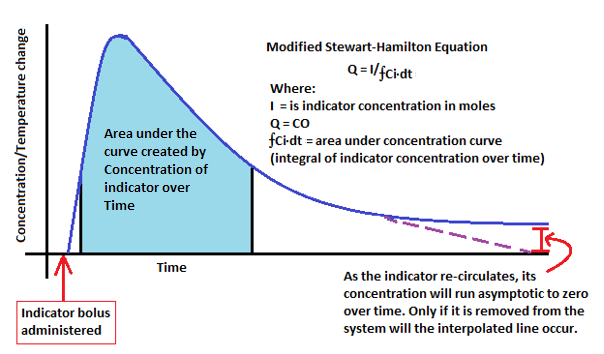
Figure 1: Modified Stewart-Hamilton equation applied in PAC Thermal dilution during CO monitoring [2].

The use of the PAC method for CO monitoring has many advantages over other techniques. Conversely, due to it being an invasive device, it does carry inherent risks including an increased possibility of dysrhythmias such as complete heart block, perforation of heart chambers, cardiac tamponade, pneumothorax, valve damage, infection, and emboli [1]. Post hoc analyses of larger studies have also reported no benefit in using the PAC method, other than in elective surgical patients [5]. However, given that elective surgical patients are often healthier than non-elective intensive care patients, queries over confounding factors within the population have been raised [5]. This demonstrates that it can be used safely, but brings into question its patient-related value in situations arising from the critical care environment [6]. Despite the relatively high reliability of this device, debate still exists around whether it actually improves outcomes in various patient groups [6]. Regardless, its clinical benefit in monitoring both undifferentiated, multi-factorial shock states and cardiac cases is well documented [6].

Minimally invasive methods: CO2 re-breather, Doppler ultrasound, and bio-impedance

Indirect Fick principle (CO2 re-breather)

The pulmonary circulation is the part of the cardiovascular system that involves deoxygenated blood flowing from the right heart, through the lungs, and back into the left side of the heart (now oxygenated blood). This circulation can be used to estimate CO via the use of a mathematical equation called the “Fick” principle [7]. The Fick principle is a mathematical interpretation of gasses, based on the conservation of mass, which allows the calculation of blood flow to an organ based on the uptake of a specific marker substance [2]. In more clinical terms, it grants the ability to observe the amount of gas release, classically oxygen (O2) or carbon dioxide (CO2), that occurs in the pulmonary circuit, via the alveolar blood flow, and the difference in gas concentration in the arterial and venous circulation (by collecting venous and arterial blood samples) [7]. By measuring alveolar blood flow via the rate of carbon dioxide volume (VCO2) produced from the lungs and body over the arterio-venous carbon dioxide gradient of arterial carbon dioxide partial pressure (PaCO2) and venous carbon dioxide partial pressure (PvCO2), one can extrapolate CO from the difference between these CO2 gradients [8]. Mathematically, with Q representing CO and VO2 being volume of oxygen consumed, the generic formula is as follows:

VO2 = Q (PaCO2 – PvCO2) [8]

For example, if the concentration of CO2 being delivered to an organ is known, and the amount this concentration increases after perfusing the organ (venous and arterial difference) is also known, it is possible to divide the two in order to determine the “flow” of gas, and therefore blood, to that organ [7]. In CO monitoring, the use of the pulmonary circuit to represent “the body”, with a sample taken before (venous) and after (arterial) allows us to determine the change in CO2 concentration as the blood flows to the heart. Concurrently, the patient in which CO is being measured must be intubated and under mechanical ventilation in order to control this gas exchange and its volume parameters [8].

The advantages of using the Fick technique are largely associated with it being a minimally invasive technique (such as relatively fewer contraindications and adverse outcomes than comparable methods) [7]. However, this method has limitations in that it has a lower level of accuracy than invasive methods, and requires the patient to undergo intubation [7]. Additionally, it was found that in the determination of CO for patients undergoing cardiac surgery, an underestimation preoperatively and an overestimation postoperatively commonly occurred [9]. This brings into question its clinical reliability in the vital perioperative setting [9]. Thus, although it has clinical advantages in patients who cannot tolerate more invasive methods, its reliance clinically is not as consistent as that of PAC.

Doppler ultrasound: aortic and echocardiography

Doppler ultrasound operates on the principle that the shift in frequency of a wave is directly proportional to the velocity of the moving plasma [10]. By measuring plasma velocity, Doppler ultrasound can be used to estimate CO. Typically, Doppler ultrasound calculates CO by measuring the velocity of blood plasma at the level of the thoracic aorta, generating an estimation of blood flow (stroke volume) by measuring plasma flow across the cross-sectional area of the aortic vessel or valve. This velocity can then be multiplied by the heart rate to provide an estimation of CO [7]. This technique is commonly combined with echocardiography, which measures left-side cardiac filling pressures via a two-dimensional ultrasound, and Doppler measurement of the aortic annulus diameter [11]. Additional benefits derived from this method include the provision of clinically relevant information on the global structure of the heart, in addition to valvular anatomy and presence of pericardium pathology (including tamponade or constrictive pericarditis) [11]. This allows for accurate, non-invasive measurement of left ventricular diastolic dysfunction, which is predictive of mortality in hospitalised patients [10]. Furthermore, echocardiography can also measure the response of stroke volume to both fluid bolus and diuretic therapy, while monitoring left (trans-mitral) and right (vena cava) arterial pressure [12]. This may be critical in patients at risk of both systolic and diastolic heart failure [12].  Importantly, these changes can be monitored serially after interventions have been performed.

Significant drawbacks of this method include its inability to provide continuous monitoring and difficulty in measuring sample volume placement; probe placement and beat-to-beat variation in stroke volume both impact accuracy [11]. Additionally, in the setting of decompensated systolic heart failure, tissue Doppler imaging can be inaccurate for monitoring filling pressures [11]. Since this technique also requires particular placement of the probe, as this is an important variable for reproducible results, significant operator training and skill is required [12]. Therefore, while this method is both reliable and has multiple reasons for use in the clinical setting, consideration needs to be given to operator skill and reproducible monitoring.  An additional limitation is that echocardiography requires considerable skill to perform well.

Finally, while not commonly used in Australia, oesophageal and transthoracic Doppler methods also exist. As with Doppler echocardiography, monitoring has a similar level of reliability as more invasive methods, but is predicated upon three factors: the aortic cross-section accuracy, the parallel placement of the transducer, and the maintenance of constant beam direction between measurements [7]. Even controlling for these variables, inherent factors limit this technique, including physiological expansion of the aorta during systole, noted to be approximately 12% [10]. Furthermore, the use of a nomogram (a pre-determined graph using three or more logarithmic scales to show a relation between them, such as occurs in weight and height charts to calculate body mass index) introduces potential measurement error secondary to variation with vascular tone and volume status that needs to be considered for all measurements [10].

Bio-impedance

CO can also be estimated via bio-impedance in a technique known as impedance cardiography. This technique measures the electrical impedance of the thoracic cavity generated during systole and left ventricular outflow into the aorta through the conductive properties of blood via four electrodes placed around the thorax; two placed on the left neck and another two upon the lower thorax, focus alternating current toward the ascending and descending aorta, within which blood is the most conductive material [13]. Put simply, the electrodes emit a small electrical current that is then “bounced” off conductive tissue, of which blood produces the strongest “bounce”. This current is then received by the electrode, wherein the change of impedance (current “bounced” from the blood tissue and received by the electrode) correlates to stroke volume [1].

Similar to all minimally invasive techniques, impedance cardiography nullifies the risk of infection and haemorrhage, however, it remains particularly sensitive to movement and is unsuitable for use in patients with arrhythmias or who are haemodynamically unstable [7]. This limits its use in Australian medical settings and so it is not commonly seen in practice [7,13].

Semi-invasive: pulse contour analysis

The final, commonly used method of estimating CO is pulse contour analysis. Pulse contour analysis utilises arterial waveforms, obtained either from an arterial catheter (invasive) or a peripheral finger probe (minimally invasive), in order to extrapolate stroke volume and systemic vascular resistance [2]. This technique relies on measuring pulse pressure, that is, the difference between diastolic and systolic pressure (as proxy to volume), in order to create a picture or “wave form” that can be analysed mathematically to find the area under the curve. Initially, this method assumed compliance of aortic wall to be uniform, regardless of patient demographics and co-morbidities (as the compliance of a vessel alters its distensibility). However, via the use of an algorithm developed by Wesseling et al. [14], it become possible to gauge stroke volume by integrating the area under the curve measured during systole to minimise this error. Concurrently, the estimated stroke volume can then be multiplied by heart rate to give CO [7,14,15]. Many devices rely on the principle of pulse contour analysis, such as Pulse Contour Cardiac Output Monitoring (Pulsion Medical Systems, Munich, Germany) and Lithium Chloride Dilution (LiDCO Ltd., Cambridge, UK), are invasive, needing arterial lines. However, the Vigileo™/FloTrac™ system (Edwards Lifesciences, Irvine, CA, USA) and the Nexfin® system (BMEYE, Amsterdam, Netherlands), which uses a finger cuff to measure pulse pressure of the digital arteries, are non-invasive options. Currently, all of these devices are limited by the fact that they require inter-patient calibration [16].

As in the bio-impedance method, this technique is limited in patients with cardiac arrhythmias, aortic regurgitation and intra-aortic balloons as they alter measurement accuracy [4]. Equally, the use of arterial catheters introduces an additional risk of infection and haemorrhage. Despite this, a recent meta-analysis performed by Mayer et al. determined that a strong positive correlation exists between the contemporary pulse contour analysis devices (FloTrac/Vigileo) and the PAC method when measuring CO [16]. Concurrently, this method possesses a high clinical applicability with significant potential for future development.

In conclusion, accurate CO assessment and monitoring in the perioperative patient remains critically important. While PAC remains the most accurate and reliable method, its invasiveness and the subsequent risk of complications makes it unsuitable for specific patient subsets. Conversely, while minimally invasive techniques reduce the risk of procedural complications, this is often at the expense of reliability. Of these methods, pulse contour analysis is the most extensively studied, with precision being similar, if not equivalent to, PAC. Until definitive, outcome-based comparison studies have been completed, the selection of the most appropriate CO measurement modality remains the decision of the treating clinician, the patient, and faculty clinical guidelines.

Acknowledgements

Dr M. Peach.

Conflicts of Interest

None declared.

References

[1]  Wigfull J, Cohen AT. Critical assessment of haemodynamic data. Contin Educ Anaesth Crit Care Pain. 2005 Jun 1;5(3):84-8.

[2]  Scheer BV, Perel A, Pfeiffer UJ. Clinical review: complications and risk factors of peripheral arterial catheters used for haemodynamic monitoring in anaesthesia and intensive care medicine. Crit Care. 2002 Apr 18;6(3):199.

[3] Calzia, E, Iványi, Z, Radermacher P. Functional haemodynamic monitoring. Berlin: Springer Berlin Heidelberg; 2005. Determinants of blood flow and organ perfusion; p.19-32.

[4] Lavdaniti M. Invasive and non-invasive methods for cardiac output measurement. Int J Caring Sci. 2008 Sep 1;1(3):112.

[5] Harvey SE, Welch CA, Harrison DA, Rowan KM, Singer M. Post hoc insights from PAC-Man—the UK pulmonary artery catheter trial. Crit Care Med. 2008;36(6):1714-21.

[6] Harvey S, Harrison DA, Singer M, Ashcroft J, Jones CM, Elbourne D et al. Assessment of the clinical effectiveness of pulmonary artery catheters in management of patients in intensive care (PAC-Man): a randomised controlled trial. Lancet. 2005 Aug 6;366(9484):472-7.

[7]  Drummond KE, Murphy E. Minimally invasive cardiac output monitors. Contin Educ Anaesth Crit Care Pain. 2011 Oct 3:mkr044.

[8]  Chaney JC, Derdak S. Minimally invasive hemodynamic monitoring for the intensivist: current and emerging technology. Crit Care Med. 2002 Oct 1;30(10):2338-45.

[9]  Alhashemi JA, Cecconi M, Hofer CK. Cardiac output monitoring: an integrative perspective. Crit Care. 2011 Mar 22;15(2):214.

[10] Dark PM, Singer M. The validity of trans-esophageal Doppler ultrasonography as a measure of cardiac output in critically ill adults. Intens Care Med. 2004 Nov 1;30(11):2060-6.

[11] Porter TR, Shillcutt SK, Adams MS, Desjardins G, Glas KE, Olson JJ et al. Guidelines for the use of echocardiography as a monitor for therapeutic intervention in adults: a report from the American Society of Echocardiography. J Am Soc Echocardiog. 2015;28(1),:40-56.

[12] Konstadt SN, Shernan SK, Oka Y, editors.  Clinical transesophageal echocardiography: a problem-oriented approach. Philadelphia: Lippincott Williams & Wilkins; 2003.

[13] Mehta Y, Arora D. Newer methods of cardiac output monitoring. World J Cardiol. 2014 Sep 26;6(9):1022.

[14] Wesseling KH, Jansen JRC, Settels JJ, Schreuder JJ. Computation of aortic flow from pressure in humans using non-linear, three element model. J Appl Physiol, 1993; 74:2566-73.

[15] Mathews L, Singh KR. Cardiac output monitoring. Ann Card Anaesth. 2008 Jan 1;11(1):56.

[16] Mayer J, Boldt J, Poland R, Peterson A, Manecke GR. Continuous arterial pressure waveform–based cardiac output using the FloTrac/Vigileo: a review and meta-analysis. J Cardiothor Vasc An. 2009 Jun 30;23(3):401-6.

Categories
Feature Articles

Forget everything you thought you knew: how your assumptions are impacting the health outcomes of your patients.

Modern health professionals are well versed in the value of person-centred care for their patients. However, the way we are taught to view our patients through a problem-based lens is counterintuitive to this person-centred approach. Medical professionals have learned to consider the diverse sociocultural contexts of patients as a “risk” to their overall wellbeing, rather than acknowledging the unique strengths of all individuals and communities. This focus entrenches assumptions into the way we approach patients of diverse backgrounds. These assumptions and the subsequent expectations that we hold of our patients have been evidenced to serve as powerful self-fulfilling prophecies for an individual’s overall health and wellbeing. Individuals will internalise negative health identities and have poorer health outcomes if health professionals hold low expectations of them based on their sociocultural “risks”. Strengths-based practice recognises resilience and focuses on the strengths, abilities, knowledge, and capacities of all individuals, rather than on their deficits, limits, or weaknesses. It provides a framework for health professionals to better support their patients in achieving their best health outcomes. A strengths-based approach has the ability to shift the broader deficits-based discourse that exists around the diverse sociocultural groups that exist in Australia. Changing this conversation is of immeasurable importance if we are to improve the health outcomes and agency of our patients and mitigate the persistent health inequities that exist within the Australian health system.

As modern health professionals in training, we have been well conditioned to consider all biological, psychological, and sociocultural factors that may contribute to poorer health outcomes in any given patient. We are familiar with the World Health Organisation’s definition of health as “a state of complete physical, mental, and social wellbeing, and not merely the absence of disease or infirmity [1]”. But how does this translate into our practice? High patient caseloads and the sheer breadth of medicine compel us to streamline history-taking processes and problem formation through pattern recognition. We are taught to cluster “risk” factors – to make assumptions about disease, prognosis, compliance, and life expectancy, based upon sociocultural “risks”. We tick boxes. “Now, this is a question that we have to ask everyone, but do you have a history of previous intravenous drug use?”, “Do you identify as an Aboriginal or Torres Straight Islander?”, “Are you from a refugee or migrant background?”, and “Do you live in a rural or remote area?” We turn our attention to “vulnerabilities” and “high-risk groups” without much consideration of the impact of this focus and our subsequent assumptions on individual health identity.

The expectations we consciously or subconsciously hold of our patients, and the language we use toward or about them, inherently impact their health outcomes. In medical school, we are often taught to view our patients through a lens of deficit, by focusing on health problems rather than the opportunities that come from realising patients’ individual strengths. Strengths-based practice can shift this problem-based approach, and is a means of acknowledging the importance of our patients’ environments and diverse sociocultural contexts in the trajectory of their health attainment [2]. Strengths-based practice is an ecological approach to individuals, families, and communities that recognises resilience and focuses on the strengths, abilities, knowledge, and capacities of all individuals, rather than on their deficits, limits, or weaknesses [2]. A strengths-based approach is of immeasurable importance if we are to improve the health outcomes and agency of our patients, and mitigate the persistent health inequities that exist within the Australian health care system. This article will examine the impact of assumptions on patient health attainment and identity, the value and practicality of a strengths-based approach in the clinical setting, and the broader implications of our deficits discourse in the Australian public health arena.

Expectations and health identity

The expectations we hold of our patients can serve as powerful self-fulfilling prophecies for their overall health outcomes and identity. More than once I have heard a doctor use the phrase, “We save smart solutions for smart people”. This approach, however well intended, is damaging. Our clinical decision-making should not be influenced by expectations of noncompliance or assumptions of deficit. This is because health often exists at the nexus of societal expectations and our subsequent internalised perception of self. Essentially, if health professionals have high expectations of their patients, their patients are likely to have higher expectations of themselves, and subsequently experience better overall outcomes. This phenomenon is known in the behavioural psychology sphere as the Rosenthal effect, whereby our interpersonal expectations have been shown to significantly impact the learning, abilities, and health attainment of the subjects of our expectations [3,4]. We subconsciously facilitate “warmer” socio-emotional environments for individuals of whom we have higher expectations [3]. We have also been shown to input more effort into maximising the outcomes of individuals we see as having greater potential [3]. These subtle changes in the behaviour of care providers are internalised by patients and shape the expectations an individual holds of themselves [3]. Negative internalised expectations have been shown to directly lead to poorer mental and physical health outcomes in patients [5]. Internalised expectations may also directly act to motivate or discourage patients in their personal attainment of better health outcomes [5]. Essentially, negative health care provider assumptions and expectations demoralise an individual’s health identity, and this in turn impacts the mental and physical health outcomes of patients [3,6].

Our clinical approach to Indigenous patients in Australia offers an obvious example of how health care provider expectations may demoralise an individual’s health identity. At the beginning of any consultation, we are taught to ask all patients if they identify as Aboriginal or Torres Straight Islander. Identification of Aboriginal or Torres Straight Islander descent is essential in creating safety for our Indigenous patients. Too often however, doctors aren’t asking about Indigenous identity in order to practice their (often limited) cultural competencies, but rather to unfold a new list of differential diagnoses, and to remember to ask about smoking, alcoholism, substance use, diet, and exercise. On examination of Indigenous patients, we are also taught to check specifically for signs of cardiovascular disease, hypertension, type-2 diabetes, and chronic renal disease. While such a comprehensive approach to all patient consultations is desirable, our underlying assumptions relating to our Indigenous patients’ health-seeking behaviours, and our expectations for their health prognoses, is problematic. These assumptions are one of the means by which racism is maintained within our health system. As health professionals who work to support others in achieving their best possible health outcomes, we are terrified to talk about racism, or to consider that we might be contributing to its perpetuation in the Australian health system. However, it is important to define what racism in our health system actually means, in order to understand our role in it.

There are three levels of racism that contribute to poor health outcomes for Indigenous people: institutional, interpersonal, and internalised racism [7-12]. Institutional racism is often established in political systems and sustained by the policies of governments and health institutions that discriminate against Indigenous peoples [8]. Interpersonal racism in the health setting occurs when a health care provider makes assumptions about a patient on the basis of their Indigenous identity, or discounts Indigenous beliefs and practices [9]. These discriminatory interactions may be communicated to patients through non-verbal or verbal means, and often alter the course of care for an Indigenous patient [8,9]. Internalised racism occurs when an Indigenous patient accepts the stereotypes of interpersonal and institutional racism, and allows these stereotypes to shape their health identity. Institutional and interpersonal racism are often not intentional, but remain uninterrogated and largely invisible in our health system. Sometimes it is highly visible, but still unchallenged and unchanged. By not acknowledging or confronting the racism that exists within our health institutions, we are reinforcing negative internalisation among Indigenous individuals, leading to negative mental and physical health outcomes [6,8,9]. If a patient is conscious of interpersonal racism, this has been shown to influence their participation in unhealthy behaviours, and directly contribute to the long-term development of cardiovascular disease, hypertension, renal disease, and alter some of the neurochemical processes involved in diabetes [6,8]. The high prevalence of these chronic conditions within Indigenous populations is therefore something that is perpetuated, rather than mitigated, through our current approach to Indigenous health. As health professionals, we must challenge our conventional health paradigms and disrupt the processes that blame such systemic problems on the “unhealthy behaviours” of an entire culture [9].

By utilising reductionist techniques to simplify care provision for our patients, we are limiting our patients’ ability to attain their highest standard of health, as well as restricting their agency and self-aspiration [13]. An individual’s health is more than the sum of their medical issues – it is also determined by their personal resources. In medical school, we are not taught to identify the inherent strengths of all individuals, but rather to focus on health risks, problems, and limitations. That is, instead of seeing the potential that exists in celebrating the diverse sociocultural contexts of our patients, we regularly view sociocultural identity as a “risk” to overall wellbeing. This deficits-based understanding of health identity and our subsequent interpersonal communication is internalised by our patients, shaping their health behaviour and outcomes. We have the opportunity to change the conversation.

The strengths-based approach

Strengths-based practice is a well-evidenced approach in ensuring people have agency in their own health outcomes and identity. Strengths-based practice appreciates the centrality of people’s environments and sociocultural contexts in the attainment of their optimal health outcomes, and builds upon these strengths to reinforce health identity [2]. A strengths-based practice framework involves six core principles [2]:

  1. All individuals, families, groups, and communities have strengths, and the emphasis is on these strengths rather than on pathologies
  2. Communities are an abundant source of resources
  3. Interventions are built on the self-determination of the patient
  4. Collaboration is key, and a positive practitioner-patient relationship essential
  5. Outreach is utilised as a preferred mode of intervention, and
  6. All people have the inherent capacity to learn, grow, and change.

While strengths-based practice has not been formally implemented in the medical system, analysis of its feasibility in the social work setting may inform its rollout across the broader health sector. There are three developmental stages of health professional learning: socialisation, internalisation, and identification [2]. Socialisation involves health professionals learning how to enable a strengths-based dialogue among their colleagues, so that their colleagues may then develop the knowledge and skills to empower others [2]. Internalisation in this context is the process whereby health professionals internalise strengths-based principles in order to counter any barriers to enabling patients to see their strengths [2]. Identification involves the recognition of tacit assumptions about patients, and the impact of these assumptions on health provider practice and their patient’s cultural context of empowerment [2]. To shift our deficits-based approach to health care in Australia, health professionals must first be socialised to the concept of strengths-based practice, before we can then internalise its importance, and address any negative expectations we inadvertently hold of our patients.

The language we use to converse with our patients is often a product of the expectations and assumptions we hold of them. Paradoxically, our expectations are shaped by the broader public health discourse and problem-based learning that is indoctrinated into many doctors throughout their training. The rigidity of our health and medical education systems that institutionalise this deficits-based discourse make it difficult to universally adopt strengths-based practice across Australia. However as health professionals, we are still able to begin shifting this conversation and challenging the assumptions that we usually accept of our patients. Indigenous peoples in Australia are well versed in the power of strengths-based practice, and have identified three crucial ways we might enable positive change and start shifting our health discourse away from a mindset of deficit [13]:

  1. Create safety: enable a space and process for robust discussion.
  2. Challenge mindsets, habits, and conversations: take responsibility, find courage, and lead by example.
  3. Co-create transformative pathways: engage with community groups to develop change and spread the word to engage in a national dialogue.

A deficits-based health discourse extends beyond interpersonal interactions of doctor and patient. As health professionals, our opinions are respected and hold legitimacy in public health discourse. How we talk about people matters because it plays a major role in shaping the public dialogue, and subsequently assists in setting a national health agenda for our politicians to action. We should be engaging in strengths-based health rhetoric and promoting the wellbeing of all individuals, rather than focusing on their limitations. As a result of our privilege, we have a duty to amplify the voices of individuals and communities who are working hard to shift our national dialogue to a narrative of strength, resilience, and opportunity.

Changing the conversation

Over recent decades, an emerging theme in the public health discourse has been a focus on health disparities between your “average Australian” and specific sociocultural groups. Arguably, this well-intentioned advocacy has been successful in fostering the next generation of compassionate and socially conscious health professionals. Many of my fellow students would attest that they entered their medical degree because they saw it as an effective means of helping people they perceived to be marginalised in our society. This motivation is exciting and provides fertile ground to generate unprecedented change to the inequities that persist in the Australian health system. But the fundamental assumption that our current deficits-based medical curriculum will enable us to effect positive change for marginalised groups is flawed. We assume that a medical degree, taught through problem-based learning, will provide us with the knowledge, skills, and sensitivity to offer the help that is needed. However, without realising the strengths of all individuals and communities, we are missing out on an enormous opportunity to celebrate resilience, reinforce positive health identities, and improve health outcomes for all.

Attention to sociocultural determinants of health has allowed us to raise awareness of persistent inequalities in our health system. However, focusing solely on deficits is detrimental to the broader narrative of the diverse sociocultural groups in our society. Drawing back upon the example of Indigenous health in Australia, large health promotion campaigns have been incredibly valuable in shedding light on the inequalities that persist between Indigenous and non-Indigenous Australians. These campaigns have also ensured that Indigenous health and education remain on political agendas, and they have secured funding for important programs. However, these awareness-building education and health promotion strategies have inherently focused on the “gaps” experienced by Indigenous people, and are an ineffective substitute for a whole government commitment to address the broader social determinants of health and shift our discourse away from deficit [9]. If we constantly emphasise life expectancy “gaps” in our public discourse, without closely examining our role in the discriminatory policies and practices that maintain these “gaps”, we will only perpetuate the inequities that exist between Indigenous and non-Indigenous Australians. By continuing to allow deficits to eclipse individual strengths, we are doing our patients, their communities, and our broader society, a colossal disservice.

Our health discourse does not exist in a vacuum. As modern health professionals, we have an obligation to celebrate the individual strengths of each of our patients, and a duty to use our respected voice to shape the rhetoric that currently marginalises the diverse sociocultural groups that exist in Australia. So challenge expectations, transform mindsets, and check your assumptions at the door – together we can ensure better health outcomes for all.

Acknowledgements

Scott Gorringe, for his patience, persistence, resilience, strength, and friendship.

Nicholas Fava, for his invaluable assistance in proofreading this piece.

Conflicts of interest

None declared.

References

[1] World Health Organization. Constitution of the World Health Organization. Geneva, Switzerland: 2006 October.

[2] Scerra N. Strengths-Based Practice: The Evidence. Parramatta, Australia: Uniting Care, Social Justice Unit; 2011 July.

[3] Rosenthal R. Interpersonal Expectancy Effects: A 30-Year Perspective. Curr Dir Psychol. 1994;3(6):176-9.

[4] Learman LA, Avorn J, Everitt DE, Rosenthal R. Pygmalion in the nursing home the effects of caregiver expectations on patient outcomes. J Am Geriatr Soc. 1990;38(7):797-803.

[5] Mondloch MV, Cole DC, Frank JW. Does how you do depend on how you think you’ll do? A systematic review of the evidence for a relation between patients’ recovery expectations and health outcomes. Can Med Assoc J. 2001;165(2):174-9.

[6] Pascoe EA, Richman LS. Perceived discrimination and health: a meta-analytic review. Psychol Bull. 2009;135(4):531-54. PubMed PMID: PMC2747726.

[7] Paradies Y. A systematic review of empirical research on self-reported racism and health. Int J Epidemiol. 2006;35:888-901.

[8] Larson A, Coffin J, Gilles M, Howard P. It’s enough to make you sick: the impact of racism on the health of Aboriginal Australians. Aust NZ J Public Health. 2007;31.

[9] Durey A, Thompson SC. Reducing the health disparities of Indigenous Australians: time to change focus. BMC Health Serv Res. 2012;12(1):1-11.

[10] Kelaher MA, Ferdinand AS, Paradies Y. Experiencing racism in health care: the mental health impacts for Victorian Aboriginal communities. MJA. 2014;200:1-4.

[11] Williams DR, Mohammed SA. Discrimination and racial disparities in health: evidence and needed research. J Behav Med. 2009;32(1):20. PubMed PMID: PMC2821669.

[12] Paradies Y, Harris R, Anderson I. The impact of racism on Indigenous health in Australia and Aotearoa: towards a research agenda. Casuarina, Australia: Flinders University, 2008 March.

[13] Gorringe S, Ross J, Fforde C. ‘Will the Real Aborigine Please Stand Up’: Strategies for breaking the stereotypes and changing the conversation. Canberra, Australia: AIATSIS, Research Program; 2011 January.

Categories
Feature Articles

Indigenous health: what they don’t teach you in medical school

Indigenous health education is an important part of the curriculum for medical students. However, there are limited opportunities within the course for students to interact with patients from an Indigenous background. Following an Indigenous health placement in a remote community in the Eastern Kimberley region, a final year medical student reflects on how her medical education did not prepare her for the overwhelming social issues impacting on the health of Indigenous Australians. This article explores how, in the author’s experience, the current medical curriculum offers limited opportunities for first-hand exposure to Indigenous health, with a large proportion of Indigenous health content delivered in a lecture format. This style of teaching does not allow students to discuss, explore, and understand the many complex social issues that contribute to the health of Indigenous Australians. This combination of suboptimal teaching style and little or no first-hand experience results in medical students that are not well prepared to deal with these issues as junior doctors. It is necessary to review the current teaching methods and consider implementing interactive workshops to improve Indigenous Health education, which will in turn improve the health of Indigenous Australians.

This year, I spent my six-week elective rotation in Kununurra, a small town in the Eastern Kimberley region with a population of about 8,000 people [1]. Kununurra is a remote town situated approximately 500 km, 800 km, 1,000 km and 3,000 km from Katherine, Darwin, Broome and Perth respectively, which are the nearest major centres. There is a relatively large Indigenous population, with 34.8% of residents identifying as either Aboriginal or Torres Strait Islander [1]. The traditional owners of the Kununurra area are the Miriwoong Gajerrong peoples, and Kununurra is an anglicised version of Goonoonoorrang, the name of the Ord River that flows west of Kununurra. Colonisation has led to much disruption for this community, having serious impacts on health and wellbeing [2].

My placement was with an Aboriginal Health Service, a GP clinic with visiting allied health and specialist services, so the vast majority of patients that I saw were Aboriginal. I was unprepared for what I observed in this placement in remote Western Australia, in particular the social and environmental determinants of health that contribute significantly to the health of Indigenous Australians. The variety of issues that I experienced in Kununurra brought into relief the difference between my understanding of Indigenous Health from the taught medical curriculum, and the reality in Kununurra. This essay explores this gap by illustrating with anecdote some of my personal experiences of social determinants of Indigenous health, with comment on how student understanding might be improved in the teaching of medical students. The examples relate to my experiences in Kununurra only; it is important to note that each community has its own unique issues and that what is pertinent in Kununurra may not accurately reflect Indigenous health in a broader sense.

Prior to my brief placement in Kununurra, I had very little exposure to Indigenous health other than what I had learnt in the classroom. My lack of knowledge and experience became immediately apparent; I felt completely out of my depth with regard to the many complex medical and social issues that I was seeing, hearing, and learning about. One particular morning, as I left my house at quarter to eight, there was a man walking down the street with his young family; he had a bottle of wine in one hand and was pushing a stroller with the other. When I arrived at the clinic I jumped into the 4WD with one of the nurses and we drove to a community over 90 minutes away. This outreach clinic used to be serviced regularly by staff from Kununurra, but due to staff shortages it was months since the clinic had been open. In near 40-degree heat, bare-footed patients walked down the road to the clinic, which itself was covered in dirt, dust, and cobwebs, with overgrown weeds outside, broken windows, and blocked plumbing. Dead frogs were swept out from behind toilets and under desks. It was certainly not something I ever thought I would see in Australia, yet the patients were just grateful to have the clinic up and running again. We went to the school to see if any of the 11 students required immediate attention; as there was no doctor with us, anything less urgent would have to wait. While we were there, we provided dressings and antibiotics for a child with boils (a very common complaint) and removed a foreign body from a boy’s ear that thankfully turned out to be paper rather than a maggot.

Over the course of my stay in Kununurra, I saw a heavily-pregnant woman who smoked 80 cigarettes a day with no intention of stopping, a lady with a total thyroidectomy who had stopped taking thyroxine, a man who refused treatment and further investigation for a cryptococcal infection because he “felt fine”, and patients in their forties on dialysis. I had patients tell me they share a three-bedroom house with 20 people and saw children who had not attended school for weeks, and who wandered the streets with their friends at night, completely unsupervised. I had a 17 year-old boy openly tell me that he smoked cigarettes and “gunja” (marijuana), but who vehemently denied drinking alcohol because “it only causes violence”; and for the first three weeks of my placement, there was at least one person every week who presented after an attempted hanging, the youngest of which was 12 years-old. This was also highlighted in a speech given by Dr Marion Scrymgour, the CEO of the Wurli Wurlinjang Health Service and Chair of the Aboriginal Medical Services Alliance NT [3]. After six weeks in a remote Indigenous community, I was only just beginning to understand the complexity of a problem for which there is no simple solution. I began to reflect on why it had been so overwhelming; was there something missing from my education that could have better prepared me for this experience?

To understand what is lacking, we must first identify what is included in the Indigenous health curriculum. In a packed medical course, my education skimmed the surface, with lectures on culture and history that included topics such as the Stolen Generation, traditional medicine, and the different family structure of Aboriginal communities. I learned that the burden of chronic disease is much higher in the Aboriginal population, and that this is the primary reason that the life expectancy is, on average, ten years less for Aboriginal people than the rest of the Australian population. While this is worthwhile knowledge, it is an inadequate summary that only superficially touches on Indigenous health issues because it does not allow us to delve into the much more important discussion of why these discrepancies exist and how they can be rectified.

In 2004, the Committee of Deans of Australian Medical Schools (CDAMS) audited existing Indigenous health content and released the Indigenous Health Curriculum Framework, now called the Medical Deans Indigenous Health Project, which has become part of the accreditation requirements for all Australian medical schools [4]. This framework is comprehensive and meticulously developed by people with a significant amount of experience in the field. It is important to acknowledge that Indigenous health teaching is not the same across all universities and has been constantly evolving since I started my degree five years ago. However, based on my own education, I still felt very under-prepared.

There are numerous and varied reasons as to why current Indigenous health education is inadequate preparation for medical practice. Firstly, as outlined by the Indigenous Health Curriculum Framework, Indigenous people are a “non-homogenous population” with a “diversity of cultures, experiences, histories and geographical locations” [5]. It is difficult to convey that the issues encountered by urban, rural, and remote Indigenous communities seeking health care can be so vastly different. For example, the education that I received gave me essential background knowledge and an understanding of Indigenous health issues in urban areas, but failed to convey the difference between this and the difficulties faced by Indigenous Australians in remote areas. Furthermore, even within a particular setting (for example, a remote setting), there is a large variation between communities, so that the issues seen in one remote community may not be the same as those seen in other nearby communities. Secondly, thorough assessment of even the most well-constructed learning objectives is difficult to achieve. Assessment, often in the form of assignments and examinations, determines if students are meeting the desired outcomes, and from this we can extrapolate about the quality of teaching. Without this data, we are unable to accurately identify the effectiveness of teaching strategies, and are therefore unable to make appropriate changes and improvements.  Professor Richard Murray, a doctor with 14 years of clinical experience in Aboriginal health services in the Kimberley region, and the current Dean of James Cook University Medical School, illustrated this point beautifully when he said in a personal email addressing my enquiries about the Indigenous Health curriculum, “course or subject outcomes are too high level and abstract to satisfy” [6]. Therefore, not only is the curriculum difficult to implement, it is also difficult to assess its effectiveness.

One of the biggest challenges associated with Indigenous Health education is that many of the learning objectives and concepts are difficult to grasp in a classroom, especially with regard to the social determinants of health. One of the guiding principles of the Indigenous Health Curriculum Framework states that the health of Indigenous people is more associated with “historical and social determinants of health than with inherent Aboriginality” [5]. My experiences demonstrate this as well; the previously-mentioned examples are all symptoms of many complex underlying issues affecting the health of people in these communities. Many of the cases I saw would not occur if it were not for the effects of low socioeconomic status and deficits in the other social determinants of health seen across the globe: inadequate access to health care and education, health literacy and compliance issues, alcohol and drug dependence, physical and sexual assault, mental health issues and suicide, poor living conditions, and overcrowding.

For example, only 76% of Indigenous children aged 5-14 years in the Northern Territory have access to schooling, as compared to 95% of non-Indigenous children of the same age [7]. Indigenous people are less likely to drink alcohol, but twice as many drink at levels considered to be risky or high-risk for long-term harm compared to their non-Indigenous counterparts. 15% of homes with Indigenous people are considered overcrowded, which increases up to 42% in very remote areas, as opposed to 4% of other households. Furthermore, some of these houses do not have reliable water and electricity, or adequate sewerage [7]. Despite these appalling statistics, Indigenous Australians have the oldest, continuous living culture in the world, showing an incredible ability to adapt to change [8]. As a student, it is very difficult to imagine the enormity of the impact that all these factors have on health. As an example, it is difficult to explain complex health issues and the importance of treatment to patients with little or no education and health literacy.  It is even more challenging to treat and prevent the spread of communicable diseases. Consider the management of scabies or head lice which often require the entire household to be treated concomitantly; it is near impossible to achieve this in communities where overcrowding is rife. True understanding of this only comes from seeing it first-hand [9]. However, providing every student with the opportunity to go to an Indigenous or remote community is not a financially viable option for most universities, and accommodating large numbers of health science students is not necessarily feasible (or wanted) in Indigenous communities. This is one of the largest obstacles to providing the educational opportunities required to meet the outcomes outlined in the curriculum framework. Without the ability to provide the correct learning environment, it is unreasonable to expect that graduates will have an in-depth understanding of the complex interplay between these particular social determinants and health outcomes.

It is not the quality of the learning objectives and curriculum framework that limit Indigenous Health education, but rather the difficulties associated with the implementation and assessment of these objectives. The best teaching format, particularly with regard to the social determinants of health, is experience-based learning. While this is not feasible for every student, the lecture based format of Indigenous health education is suboptimal. The content is often too robust to really impart understanding of the complex health and social issues that many Indigenous people face. Other options must be considered and implemented; for example, workshops may allow educators to facilitate the discussion required to dissect more complex issues, such as why these disparities exist and how they may be overcome. Indigenous community members speaking to small groups of students about their life experiences, medical issues, and involvement with healthcare services would give students the opportunity to interact and ask questions, whilst fostering understanding and practising cultural sensitivity. Regardless of how the content is delivered, it is imperative that the social issues are emphasised within the curriculum, so that medical students graduate considering the patient in the context of their community and surroundings, because it is difficult to improve health without first improving the foundations. Students must learn that it is not enough to simply treat the presenting complaint; even our best medical efforts are not nearly as effective in promoting positive health outcomes as advocating to ensure the basic social needs (safety from violence, basic education, and adequate housing and nutrition) of our patients and communities are being met. Only when this is achieved will we see significant change in the health outcomes of Indigenous Australians. This is perhaps the biggest lesson omitted from the Indigenous health curriculum, and it is unlikely to be learnt in a classroom.

Acknowledgements

I would like to acknowledge Professor Michelle Leech and Professor Richard Murray for their helpful advice during the information gathering stages. I would like to thank Associate Professor Karen Adams, who suggested resources and ensured the article was written in a culturally-sensitive manner. I would also like to acknowledge the Deans and their representatives of all the Australian medical schools who provided me with their Indigenous health objectives or other resources. Thanks also to Dr Alexandra Van Rijn, who provided advice during the drafting stages of this project.

Conflicts of interest

None declared.

References

[1] Australian Bureau of Statistics. National regional profile : Kununurra (Statistical Area Level 2) [Internet]. Abs.gov.au. 2016 [cited 2016 Mar 28]. Available from: http://www.abs.gov.au/AUSSTATS/abs@nrp.nsf/Previousproducts/508041208Population/ People12007-2011?opendocument&tabname=Summary&prodno=508041208&issue=2007-2011&num=&view=.

[2] Mills V. How Kununurra got its name – ABC Kimberley WA – Australian Broadcasting Corporation [Internet]. Abc.net.au. 2016 [cited 2016 Apr 23]. Available from: http://www.abc.net.au/local/audio/2011/02/10/3135392.htm.

[3] Scrymgour M. Marion Scrymgour: The NT Intervention disinformation campaign – from allegations of child sexual abuse to moral panic – The Northern Myth [Internet]. Blogs.crikey.com.au. 2016 [cited 2016 Apr 23]. Available from: http://blogs.crikey.com.au/northern/2014/10/09/marion-scrymgour-the-nt-intervention-disinformation-campaign-%E2%80%93-from-allegations-of-child-sexual-abuse-to-moral-panic/.

[4] Medical Deans Australian and New Zealand. Indigenous Health Curriculum Framework [Internet]. Medicaldeans.org.au. 2016 [cited 2016 Mar 28]. Available from: http://www.medicaldeans.org.au/projects-activities/indigenous-health/cdams-indigenous-health-curriculum-framework/.

[5] Phillips G. CDAMS Indigenous Health Curriculum Framework [Internet]. 1st ed. Melbourne: VicHealth Koori Health Research and Community Development Unit; 2004 [cited 2016 Mar 28]. Available from: http://www.medicaldeans.org.au/wp-content/uploads/CDAMS-Indigenous-Health-Curriculum-Framework.pdf.

[6] Murray R. Indigenous Health Project. 2016.

[7] Wakelin B. Many ways forward [Internet]. 1st ed. Canberra: The Parliament of the Commonwealth of Australia; 2004 [cited 2016 Mar 28]. Available from: https://www.aph.gov.au/binaries/house/committee/atsia/indigenouscommunities/report/report.pdf.

[8] Cultural Heritage – Australian Museum [Internet]. Australianmuseum.net.au. 2015 [cited 2016 Apr 23]. Available from: http://australianmuseum.net.au/indigenous-australia-cultural-heritage.

[9] Benson J, Ryder C, Gill M, Balabanski A. A brief experience for medical students in a remote Aboriginal community [Internet]. Racgp.org.au. 2015 [cited 2016 Mar 28]. Available from: http://www.racgp.org.au/afp/2015/october/a-brief-experience-for-medical-students-in-a-remote-aboriginal-community/.