Categories
Case Reports Articles

An unusual case of bowel perforation in a 9 month old infant

In Australia, between 2009 and 2010 almost 290 000 cases of suspected child abuse and neglect were reported to Australian state and territory authorities. Child maltreatment may present insidiously, not allowing signs of the maltreatment to be elicited until after a culmination of events. Ms. LW, a 9-month- old Indigenous female, presented to the Alice Springs Hospital emergency department (ED) with complaints of bloody diarrhea. A provisional diagnosis of viral gastroenteritis was suggested and she was managed with fluids to which her vitals responded positively. She was discharged six hours post presentation but presented three days later in a worsened condition with a grossly distended abdomen. Exploratory laparotomy found a perforated jejunum, which was deemed as a non-accidental injury. This case outlines the pitfalls in collateral communication in which we discuss the lack of use of an interpreter or Aboriginal health worker. We also emphasise the onus on junior doctors to practice in a reflective manner with the burdens of ED, so that they do not miss key diagnostic clues. Early detection of chronic maltreatment is important in the prevention of toxic stress to the child, which has been shown to contribute to a greater burden on society in the form of chronic manifestations later in life.

Introduction

Maltreatment, especially that of children can be insidious in nature, whose signs may not be evident until a culmination of unfortunate events. In Australia, during 2010-2011, there were 286,437 [1] reports of suspected child abuse and neglect made to state and territory authorities with a total of 40,466 substantiations (Figure 1). These notifications include four maltreatment types: physical abuse, sexual abuse, emotional abuse and neglect (Figure 2). As of 30 June 2010, there were 11,468 Aboriginal and Torres Strait Islander children in out-of-home care as a result of this. The national rate of Indigenous children in out-of-home care was almost ten times higher than for non- Indigenous children. [1]

Child protection statistics shown above tells us how many children have come into contact with child protection services; however, they do not take in to account the silent statistics of those who suffer without seeking aid. In all jurisdictions in 2010-11, girls were much more likely than boys to be the subject of a substantiation of sexual abuse. In contrast, boys were more likely to be subject to physical abuse than girls in all jurisdictions except Tasmania and the Northern Territory. [1]

Unfortunately it is difficult to obtain accurate statistics regarding the number of children who die from child abuse or neglect in Australia, as currently comprehensive information is not collected in every jurisdiction. Taking this into account however latest data recorded indicated that in 2006, assault was the third most common type of injury causing death for Australian children aged 0-14 years, [2] and totaled 27 children mortalities in 2006-07. Medical practitioners must be aware of the signs of child maltreatment and their long- term consequences, as they possess the opportunity to intervene and change the consequences of this terrible burden on afflicted children.

Case Presentation

Ms. LW, a nine month old Indigenous female and her mother presented to the Alice Springs ED at 2100, with complaints of bloody diarrhea. Emergency department staff noted that on presentation the infant was notably uncomfortable and tearful. She was afebrile, with mild tachypnoea (50 respirations per min) all other vitals were normal. Examination of the infant revealed discomfort in the epigastric region with no other significant findings including no organomegaly or distention. No other abdominal signs in particular signs such as guarding or rigidity were noted on admission. Systemic review did not show any significant findings. Past medical history included recurrent chest infections with the last episode two months prior. No immunisation history was available. The staff had difficulty examining the child because she was highly irritable. It was also difficult to elicit a comprehensive history from the mother as she spoke minimal English and was relatively dismissive of questions. No interpreter was used in this setting.

The patient was diagnosed with viral gastroenteritis and treated conservatively by the administration of intravenous fluids to maintain hydration. After six hours of observation and a slight improvement in Ms. LW’s vitals she was sent home in the early morning hours after intense pressure from the family. No other treatments and investigations were done and the staff discharged her with the recommendation of returning if the symptoms worsened over the next day.

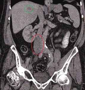
The patient returned three days later to ED with symptoms clearly of a different nature and not that of the previous diagnosis of gastroenteritis. On general observation the patient appeared unwell, irritable and was crying weakly. On examination she was found to be febrile (40°C) and toxic with tachycardia (168 bpm) tachypnoea (60 respirations per minute), and gross distention of her abdomen (Figure 3).

The case was referred to the on-call surgeon, who gave a provisional diagnosis of perforated bowel and decided to perform a laparotomy. She was immediately started on intravenous broad-spectrum antibiotics, ampicillin (200mg /6hourly), metronidazole (30mg /12 hourly) and gentamicin (20 mg/daily) before surgery.

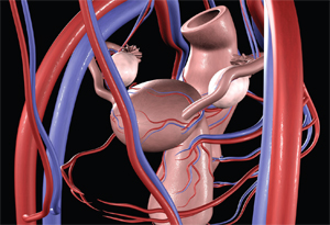
Emergency laparotomy was performed, and on initial exploration it was found that the peritoneum contained foul smelling serous fluid with a mixture of blood and faecal matter. Further exploration found perforation of the jejunum with the mesentery torn from the fixed end of the jejunum (Figure 5). The surgeons resected the gangrenous portion of the jejunum and performed an end-to-end anastomosis of small bowel.

The abdomen was lavaged with copious amounts of warm saline and the abdominal wall was closed in interrupted layers. Post surgery the child remained intubated, ventilated and was admitted to the ICU. After 24 hours post surgery the infant was extubated successfully and oral feeding was commenced after 48 hours post surgery. The patient made an uneventful recovery and was later transferred to the paediatric ward.

The surgeons commented that the initial perforation to the jejunum fixed to the mesentery caused de-vascularisation of this portion, leading to the further degradation and gangrenous state of the intestine and thus worsening the child’s condition.

As the surgeons had indicated that this injury was of a non-accidental nature the parents of the infant were brought in to be interviewed by the consultant, with the aid of an interpreter. The parents denied any falls or injuries sustained in the events leading to the presentation, which the surgical team had already exclude, due to the absence of associated injuries and symptoms. The consultant noted that both parents were not forthcoming with information even with the aid of an interpreter. Further questioning from the allied health team finally led to an answer. The father admitted that on the morning of the initial presentation while he was sitting on the ground his daughter pulled his hair from behind him to which he responded by elbowing her in the mid-region of her abdomen. Upon obtaining this information a skeletal survey was undertaken, in which a hairline fracture of the shaft of the left humerus and minor bruising in this region was found.

Case resolution

The infant was assumed into care under the basis of neglect and the case was mandatorily reported to Child Protective Services. The parents were then reported to the police for further questioning and probable court hearings. Once the patient was stable, she was discharged into the care of her grandmother, with a further review to be made by Child Protective Services at a later date.

Discussion

Child abuse is still a cause for concern in Australia although there has been a decrease in substantiations since 2007. [3] Although the total substantiations have decreased, on a state level, Victoria, South Australia, Western Australia, Tasmania and the Northern Territory have recorded an increase in the number of abuse substantiations. The most common abuse type reported in the 2010-2011 was of emotional abuse (36%) followed by neglect (29%), physical abuse (22%) and sexual abuse (13%).

Children who suffer through maltreatment not only have physical burdens placed on them, they often have many associated long-term problems. [4] The term recently coined is ‘toxic stress’, which results from sustained neglect or abuse. Children are unable to cope and hence activate the body’s stress response (elevated cortisol levels). When this occurs over a prolonged period of time it can lead to permanent changes in the development of the immune and central nervous systems (e.g. hippocampus). [5] This combination results in cognitive deficits that result in unwanted manifestations during adult life including poor academic performance, substance abuse, smoking, depression, eating disorders, risky sexual behaviors, adult criminality and suicide. [6] These health issues contribute to a significant proportion of society’s health burden.

Medical practitioners and especially those working in ED, are in an advantageous position to be able to intervene in child toxic stress. It is important to be aware of signs or ‘red flags’ that may point to maltreatment including: failure to thrive, burn marks (cigarette), unusual bruising and injuries, symptoms that do not match the history, recurrent presentation to health services, recurrent vague symptoms, child being cold and withdrawn, lethargic appearance, immunodeficiency without specific pathology and less commonly Munchausen syndrome by proxy. [7]

Previously we alluded to the fact that the child protection data only reflects those reported to the child protective services. Economically disadvantaged families are more likely to come into contact with and be under the scrutiny of public authorities. This means that it is more likely that abuse and neglect will be identified in the economically disadvantaged, [4] however child abuse may occur in all socioeconomic demographics.

This case illustrates the common pitfalls in the clinical setting, one of these being the lack of a clear history obtained at initial presentation. It was mentioned that there was poor communication between the patient’s mother and the attending to gain any meaningful information, yet there was no use of an interpreting service or Aboriginal health workers. As Aboriginal health workers have usually lived in the community they work in and most have developed lasting relationships with the community and with the various government agencies. [8] This makes them experts at bridging the communication gap between the patient and the doctor.

Another clinical pitfall demonstrated by this case was the poor examination of this infant, and the failure to recognise important signs such as guarding and rigidity – highly suggestive of insidious pathology. These finding would lead a clinician to perform further investigations such as a CXR or CT-scan which would have determined the underlying pathology. Additionally, no systemic examination was conducted in the haste to discharge the patient from ED. However, this meant, another important sign of abuse – the bruising on the infant’s left arm, was missed. Additionally, no investigations were performed when the infant initially presented to ED and hence, the diagnosis of viral gastroenteritis was not confirmed. Furthermore, bacterial gastroenteritis was not properly excluded although it is highly likely in the context of bloody diarrhoea.

Emergency department physicians have many stressors and constant interruptions during their shifts and this combination is known to cause breaks in routine tasks. [9] In 2008, the Australian Medical Association conducted a survey of 914 junior doctors and found that the majority of individuals met well established criteria for low job satisfaction (71%), burnout (69%) and compassion fatigue (54%). [10] These factors indirectly affect patient outcomes and in particular, can lead to overlooking key diagnostic clues. With the recent introduction of the National Emergency Access Target (NEAT), also know as the ‘4 hour rule’, statistics have shown that there has been no change in mortality. [11,12] However, this is a recent implementation and there is a possibility that with junior doctors and nursing staff pushed for a high turnover of patients, that child maltreatment may be missed.

Recommendations

1. Early recognition of child abuse requires a high index of suspicion.

2. Be familiar with mandatory reporting legislation as it varies between state/territories.

3. As junior doctors it is imperative that we use all hospital services such as the interpreting services and the Aboriginal health workers. We can thus enhance optimum history taking.

4. It is important to practice in a reflective manner to prevent inexperience, external pressures and job dissatisfaction from affecting patient quality of care.

5. Services should be encouraged to have Indigenous social/case workers available for consultation.

Conclusion

Paediatric presentations within a hospital can be very challenging, and as junior doctors have the most contact with these patients, they must be aware of important signs of abuse and neglect. We have outlined the importance in communicating with Indigenous patients and the related pitfalls if this is done incorrectly. Doctors are in a position to detect child abuse and to intervene before the long-term consequences manifest.

Conflict of interest

None declared.

Consent declaration

Informed consent was obtained from the next-of-kin for publication of this case report and all accompanying figures.

Correspondence

M Jacob: matt.o.jacob@gmail.com


Categories
Case Reports Articles

Dengue fever in a rural hospital: Issues concerning transmission

Introduction: Dengue is either endemic or epidemic in almost every country located in the tropics. Within northern Australia, dengue occurs in epidemics; however, the Aedes aegypti vector is widespread in the area and thus there is a threat that dengue may become endemic in future years. Case presentation: An 18 year old male was admitted to a rural north Queensland hospital with the provisional diagnosis of dengue fever. No specific consideration was given to the risk that this patient posed to other patients, including a 56 year old male with chronic myeloid leukaemia and prior exposure to dengue. Discussion: Much media and public attention has been given to dengue transmission in the scope of vector control in the community. Hospital-based dengue transmission from patient-to-patient requires consideration so as to minimise unnecessary morbidity and mortality. Vector control within the hospital setting appears to be an appropriate preventative measure in the context of the presented case. Transfusion and transplantation-related transmission of dengue between patients are important considerations. Vertical dengue infection is also noted to be possible. Conclusion: Numerous changes in the management of dengue-infected patients can be made that are economically feasible. Education of healthcare workers is essential to ensure the safety of all patients admitted to hospitals in dengue-affected areas. Bed management in particular is one area that may benefit from increased attention.

Introduction

Dengue is diagnosed annually in more than 50 million people worldwide and represents one of the most important arthropod-borne viral infections. [1-4] Estimates suggest that the potentially lethal complication of dengue haemorrhagic fever occurs in 500 000 people and an alarming 24 000 deaths result from infection annually. [1,2,4] Coupled with the increasing frequency and severity of outbreaks in recent years, dengue has been identified as a major and escalating public health concern. [2,4,5]

Whilst most of the burden of dengue occurs in developing countries, northern Australia is known to have epidemics. Suggestions have been made that dengue may become endemic in this region in future years based on increasing migration, international travel, population growth, climate change and widespread presence of vectors. [6-12] The vast majority of studies have focused on vector control in the community setting. [2,4,5,9] The purpose of this report is to discuss the risks of transmission of dengue in a hospital setting and in particular, patient- to-patient transmission. Transmission of dengue in a hospital is important to consider as immunological responses and health status of hospitalised patients can be poor. Inadequate management of dengue- infected patients may ultimately threaten the lives and complicate treatment of other patients, creating unnecessary economic costs and demands on healthcare. [12-14]

This case report highlights the difficulties of handling a suspected dengue-infected patient from the perspective of an Australian rural hospital. Recommendations are made to improve management of such patients, in particular, embracing technological advancements including digital medical records that are likely to become available in future years.

Case report

An 18 year old male, patient 1, presented to a rural north Queensland hospital emergency department with a four day history of fever, generalised myalgia and headache. He resided in an area that was known to be in the midst of a dengue outbreak. He had no past medical or surgical history and had never travelled. On examination, the patient’s tympanic temperature was 38.9°C and he had dry mucous membranes. No rash was observed and no other abnormal findings were noted. Laboratory investigations, which included dengue PCR and dengue serology, were taken. He was admitted for observation and given intravenous fluids. A provisional diagnosis of dengue fever was made.

The patient was subsequently placed in a room with four beds. Whilst two of the beds in the room did not have patients in them, the remaining bed was occupied by patient 2, a 56 year old male with chronic myeloid leukaemia (CML), who had been hospitalised the previous day with a lower respiratory tract infection. The patient’s medical history was notable for a past episode of dengue fever five years previously following an overseas holiday.

The patient with presumed dengue fever remained febrile for two days. He walked around the ward and went outside for cigarettes. He also opened the room window, which was unscreened. Tests subsequently confirmed that he had a dengue viral infection.

Whilst no dengue transmission occurred, the incident raised a number of issues for consideration, as no concerns regarding transmission was raised by staff or either patients.

Discussion

The dengue viruses are single positive-stranded RNA viruses belonging to the Flaviviridae family, with four distinct serotypes described. [4,12] Infection can range from asymptomatic, to a mild viral syndrome associated with fever, malaise, headache, myalgia and rash, or an eventual severe presentation characterised by haemorrhage and shock. [3,9] Currently the immunopathogenesis of severe dengue infection, which occurs in less than 5 percent of infections and includes dengue haemorrhagic fever and shock syndromes, is poorly defined. [2,3]

Whilst primary infection in the young and well nourished has been associated with the development of severe infection, the major aetiology of severe infection is thought to be secondary infection with a different serotype. [3,9] This has been hypothesised to be as a result of an antibody-mediated enhancement reaction, although authors also suggest that other factors are likely to contribute. [3,4,9] Untreated dengue haemorrhagic fever is characterised by increased capillary permeability and haemostatic changes and has a mortality rate of 10-20 percent. [2,3,5] This complication can further deteriorate into dengue shock syndrome. [3] Whilst research shows that the serious complications of dengue infection occurs mainly in children, adults with asthma, diabetes and other chronic diseases may be at increased risk and secondary dengue infections could be life threatening in these groups. [4,5,15]

The most commonly reported route of infection is via the bite of an infected Aedes mosquito, primarily Aedes aegypti. [2-14] This vector feeds during the day, prefers human blood and breeds in close proximity to humans. [5,12,13] The transmission of dengue has been widely reported in the urban setting and has a geographical distribution including more than 100 countries. [3,13] However, only one study has reported dengue vector transmission from within a hospital. [16] Kularatne et al. (2007) recently described a dengue outbreak that started within a hospital in Sri Lanka and was unique such that a building site next to the hospital provided breeding sites for mosquitoes. [16] Dengue infection was noted to cause significant cardiac dysfunction, and of particular note was that medical students, nurses, doctors and other hospital employees were the main targets. [16] The authors report that at the initial outbreak one medical student died due to shock and severe pulmonary oedema as a result of acute viral myocarditis. [16] This case highlights the risk of dengue transmission within a hospital setting.

In addition to the vector-borne transmission, dengue can be also be transmitted by other routes, including transfusion. [17,18] The incidence of blood transfusion-associated dengue infection has been one area of investigation that has primarily been reported in endemic countries. In one study conducted in Hong Kong by Chuang et al. (2008) the prevalence of this mode of transmission was 1 in 126. [17] Whilst rare in Australia, an investigation undertaken during the 2004 outbreak in Cairns, Queensland calculated the risk of transfusion- related dengue infection by mathematical modelling and reported the risk of collecting a viraemic donation as 1 in 1028 persons during the course of the epidemic. [18] Donations from the affected areas were not used for transfusion. [18]

Case reports have also been published demonstrating that transplantation can represent a route of dengue infection between hospitalised patients. [19,20] Rigau-Pérez and Laufer (2006) described a six year old child who developed fever four days post-bone marrow transplantation and subsequently died. [19] Dengue virus was isolated from the blood and tissues of the child and the donor was subsequently known to have become febrile with tests for dengue being found to be positive. [19] Dengue infection resulting from solid organ transplantation has also been described in a 23 year old male with end-stage renal failure. [20] The donor of the transplanted kidney had dengue fever six months prior to the transplant and the recipient of the organ had dengue fever five days postoperatively. [20] The recipient had a complicated recovery and required an emergency laparotomy and blood products to ensure survival. [20] The authors of this case report further discuss the fact that the patient in question had resided in a dengue-endemic region and therefore could not exclude the usual mode of infection. [20]

Whilst not applicable to the presented case, vertical transmission of dengue has also been noted to be an important consideration in hospitalised patients. Reports from endemic countries have suggested that transmission can occur if infection of the mother occurs within eight days of delivery. [9,21] One neonatal death has been reported as a result of dengue infection and a number of studies have reported peripartum complications requiring medical treatment in other neonates. [21,22] Interpretation of this result should be viewed with caution due to difficulties cited in the clinical diagnosis of dengue in neonates, as it is possible that vertical transmission may be underreported. [22]

Taking into account the reported case study and presented evidence, it is clear that patient 1 presented a risk to patient 2. It is essential to acknowledge that dengue transmission can occur within a hospital setting. Whilst only one study has reported vector transmission of dengue within a hospital, it does define the real possibility of transmission associated with close contact and a competent vector. [16] There is also a need to emphasise the fact that patient 1 walked outside the hospital on numerous occasions and that unscreened windows were open within the hospital ward room. Consequently, it can be stated that patient-to-patient dengue infection would have been possible not only for patient 2, but also other admitted patients. Additionally, healthcare workers and community members that lived within the area surrounding the hospital were also at risk.

In acknowledging that vector transmission within a hospital is the most important hazard in regards to transmission of dengue from patient-to-patient, numerous control measures can be implemented to decrease the risk of transmission. Infrastructure plans within hospitals are important, as screened windows would decrease the ability of mosquitoes to enter hospitals. In those hospitals where such changes may not be economically feasible, studies have reported that having patients spend as much time as possible under insecticide treated mosquito nets, limiting outdoor time for infected patients, wearing protective clothing and applying insecticide numerous times throughout the day may decrease the possibility of dengue infection within hospitals. [23-25]

Educational programs for healthcare professionals and patients also warrant consideration. Numerous programs have been established primarily in the developing world and have proven to be beneficial. [26,27] It is important to create innovative education programs aimed at educating those healthcare workers that care for suspected dengue- infected patients as well as members of the public. This is one area that needs to be explored in future years.

Additionally, this case study demonstrates that current protocols in bed management do not consider a past medical history of dengue infection when assigning patients to beds. This report draws attention to the importance of identifying those patients at risk of secondary infection with dengue. As electronic patient records are implemented in many countries throughout the world, a past history of confirmed dengue infection needs to be considered. This may mean when resources are available, that patients are not placed in the same room thereby avoiding unnecessarily placing patients at risk. Whilst this would not completely exclude the possibility of dengue transmission in a hospital, it may set the trend for improved protocols in infection control particularly when secondary infection is associated with poorer outcomes. [2-5,9]

Conclusion

Infection control is often targeted in tertiary referral centres. This report clearly highlights the importance of appreciating infection control within a rural setting. Dengue infection between patients is a possibility with available evidence suggesting that this is most likely to be from exposure of an infected individual to a competent vector. Numerous changes have the potential to decrease the likelihood of dengue infection. Healthcare worker education is a critical component of these changes so that suspected dengue infected patients may also be educated regarding the risk that they represent to members of the public. The utilisation of screened windows, insecticide treated mosquito nets, and patient measures such as wearing protective clothing and applying insect repellents are all preventative measures that need to be considered. Future research is likely to develop technological aides for appropriate bed assignment. This will ensure that unnecessary morbidity and mortality associated with dengue infection are avoided.

Consent declaration

Informed consent was obtained from the patients for publication of this report.

Conflict of interest

None declared.

Correspondence

R Smith: ross.smith@my.jcu.edu.au


Categories
Case Reports Articles

Use of olanzapine in the treatment of acute mania: Comparison of monotherapy and combination therapy with sodium valproate

Introduction: The aim of this article is to review the literature and outline the evidence, if any, for the effectiveness of olanzapine as a monotherapy for acute mania in comparison with the effectiveness of its use as a combined therapy with sodium valproate. Case study: GR, a 55 year old male with no previous psychiatric history was assessed by the Consultation and Liaison team and diagnosed with an acute manic episode. He was placed under an involuntary treatment order and was prescribed olanzapine 10mg once daily (OD). After failing to respond adequately to this treatment, sodium valproate 500mg twice daily (BD) was added to the regimen. Methods: A literature search was conducted using Medline Ovid and NCBI Pubmed databases. The search terms mania AND olanzapine AND valproate; acute mania AND pharmacotherapy and olanzapine AND mania were used. Results: Two studies were identified that addressed the efficacy and safety of olanzapine for the treatment of acute mania. Both studies confirmed the superior efficacy of olanzapine in the treatment of acute mania in comparison to placebo. There were no studies identified that directly addressed the question of whether use of combination therapy of olanzapine and sodium valproate was more efficacious than olanzapine monotherapy. Conclusion: There is no evidence currently available to support the use of combination olanzapine/ sodium valproate as a more efficacious treatment than olanzapine alone.

Case report

GR is a 55 year old Vietnamese male with no previous psychiatric history who was seen by the Consultation and Liaison Psychiatry team at a Queensland hospital after referral from the Internal Medicine team. He was brought into the Emergency Department the previous day by his ex-wife after noticing increasing bizarre behaviour and aggressiveness. He had been discharged from hospital one week earlier after bilateral knee replacement surgery twenty days prior to his current admission. GR was assessed thoroughly for delirium caused by a general medical condition, with all investigations showing normal results.

GR was previously working as an electrician, but is currently unemployed and is on a disability benefit due to a prior back injury. He currently acts as a carer for his ex-wife who resides with him at the same address. He was reported to be irritable, excessively talkative with bizarre ideas, and sleeping for less than two hours each night for the past four nights. He has no other past medical history apart from hypertension which is currently well controlled with candesartan 10mg OD. He is allergic to meloxicam with an unspecified reaction.

On assessment, GR was dressed in his nightwear, sitting on the edge of his bed. He was restless and erratic in his behaviour with little eye contact. Speech was loud, rapid and slightly pressured. Mood was unable to be established as GR did not provide a response on direct questioning. Affect was expansive, elevated and irritable. Grandiose thought was displayed with flight of ideas. There was no evidence of perceptual disturbances, in particular any hallucinations or delusions. Insight and judgement was extremely poor. GR was assessed to have a moderate risk of violence. There was no risk of suicide or self harm or risk of vulnerability.

After a request and recommendation for assessment, GR was diagnosed with an acute manic episode in accordance with Diagnostic and Statistical Manual of Mental Disorders, 4th Edition, Text Revision (DSM-IV-TR) criteria and placed under an involuntary treatment order. He was prescribed olanzapine 10mg OD. After failing to respond adequately to this treatment, sodium valproate 500mg BD was added to the regimen. Improvement with the addition of the new medication was seen within a number of days.

Introduction

A manic episode, as defined by the DSM–IV-TR, is characterised by a distinct period of abnormally and persistently elevated, expansive or irritable mood lasting at least one week (or any duration if hospitalisation is required) and is associated with a number of other persistent symptoms including grandiosity, decreased need for sleep, talkativeness, distractibility and psychomotor agitation, causing impaired functioning and not accounted for by another disorder. [1] Mania tends to have an acute onset and it is these episodes that define the presence of bipolar disorder. Bipolar I Disorder is characterised by mania and major depression, or mania alone, and Bipolar II Disorder is defined by hypomania and major depression. [1] The pharmacological management of acute mania involves primary treatment of the pathologically elevated mood. A number of medications are recommended including lithium, anti-epileptics either sodium valproate or carbamazepine and second generation antipsychotics such as olanzapine, quetiapine, risperidone, or ziprasidone. [2] Suggested approaches to patients with mania who fail to respond to a single medication include optimising the current drug; switching to a different drug or using drugs in combination. [2] GR was initially managed with olanzapine 10mg OD and then after failing to respond adequately, sodium valproate 500mg BD was added. This raises the following question: Is the use of combination therapy of olanzapine and sodium valproate more efficacious than olanzapine monotherapy?

Objective

The objective of this article was to review the literature and outline the evidence that is available, if any, for the effectiveness of olanzapine as a monotherapy for acute mania in comparison with the effectiveness of its use as a combined therapy with sodium valproate. The issue of long term outcome and efficacy of these two therapies is outside the scope of this particular report.

Data collection

In order to address the question identified in the objective, a literature search was conducted using Medline Ovid and NCBI Pubmed databases with limits set to only include articles that were written in English and available as full text journals subscribed to by James Cook University. The search terms mania AND olanzapine AND valproate; acute mania AND pharmacotherapy AND olanzapine AND mania were used. A number of articles were also identified through the related articles link provided by the NCBI Pubmed Database. A number of articles including randomised controlled trials (Level II Evidence) and meta-analyses (Level I Evidence) were reviewed, however no study was found that compared the use of olanzapine as a monotherapy with the use of combined therapy of olanzapine and sodium valproate.

Discussion

Efficacy of olanzapine as a monotherapy

Two studies were identified that addressed the efficacy and safety of olanzapine for the treatment of acute mania. The first, by Tohen et al. in 1999 [3], was a random assignment, double blind, placebo controlled parallel group study involving a sample of 139 patients who met the DSM-IV-TR criteria for either a mixed or manic episode with 70 assigned to olanzapine 10mg OD and 69 to placebo. Both treatment groups were similar in their baseline characteristics and severity of illness with therapy lasting for three weeks. After the first day of treatment, the daily dosage could be increased or decreased by 5mg each day within the allowed range of 5-20mg/day. The use of lorazepam as a concurrent medication was allowed up to 4mg/day. [3] Patients were assessed at baseline and at the end of the study. The Young Mania Rating Scale was used as the primary efficacy measure with a change in total score from baseline to endpoint.

The study found those treated with olanzapine showed a greater mean improvement in total scores on the Young Mania Rating Scale with a difference of -5.38 points (95% CI -10.31-0.93). [3] Clinical response (decrease of 50% or more from baseline score) was also seen in 48.6% of patients receiving olanzapine compared to 24.2% of those assigned to placebo. [3] Improvement was also seen in other measures such as the severity of mania rating on the Clinical Global Impression – Bipolar version and total score on the Positive and Negative Symptom Scale. [3]

A second randomised, double blinded placebo controlled study was conducted by Tohen et al. in 2000. [4] This four week trial had a similar methodology with identical criteria for inclusion, primary efficacy measure and criteria for clinical response. It was, however, designed to also address some of limitations of the first trial, particularly the short treatment period, and to further determine the efficacy and safety of olanzapine in the treatment of acute mania. [4] The study design, method and assessment were clearly outlined. The study involved 115 patients and experienced a -6.65 point mean improvement in the Young Mania Rating Scale score and also showed a statistically significant greater clinical response in the olanzapine group compared to the placebo group. [4] Both studies confirmed the superior efficacy of olanzapine in the treatment of acute mania in comparison to placebo in a number of subgroups including mania versus mixed episode and psychotic-manic episode versus non-psychotic. [3,4]

The efficacy of olanzapine as monotherapy has also been compared to a number of other first line medications including lithium, haloperidol and sodium valproate. Two studies were identified that evaluated the efficacy of olanzapine and sodium valproate for the treatment of acute/mixed mania. Both demonstrated olanzapine to be an effective treatment. [5,6] Tohen et al. (2002) [5] showed olanzapine to have a superior improvement in mania rating scores and clinical improvement when compared to sodium valproate, however, this may have been affected by differences in dosage regimens between the study and mean model dosages. [7] Zajecka (2002) [6] described no significant differences between the two medications. In comparison to lithium, a small trial by Beck et al. in 1999 [8] described no statistically significant differences between the two medications. Similar rates of remission and response were shown in a twelve week double blinded study comparing olanzapine and haloperidol for the treatment of acute mania. [9]

The evidence presented from these studies suggests olanzapine at a dosage range of 5-20mg/day is an efficacious therapy in the treatment of acute manic episodes when compared to placebo and a number of other medications.

Efficacy of combination therapy of olanzapine and sodium valproate

As mentioned previously, there was no studies identified that directly addressed the question of whether use of combination therapy of olanzapine and sodium valproate were more efficacious than olanzapine monotherapy. One study by Tohen et al. in 2002 [10] was identified that investigated the efficacy of olanzapine in combination with sodium valproate for the treatment of mania, however this was in comparison to sodium valproate monotherapy rather than olanzapine.

This study was a six week double-blind, placebo controlled trial that evaluated patients with failure to respond to two weeks of monotherapy with sodium valproate or lithium. 344 patients were randomised to receive either combination therapy with olanzapine or continued monotherapy with placebo. [10] Efficacy was measured by use of the Young Mania Rating Scale with results showing combination therapy with olanzapine and sodium valproate showed greater improvement in total scores as well as clinically significant improved clinical response rates when compared to sodium valproate monotherapy. [10] This improvement was demonstrated by almost all measures used in the study. However, assignment to valproate or lithium therapy was not randomized with a larger number of patients receiving valproate monotherapy. This was noted as a limitation of the study. [10] The lack of an olanzapine monotherapy group within this study also prevents exploration of a postulated synergistic effect between olanzapine and the mood stabilisers such as sodium valproate. [10]

The study by Tohen et al. (2002) [10] does show that olanzapine when combined with the use of sodium valproate shows superior efficacy for the treatment of manic episodes than sodium valproate alone which may indicate that combination therapy may be more effective than monotherapy. Whilst suggestive that a patient not responding to initial therapy may benefit from the addition of a second medication, these study results cannot be generalised to compare olanzapine monotherapy and sodium valproate/olanzapine combination therapy.

Conclusion

When first line monotherapy for the treatment of acute manic episodes fails, the therapeutic guidelines recommend combination therapies as an option to improve response to therapy. [2] However there is no evidence currently available to support or disprove the use of combination olanzapine/sodium valproate as a more efficacious treatment than olanzapine alone. As no studies have been conducted addressing this specific question, the ability to comment about the appropriateness of the management of GR’s acute manic episode is limited.

This review has revealed a need for further studies to be undertaken evaluating the effectiveness of combination therapy for the treatment of acute manic episodes. In order to answer the question raised, it is essential that a trial be conducted with a large sample size; placebo controlled involving monotherapy with olanzapine and combination therapy in order to ascertain what approach is most effective. Another potential area for future research is for further assessment of what approach is best for those patients who fail to respond to initial monotherapy (increase current dose, change drugs or addition of medications) and then to identify whether characteristics of the patient such as whether they are experiencing a manic or mixed episode has any infl uence on the effectiveness of particular pharmacotherapies. This information would provide more evidence on which to base future recommendations.

There is clear evidence that supports the efficacy of olanzapine monotherapy in the treatment of acute mania as well as evidence suggesting combined therapy with sodium valproate is also an effective treatment; however a comparison between the two approaches to management was unable to be made. When evidence is lacking, it then becomes appropriate to consider the progress of the patient in order to assess the efficacy of the current management plan, as GR experienced considerable improvement, this may indicate that his current therapy is suitable for his condition.

Consent declaration

Informed consent was obtained from the patient for the original case report.

Conflicts of interest

None declared.

Correspondence

H Bennet: hannah.bennett@my.jcu.edu.au


Categories
Case Reports Articles

Ovarian torsion in a 22-year old nulliparous woman

Ovarian torsion is the fifth most common gynaecological emergency with a reported prevalence of 2.7% in all cases of acute abdominal pain. [1] It is defined as the partial or complete rotation of the adnexa around its ovarian vascular axis that may cause an interruption in the ovarian blood flow. [2] Ischaemia is therefore, a possible consequence and this may lead to subsequent necrosis of the ovary and necessitate resection. As symptoms of ovarian torsion are non-specific and variable, this condition remains a diagnostic challenge with potential implications for future fertility. [3] Consequently, clinical suspicion and timely intervention are crucial for ovarian salvage.

This case report illustrates the multiple diagnoses that may be incorrectly ascribed to the variable presentations of ovarian torsion. Furthermore, a conservative treatment approach is described in a 22-year old nulliparous woman, with the aim of preserving her fertility.

Case report

A 22 year old nulliparous woman presented to the emergency department in the middle of her regular 28 day menstrual cycle with sudden onset of right iliac fossa pain. The pain was post-coital, of a few hours duration and radiating to the back. The pain was described as constant, severe and sharp, and associated with episodes of emesis. Similar episodes of pain were experienced in the previous few weeks. These were, however, shorter in duration and resolved spontaneously. She was otherwise well and had no associated gastrointestinal or genitourinary symptoms. She had no past medical or surgical history and specifically was not using the oral contraceptive pill as a form of contraception. She was in a two year monogamous relationship, did not experience any dyspareunia and denied any prior sexually transmitted diseases. Her cervical smears were up to date and had been consistently reported as normal.

On examination, she was afebrile with a heart rate of 90 beats per minute (bpm) and a blood pressure of 126/92 mmHg. Her abdomen was described as “soft” but she displayed voluntary guarding particularly in the right iliac fossa. There was no renal angle tenderness and bowel sounds were present.

Speculum examination did not demonstrate any vaginal discharge and bimanual pelvic examination demonstrated cervical excitation with significant discomfort in the right adnexa.

Urinalysis did not suggest a urinary tract infection due to the absence of protein or blood in the urine sample. The corresponding urine pregnancy test was negative. Her blood tests confirmed the negative urine pregnancy test. There was a mild leukocytosis, and the CRP was normal.

Pelvic ultrasound demonstrated bilaterally enlarged ovaries that contained multiple echogenic masses measuring 31mm, 14.4mm, and 2mm on the right side, and 6mm, 17mm and 2mm on the left side. Blood supply to both ovaries was described as determined by blood flow Doppler. There was a small amount of free fluid in the pouch of Douglas. The report suggested there were no features suggestive of acute appendicitis and that the findings were interpreted as bilateral endometriomas. Initially her pain was unresponsive to narcotic analgesics but she was later discharged home with simple analgesics as her symptoms improved.

Two days later she represented to the hospital with an episode of post-coital vaginal bleeding and uncontrolled ongoing severe lower abdominal pain. She was now febrile with a temperature of 38.2°C and a heart rate of 92 bpm. Her blood pressure was 114/66 mmHg. Repeat blood tests revealed a slightly raised CRP of 110 mg/L and a WCC of 11.5 x10⁹/L. Abdominal and pelvic examinations elicited guarding and severe tenderness. On this occasion endocervical and high vaginal swabs were taken and she was treated for pelvic inflammatory disease based on her raised temperature and elevated CRP.

Subsequently, a repeated pelvic ultrasound showed bilaterally enlarged ovaries similar to the previous ultrasound. On this occasion, the ultrasound findings were interpreted as bilateral ovarian dermoids. No comment was made on ovarian blood flow, but in the right iliac fossa a tubular blind-ended, non-compressible, hyperaemic structure measuring up to 8mm in diameter was described. These latter findings were considered consistent with appendicitis.

The patient was admitted and the decision was made for an emergency laparoscopy.

Intraoperative findings revealed an 6cm diameter partially torted left  ovary containing multiple cysts, and an 8cm dark haemorrhagic oedematous torted right ovary (Figure 1). There was a haemoperitoneum of 100 mL. Of note there was a normal appearing appendix and no evidence of adhesions, infection or endometriosis throughout the pelvis.

Laparoscopically, the right ovary was untwisted and three cystic structures, suggestive of ovarian teratomas were removed intact from the left ovary. The nature of these cystic structures was confirmed by the subsequent histopathology report of mature cystic teratomas. During this time the colouration to the right ovary was re-established. Even though the ultrasound scan suggested cystic structures within the right ovary, due to the oedematous nature of this ovary and the haematoperitoneum that appeared to have arisen from this ovary, no attempt was made at this time to reduce the size of the ovary by cystectomy.

The postoperative period was uneventful and she was discharged home on the following day. She was well two weeks post-operation and her port sites had healed as expected. Due to the possibility of further cystic structures within her right and left ovary, a repeat pelvic ultrasound was organised in four months. The patient was reminded of her high risk of re-torsion and advised to represent early if there were any further episodes of abdominal pain.

The repeat ultrasound scan confirmed the presence of two ovarian cystic structures within the left ovary measuring 3.5cm and 1.3cm in diameter as well as a 5.5cm cystic structure in the right ovary. The ultrasound scan features of these structures were consistent with ovarian dermoids. She is currently awaiting an elective laparoscopy to perform bilateral ovarian cystectomies of these dermoid structures.

Discussion

Ovarian torsion can occur at any age with the greatest incidence in women 20-30 years of age. [4] About 70% of ovarian torsion occurs on the right side, which is hypothesised to occur due the longer uteroovarian ligament on this side. In addition, the limited space due to the presence of the sigmoid colon on the left side is also thought to contribute to the laterality incidence. [1] This is consistent with this case report in which there was partial torsion on the left side and complete torsion on the right side.

Risk factors for ovarian torsion include pregnancy, ovarian stimulation, previous abdominal surgery, and tubal ligation. [1,4] However, torsion is frequently associated with ovarian pathologies that result in enlarged ovaries. The most frequent encountered pathology is that of an ovarian dermoid, although other structures include parameso/tubal cysts, follicular cysts, endometriomas and serous/mucinous cystadenoma. [5] In this case report, despite the suggestion of endometriomas and tubo-ovarian masses secondary to presumed pelvic inflammatory disease, bilateral ovarian dermoids were the actual cause of ovarian enlargement. The incidence of bilateral ovarian dermoids is 10-15%. [6,7]

The diagnosis of ovarian torsion is challenging as the clinical parameters yield low sensitivity and specificity. Abdominal pain is reported in the majority of patients with ovarian torsion, but the characteristics of this pain are variable. Sudden onset pain occurs in 59-87%, sharp or stabbing in 70%, and pain radiating to the flank, back or groin in 51% of patients. [4,8] Patients with incomplete torsion may present with severe pain separated by asymptomatic periods. [9] Nausea and vomiting is common in 59-85% of cases and a low grade fever in 20%. [4,8] Other non-specific symptoms including non-menstrual vaginal bleeding and leukocytosis, reported in about 4.4% and 20% of cases, respectively. [4] In this case report the patient presented with such non-specific symptoms. These symptoms are common to many other differential diagnoses of an acute abdomen, including: ectopic pregnancy, ruptured ovarian cyst, pelvic inflammatory disease, gastrointestinal infection, appendicitis, and diverticulitis. [4] In fact, the patient was initially incorrectly diagnosed as having bilateral endometriomas and together with ultrasound scan features, appendicitis was considered.

Acute appendicitis is the most common differential diagnosis in patients with ovarian torsion. Fortunately, this usually results in an operative intervention. Therefore, if a misdiagnosis has occurred, the gynaecologist is usually summoned to deal with the ovarian torsion. Conversely, gastrointestinal infection and pelvic inflammatory disease are non-surgical misdiagnoses that may result in delayed surgical intervention. [10] Consequently, it is not surprising that in one study, ovarian torsion was only considered in the admitting differential diagnosis of 19-47% of patients with actual ovarian torsion. [4] In this present case report, the patient had variable symptoms during the course of her presentations and ovarian torsion was not initially considered.

Imaging is frequently used in the management of an acute abdomen. In gynaecology, ultrasound has become the routine investigation for potential pelvic pathologies, and colour Doppler studies have been used to assess ovarian blood supply. However, the diagnostic contribution of ultrasound scan to the diagnosis of ovarian torsion remains controversial. [2] Non-specific ultrasound findings include heterogeneous ovarian stroma, “string of pearls” sign, and free fluid in the cul de sac. [2,12] However, ovarian enlargement of more than 4cm is the most consistent ultrasound feature in ovarian torsion, the greatest risk occurring in cysts measuring 8-12 cm. [2,11]

Furthermore, the use of ultrasound scan Doppler results in highly variable interpretations and some studies disagree on its usefulness. [1,2] Because cessation of venous flow precedes the interruptions in arterial flow, the presence of blood flow on ultrasound scan Doppler studies indicates probable viability of the ovary rather than the absence of ovarian torsion. [2,13] In the presented case, both ovaries demonstrated blood flow two days prior to the patient receiving an operation to de-tort her left ovary. However, it is possible that complete ovarian torsion actually occurred after the last ultrasound was performed.

Other imaging modalities, such as contrast CT and MRI, are rarely useful when the ultrasound findings are inconclusive. Thus, direct visualisation by laparoscopy or laparotomy is the gold standard to confirm the diagnosis of ovarian torsion.

Laparoscopy is the surgical approach of choice as it has the advantages of a shorter hospital stay and reduced postoperative pain requirements. [14,15] Although laparoscopy is frequently preferred in younger patients, the surgical skill in dealing with these ovarian masses may require a laparotomy. Furthermore, in patients where there is a suspicion of malignancy, for example, a raised CA125 (tumour marker) in the presence of endometriomas, a laparotomy may be appropriate. [16] Eitan et al. reported a 22% incidence of malignancy in 27 postmenopausal patients with adnexal torsion. [16,17]

Traditionally, radical treatment by adnexectomy was the standard approach to ovarian torsion in cases of ovarian decolouration/necrosis. This was due to the fear of pulmonary embolism from untwisting of a potentially thrombosed ovarian vein. This approach obviously resulted in the loss of the ovary and potential reduction in fertility. More recently this approach has been challenged. A more conservative treatment that consists of untwisting the adnexa followed by cystectomy or cyst aspiration has been reported. [1]

Rody et al. [5] suggest conservative management of ovarian torsion regardless of the macroscopic appearance of the ovary. Their large literature review reported no severe complications, such as embolism or infection, even after the detorsion of “necrotic-looking” ovaries. In support of this, animal studies suggest that reperfusion of ischaemic ovaries even after 24 hours, with a time limiting interval of 36 hours, results in ovarian viability as demonstrated histologically. [18]

This ovary sparing approach after detorsion of ischaemic ovaries is considered safe and effective in both adults and children. [19,20] A cystectomy is usually performed on suspected organic cysts for histological examination. In the case of difficult cystectomy due to ischaemic oedematous ovary, some authors recommend a reexamination 6-8 weeks following the acute episode and secondary surgery at this later time if necessary. [5,19,20] In this case report, detorsion alone of the haemorrhagic left ovary was sufficient to resolve the pain, allowing a second laparoscopic procedure to be arranged in order to remove the causative pathology.

Summary points on ovarian torsion

1. Ovarian torsion is difficult to diagnose clinically and on ultrasound.

2. Clinical suspicion of ovarian torsion determines the likelihood of operation.

3. Laparoscopy is the surgical approach of choice.

4. Detorsion is safe and may be preferred over excision of the torted ovary.

What did I learn from this case and my reading?

1. Accurate diagnosis of ovarian torsion is difficult.

2. Suspicion of ovarian torsion should be managed, like testicular torsion, as a surgical emergency.

3. An early laparoscopy/laparotomy should be considered in order to avoid making an inaccurate diagnosis that may significantly impact on a woman’s future fertility.

Acknowledgements

The authors would like to acknowledge the Graduate School of Medicine, University of Wollongong for the opportunity to undertake a selective rotation in the Obstetrics and Gynaecology Department at the Wollongong Hospital. In addition, we would like to extend a special thank you to Ms. Sandra Carbery (Secretary to A/Prof Georgiou) and the Wollongong Hospital library staff for their assistance with this research project.

Consent declaration

Consent to publish this case report (including figure) was obtained from the patient.

Conflict of interest

None declared.

Correspondence

H Chen: hec218@uowmail.edu.au

Categories
Case Reports

IVC thrombosis: An unusual complication of metastatic prostate cancer

Figure 1. Contrast enhanced abdominal CT scan: coronal section. This image demonstrates the ovoid hypodense filling defect in the IVC distal to the renal veins. The thrombus is expanding the cava (red circle). Note also the hypodense metastatic deposit in the liver (green circle).

This case report identifies an IVC thrombosis in a patient with stage IV prostate cancer. The case demonstrates hypercoagulability as one of the many complications of malignancy. The patient presented clinically with bilateral pitting oedema to the groin and into the scrotum with dilated superficial abdominal veins. The prostate cancer was aggressive and unresponsive to anti-androgen therapy and brachytherapy. The latest staging CT and bone scans revealed diffuse disseminated disease and a caval thrombus. He is now receiving chemotherapy as an outpatient and unfortunately his prognosis is unfavourable.

Categories
Case Reports

Intra-vitreal bevacizumab in patients with Juvenile Vitelliform Dystrophy (Best Disease)

Figure 1. Right fundus of Case One, eighteen months prior to the time of presentation with decreased left visual acuity. A vitelliform macular lesion typical of Best disease is present.

Juvenile Vitelliform Dystrophy (Best disease) is a degenerative macular condition that is genetically inherited. In recent years monoclonal antibodies have been employed to help prevent the decline in vision associated with macular fluid. This report documents the use of intra-vitreal bevacizumab in two siblings (aged thirteen and fifteen) with Best Disease. This work studies the changes observed in visual acuity and macular oedema over a 39 and nineteen week period respectively.

Categories
Case Reports

Enforcing medical treatment under the Involuntary Treatment Order: An ethical dilemma?

Introduction: This case report aims to address the ethical issues and obligations of enforcing medical care onto psychiatric patients under the Queensland Mental Health Act 2000 Involuntary Treatment Order (ITO), and will also present Queensland’s legal standpoint and limitations on providing this care under the Act. Case Presentation: PF, a 47 year old male with a history of depression and recent diagnosis of Gleason 7 prostate cancer was admitted to the acute mental health unit following an intentional overdose of alprazolam. His risk to himself prompted the application of an ITO. Although PF was due for investigation of his recently diagnosed prostate cancer, he refused following his suicide attempt. Conclusion: Although an ITO allows for enforcement of psychiatric treatment, no legal allowances exist for enforcement of medical care. In situations where medical conditions may be indirectly detrimental to a person’s mental health, ethicallyappropriate techniques should be employed.

Categories
Case Reports

Ovarian hyperstimulation syndrome

This case report describes a lady who presented with abdominal pain, hypotension and multiple ovarian follicles following egg collection and embryo transfer. She was provisionally diagnosed with Ovarian Hyperstimulation Syndrome (OHSS) and managed accordingly. This case study describes her clinical presentation, investigations, progress, management and outcome. No current laboratory diagnostic/prognostic markers are available for OHSS; the condition is currently diagnosed clinically. The subsequent discussion elaborates on the epidemiology, pathophysiology, clinical features, assessment, management and risk factors of OHSS, and aims to increase awareness of this important complication of infertility treatment to assist diagnosis, prevention and early institution of treatment.

Categories
Case Reports

Use of retrograde intra-operative cholangiogram for detection and minimisation of common bile duct injury

Intraoperative cholangiogram during a laproscopic cholecystectomy

Abstract

Iatrogenic bile duct injury (BDI) is a known complication of laparoscopic cholecystectomy with serious consequences for the health of the patient. Intra-operative cholangiogram (IOC) has been shown to reduce the incidence of a major BDI, and is currently used routinely by the majority of surgeons in Queensland. This case report details the use of a ‘retrograde IOC’ for the detection of a BDI after inadvertent cannulation of the common bile duct (CBD). Application of this method has the potential to improve patient outcomes in two ways. Firstly, by limiting the degree of damage to the CBD, it may facilitate a simpler and more successful repair. Secondly, it provides a method of laparoscopic confirmation of BDI and, where laparoscopic hepaticojejunostomy is available, can entirely prevent the need for an open procedure.