Categories
Case Reports

Impact of socioeconomic status on the provision of surgical care

In Australia, there is an association between low socioeconomic status (SES) and poor health outcomes. Surgical conditions account for a large portion of a population’s disease burden. The aim was to determine the difference in provision of surgical care and patient satisfaction between low and high SES communities in Sydney, Australia. A cross sectional analytical study was conducted using questionnaire-based data. Patients were recruited from five general practice centres across low and high SES areas. Participants were eligible for this study if they had surgery performed under general anaesthesia  within  the  last  five years.  Analysis  was performed to determine whether waiting times for surgery and surgical consultations were different between low and high SES groups, and whether private health insurance impacted on waiting times. A total of 107 patient responses were used in the final data analysis. Waiting times for elective surgery were longer in the low SES group (p=0.002).The high SES group were more likely to have private health insurance (p <0.001) and were 28.6 times more likely to have their surgery in a private hospital. Private health insurance reduced waiting times for elective surgical procedures (p = 0.004), however, there was no difference in waiting times for initial surgical consults (p=0.449). Subjective patient satisfaction was similar between the two groups. In conclusion, our study demonstrates that SES does not impact on access to a surgical consultation, but a low SES is associated with longer waiting times for elective surgeries. Despite this, patients in both groups remained generally satisfied with their surgical care.

v6_i1_a18a

Introduction

In Australia, low socioeconomic status (SES) has been linked to poor health outcomes [1] with a 1.3 times greater mortality risk in low SES areas when compared to the highest SES areas. [2-3] Individuals living in more disadvantaged areas are more likely to engage in unhealthy behaviours, and their poorer health is reflected in more frequent utilisation of health care services. [4] Greater Western Sydney represents one of the lowest SES areas in Sydney, Australia [5] and according to the Socio-Economic Indexes of Areas (SEIFA), contains eight of the ten most disadvantaged areas in Sydney. [5-6] For general elective procedures, average waiting times in Greater Western Sydney hospitals varied from 23 to 93 days, compared with 4 to 36 days in other areas of Sydney. [6] Thus, timely and easily accessible provision of surgical services is a growing necessity for the expanding population of Greater Western Sydney.

Methods

The research was approved by the University of Western Sydney Human Research Ethics Committee (H9067).  The SEIFA [7] score was used to determine the areas chosen for data collection. A total of five Sydney General Practices, three located in low SES areas and two in high SES areas, were chosen randomly for patient recruitment.

The data collection tool employed was a survey which included questions relating to SES factors, health fund status, comorbidities, details of the surgical procedures undertaken, waiting times for operations,  follow-up   consultations,  post-operative   complications and patient satisfaction. The survey and written consent were offered to all General Practice waiting room patients over a period of two weeks by the authors. Patients were eligible to participate if they had undergone a surgical procedure in Sydney, performed under general anaesthesia within the last five years. The survey was anonymous with no personally identifying information recorded.

Data were analysed using Microsoft Excel 2010 and SPSS software version 22.0. Logarithmic values were calculated for all data sets and t-tests performed for analysis. Chi-squared analyses were conducted to assess the effect of private health insurance on hospital choice.

Results

A total of 107 surveys were eligible for analysis after excluding dental procedures, colonoscopies, procedures performed outside Sydney, emergency procedures, caesarean sections and respondents under 18 years of age.

Table 1 illustrates the characteristics of the sample studied. Notable differences between responses from high and low SES areas include level of education and private health insurance status. The median ages were 56 for low SES and 66 for high SES (p=0.02). Table 2 displays the types of surgical procedures that were included in the study.

v6_i1_a18b

v6_i1_a18c

Waiting times

The average waiting time for consultation with a surgeon was 2.5 weeks in the low SES group and two weeks in the high SES group (p=0.449). Private health insurance status did not influence this waiting time. Waiting times for elective surgery were on average six weeks in the low SES group and 2.5 weeks in the high SES group (p=0.002). Possession of private health insurance was associated with a decreased waiting time (p=0.004).

Private health insurance and choice of hospital

Responders with private health insurance were 28.6 times (p < 0.001) more likely to have surgery performed at a private hospital.

Patient satisfaction

Table 3 demonstrates rates of patient satisfaction between the low and high SES groups. There was an overall trend for patients in the lower SES groups to be dissatisfied with waiting times but be generally satisfied with other aspects of surgery.

v6_i1_a18d

Discussion

The study found that patients from lower SES groups had less private health insurance and longer wait times for surgery. Despite this, a high level of satisfaction was expressed across both SES groups regarding surgical outcomes and overall medical care during hospital admission.

These findings were anticipated and are consistent with previous research which has shown that patients in the public system experienced longer waiting times and were 60-95% less likely to undergo surgery than private patients. Furthermore, privately insured patients were also found to have greater access to surgical care, shorter overall length of stay and lower mortality rates. [8] This relationship creates the premise that increasing access to private care will relieve the burden on the public system and reduce waiting times. However, the converse has been shown to be the case, with an increase in waiting times for surgery when access to private hospitals is increased. [9] The trend for generally high levels of satisfaction is counter-intuitive, however, is consistent with the literature. [10-11]

The implications of longer waiting times in Western Sydney is of concern because the region’s population is expected to grow by 50% over the next 20 years, a growth of 1 million people [12], and the availability of health care services will have to expand to accommodate this increasing population. There are increasing numbers of additions to public hospital elective surgery waiting lists every year. [13] Availability and staffing of beds in public hospitals are lower in the Western Sydney region, and there is a relative lack of private hospitals compared to the wider Sydney metropolitan area [6]. Compounding the issue of access

to healthcare are lower rates of private health insurance membership and the generally poorer health of low SES populations. [6] It becomes apparent  that  there  is  a  relative  lack  of  services  available  in  low SES areas of Sydney. It is estimated that the cost of funding enough public hospital beds to accommodate a populace of this size would be a minimum of $1.29 billion a year. This poses the risk of escalating inequality in access to health services between the low SES areas of Western Sydney and the wider metropolitan area. [6] The NSW government has invested $1.3 billion from the recent health budget to upgrade existing hospitals [14], however, ongoing funding of these hospitals will need to increase to accommodate the growing demand. [6]

Data were collected from a small number of locations across only three SES regions in Sydney, providing a limited sample size for analysis. Recall bias would also have an impact on accuracy of responses, despite the criteria for a five year cut off. Future research would benefit from increasing data collection across a larger number of SES sites to reduce any possible sample bias. Furthermore, expanding data sources to include hospital databases would minimise recall bias, allowing for more objective and accurate data regarding the length of time spent on surgical waiting lists and utilisation of private health cover.

Conclusion

It is well established that a low SES is associated with poorer health. This study has found that patients from low SES areas experienced longer waiting times for elective surgery. A contributing factor to the longer waiting times was possession of private health insurance. Patients from low SES areas felt that they waited too long for their surgery; however, overall satisfaction ratings were generally high across both SES groups. The interplay between SES and the public and private health systems has created a disparity in access to timely elective surgery.

Acknowledgements

None.

Conflict of interest

None declared.

Correspondence

Z El-Hamawi: z.elhamawi@hotmail.com

References

[1] Armstrong BK, Gillespie JA, Leeder SR, Rubin GL, Russell LM. Challenges in health and health care for Australia. Med J Aust. 2007;187(9):485-489.

[2] Korda RJ, Clements MS, Kelman CW. Universal health care no guarantee of equity: Comparison of socioeconomic inequalities in the receipt of coronary procedures in patients with acute myocardial infarction and angina. BMC Public Health. 2009 14;9:460.

[3] Clarke P, Leigh A. Death, dollars and degrees: Socio-economic status and longevity in Australia. Economic Papers: 2011 Sept 3;30(No. 3): 348–355.

[4] Australian Bureau of Statistics. Health Status: Health & socioeconomic disadvantage of area. Canberra. 2006 May. Cat. No 4102.0

[5] Australian Bureau of Statistics. ABS releases measures of socio-economic advantage and disadvantage. Canberra. 2008 March.Cat. No. 2033.0.55.001

[6] Critical Condition: A comparative study of health services in Western Sydney [Internet]; Australia: Western Sydney Regional Organisation of Councils. August 2012. [cited 2013 Feb]

[7] Australian Bureau of Statistics. Census of population and housing: Socio-economic index for areas, Australia, 2011. Canberra. 2013 March. Cat. No. 2033.0.55.001

[8] Brameld K, Holman D, Moorin R. Possession of health insurance in Australia – how does it affect hospital use and outcomes? J Health Serv Res Policy. 2006;11(2):94-100.

[9] Duckett S.J. Private care and public waiting. Aust Health Rev. 2005;29(1);87-93

[10] Myles PS, Williams DL, Hendrata M, Anderson H, Weeks AM. Patient satisfaction after anaesthesia & surgery: Results of a prospective survey of 10811 patients. Br J Anaesth. 2000;84(1):6-10

[11] Mira JJ, Tomás O, Virtudes-Pérez M, Nebot C, Rodríguez-Marín J. Predictors of patient satisfaction in surgery. Surgery. 2009;145(5):536-541.

[12] New South Wales in the future: Preliminary 2013 population projections [Internet]. Australia: NSW Government Department of Planning and Infrastructure;2013 [cited 2014 Sept]

[13]  Australian  Institute of Health and Welfare. Australian hospital statistics 2011-12: Elective surgery waiting times – Summary. 2012 Oct

[14] $1.3 billion building boom for NSW hospitals [Internet].Media Release. Australia: NSW Government Budget 2014-2015; 2014. [cited 2014 Sept]

Categories
Case Reports

Adolescent-onset metabolic syndrome

Obesity is a common cause of insulin resistance (metabolic syndrome) in adults, however in recent years this has extended into much younger age groups. Associated conditions including dyslipidaemia, type 2 diabetes mellitus, and cardiovascular complications are all major components of metabolic syndrome. This case report describes a sixteen-year-old with features typical of adult-onset metabolic syndrome. The patient described in this report did not receive adequate treatment for three years after her initial diagnosis, which highlights challenges in engaging with and managing this age group. This report discusses the use of a biopsychosocial approach in managing metabolic syndrome in the adolescent population.

v6_i1_a16a

Case

DT is a sixteen-year-old female who was referred to the emergency department by her general practitioner (GP) after she was found to have a blood glucose level of 14.1mmol/L. She was commenced on intravenous saline and short-acting insulin, and transferred to the paediatric ward.

DT had been diagnosed with a cluster of health problems collectively known as the metabolic syndrome at 13 years of age, but subsequently ceased prescribed medication and failed to attend follow-up appointments. Her co-morbidities at the time included type 2 diabetes mellitus (T2DM), dyslipidaemia, obesity, and non-alcoholic fatty liver disease. She was also found to have obstructive sleep apnoea and polycystic ovarian syndrome.

She reported that most of her adult relatives were overweight, however denied any family history of T2DM or any hereditary conditions. She had never smoked, or participated in alcohol or recreational drug use.

Having emigrated from Samoa at age 11, DT said she had few friends, although she socialised within her church community. DT dealt with domestic violence in her immediate family, parental separation, and was responsible for the care of her seven siblings.

On examination, DT was severely obese with a body mass index (BMI) of 41.8kg/m2. Her vital signs were all within the normal ranges and she had no signs of diabetic ketoacidosis. Of significance was the presence of acanthosis nigricans on her neck, elbow creases, and axillae, indicating longstanding insulin resistance. She had a deep voice, but no other signs of hyperandrogenism.

DT’s investigations revealed a HbA1c of 12.2% (reference range [RR]<6.5%), fasting glucose level of 14.1mmol/L (RR: 4.0-6.0), alanine transaminase of 56 mmol/L (RR <30), aspartate amino transferase of 44mmol/L (RR <30), and gamma-glutamyl transferase of 64 mmol/L (RR  <30).  Ketones,  fasting  lipid  profile,  and  thyroid  function tests were all within the normal ranges, and no insulin autoantibodies were present. Urinalysis demonstrated glycosuria but not ketonuria.

These results confirmed the previous diagnosis of T2DM. However, due to the resolution of dyslipidaemia and her normal blood pressure, DT no longer met the International Diabetes Federation criteria for metabolic syndrome. The deranged liver function tests were consistent with her previous diagnosis of non-alcoholic fatty liver disease.

DT was managed in a multidisciplinary setting involving a paediatrician, endocrinologist, diabetes educator, dietician, and a social worker. She received ongoing care from a local GP and the paediatric endocrinology hospital outpatient service. The GP initially checked her blood glucose level weekly and adjusted the metformin dosage (1 x 850mg mane, 2 x 850mg nocte) [1] as required. The allied health team provided her with a lifestyle plan to reduce her dietary energy intake, to include incidental exercise as part of a regular exercise regimen, and distraction strategies to address overeating. DT was also booked for appointments to monitor diabetes-related complications (ophthalmology, renal, and podiatry clinics). Due to difficulty locating an appropriate interpreter, DT’s mother was not actively involved in discussions regarding her ongoing management. This made it incredibly difficult for the treating team to include DT’s family in the management plan, despite family involvement being a crucial component of care of the adolescent.

DT was involved in many discussions around her extensive management plan, however she asked upon discharge, “What if I can’t?” Her self-doubt demonstrates a normal, adolescent response to an overwhelming challenge and is worsened by a lack of family involvement in her care. It remains uncertain as to whether DT will attend any follow-up appointments.

Discussion

Adolescent obesity

With obesity rates in Australians being very high, the public eye has long been focused on the health impacts of the modern lifestyle. The 2007 Australian  National  Children’s  Nutrition  and  Physical  Activity Survey found that 17% of Australian children were overweight and 6% were obese. [2] Despite these figures, only a minority of Australian GPs  routinely perform  measurements  such  as  height,  weight,  and calculation of body mass index in children, relying on visual inspection alone to assess weight. In addition, many GPs find it difficult to raise the issue of weight management with children and their families, resulting in delayed or lack of dietary control and lifestyle modification. [3]

Adolescence is a time when the ability to learn increases and new habits are adopted yet the ability to self-regulate is not fully developed. [4] Overweight adolescents may desire the improved body image and self-esteem that weight loss might entail but lack an understanding of the practical steps that need to be undertaken in order to achieve that goal. [5]

The Metabolic Syndrome in the Paediatric Population

The metabolic syndrome is a term used to describe the co-occurrence of  a  range  of  metabolic  risk  factors  including  abdominal  obesity, hyperglycaemia, dyslipidaemia, and hypertension. [6] While the overt disease is rare in the paediatric population, adult cardiovascular disease is more common in those who exhibited metabolic syndrome traits as children compared to those who did not. [7]

The International Diabetes Federation requires the presence of central obesity as well as two other metabolic abnormalities to reach a diagnosis of metabolic syndrome (Table 1). [8] While DT did not meet the full diagnostic criteria for metabolic syndrome on her current presentation she previously fulfilled these criteria and has extensive metabolic derangements consistent with this syndrome, including cardiovascular disease, non-alcoholic fatty liver disease, chronic kidney disease, and diabetic retinopathy. [6] A New Zealand study of adolescents with a Pacific Island ethnicity (including Samoan) found that although rates of overweight and obesity were high (40% and 36%), only a small proportion had aberrant glucose metabolism. This is thought to be due to better insulin secretory reserves in these populations, and thus the fact that DT has T2DM is of particular concern given that she is at an extreme end of an already at-risk population. [9] Early onset T2DM is closely associated with hereditary risk factors such as increased BMI, lower threshold for insulin resistance, and dyslipidaemia. [6] Given the established heritability of these conditions it would be suitable to test DT’s immediate family members for T2DM and dyslipidaemia.

v6_i1_a16b

Managing metabolic risk factors

Optimal management of co-morbidities reduces both the occurrence and severity of complications. Regular monitoring should be undertaken, including assessment of blood pressure, waist circumference, fasting lipid profile, fasting blood glucose, urinalysis and renal function, HbA1c, visual acuity, and pedal sensation. [6] First line management in individuals with obesity as well as obesity-associated complications includes weight loss, as well as lifestyle interventions such as diet and exercise modification, glycaemic control, and optimisation of lipid profile. Such monitoring may present a burden on both the patient and healthcare providers but is an important secondary prevention strategy to reduce the risk of major long term complications.

The identification of risk factors for metabolic complications is crucial in adolescents for two reasons: 1) many risk factors can be modified to reduce future disease burden; [6] 2) adolescents are more likely to misjudge their weight status and thus feel either overwhelmed or unable to recognise the need to make lifestyle adjustments. [10] Clinicians play an important role in providing support and initiating lifestyle changes.

Adolescent attitudes to chronic disease management

For  DT,  the  prospect  of  dietary  restriction,  an  exercise  regime, daily medication, and multiple appointments may have appeared overwhelming. The transition from childhood to adolescence is marked  by  heightened  social  awareness  and  often  a  struggle  to form an individual identity. [11] A study of adolescent females found that deviation from the BMI norm is associated with greater social anxiety, depression, and lower self-worth, all of which affect not only the mental health of the individual but also their engagement with healthcare professionals. [11] In DT, these factors may also impact on the day-to-day management of her health.

Another study investigating the experience of adolescents with T2DM, found that three main factors influence the maintenance of health and end-health outcomes: concept of illness, adjustment to diagnosis, and motivation to maintain good health. [12] The study suggests that the adolescent’s beliefs about both the cause of the condition and the ability to adhere to advice are affected by motivation stemming from immediate and future consequences. If adolescents cannot yet fully understand the consequences, their motivation is sourced from family, health professionals, and their own perceptions of their health status. [10, 12] In DT’s case, family dysfunction and lack of continuity of care due to emigration may have contributed to her apparent lack of motivation to comply with health recommendations.

What went wrong in DT’s care?

Although several of DT’s health concerns were identified when she was thirteen years old a combination of factors, including emigration and family dysfunction, meant that DT did not have adequate support. These issues might be overwhelming to an adult, and are further amplified in an adolescent who does not yet have the understanding and motivation to adhere to treatment. She may have been prevented from ‘falling through the gaps’ if a treating team in Australia had been established by her New Zealand doctor before she emigrated. With a comprehensive handover, DT may have been better supported by a team who at least had some information about her history. The central problem however, is the family dysfunction meaning that her parents have had very little insight into her medical issues. Also considering that  she  has  seven  siblings  and  her  parents  are  estranged,  her health concerns are less likely to be managed outside of the hospital environment. This complex set of issues is difficult to address and may require support from a social worker and GP. Cultural issues including language, home life, and diet may be best evaluated with a home visit by a community nurse and the assistance of an interpreter. Cultural sensitivity is imperative to establishing rapport, so input from a Pacific Islander social worker may be beneficial.

The biospsychosocial approach

When addressing chronic disease, the biopsychosocial approach is appropriate for individuals of any age. This involves consideration of the medical aspects, which for DT includes medication and specialist reviews, as well as consideration of the psychological and social factors that influence attitudes and behaviours. Traditionally, the focus has been on addressing lifestyle factors in the individual, when there are perhaps better long-term outcomes by addressing wider, societal issues. [13] Family-centred models are the current mainstay of treatment and in DT’s case, will require consideration of culturally appropriate ways to engage with her family such as with social workers, interpreters, ethnic health workers, and members of her church community.

By addressing her individual concerns, which may include self-esteem and self-confidence, and by improving communication with her healthcare providers, DT may be given a better chance at improving her long-term health outcomes. As mentioned previously, by improving self-efficacy, adolescents such as DT are given the confidence in their own ability to manage their health, and thus are more likely to be able to sustain a healthy lifestyle.

It is important to consider DT’s Samoan origin, as factors such as family commitments, roles within the community, and societal expectations will influence her motivation and ability to improve her health. An investigation into the facilitators of healthy lifestyles in the Pacific Islands found that supportive role models and making physical activity more enjoyable were the most effective ways in which the health of communities could be improved. [14] These utilise the existing social structures of Pacific Island populations to provide motivation to make positive lifestyle choices and also support for long-term maintenance. Interventions should therefore focus on improving self-efficacy and providing realistic strategies. Motivational interviewing could be used by a GP to identify key goals for the individual patient to be achieved through a lifestyle plan. [4]

The increasing occurrence of typically adult-onset metabolic syndrome in children is a public health concern and DT is a prime example of the potential  for  patients  to  ‘slip  through  the  gaps’.  While  there are multiple  public  campaigns  aimed  at  improving  the  modifiable risk factors  in  the  paediatric  population, the  rates  of  obesity  and associated complications remain high. Another concern involves the many challenges unique to adolescent medicine, as the patients are not only dealing with chronic health issues but the individual changes in body and mind that are characteristic of that stage of life. This case demonstrates that a multi-faceted approach aimed at engaging, motivating, and empowering adolescents is required to optimise health outcomes in this population.

Acknowledgements

Special thanks to Dr Datta Joshi – Consultant Paediatrician, Monash Health

Consent declaration

Informed  consent  was  obtained  from  the  patient  and  parent  for publication of this case report

Conflict of interest

None declared.

Correspondence

N Ngu: natalielyngu@gmail.com

References

[1] Metformin hydrochloride: Australian Government: Department of Health; 2014 [31/05/14]. Available from: http://www.pbs.gov.au/medicine/item/1801T.

[2] Health TDo. 2007 Australian national children’s nutrition and physical activity survey – Key findings: Australian Government; 2007 [23/09/14]. Available from: http://www.health. gov.au/internet/main/publishing.nsf/Content/phd-nutrition-childrens-survey-keyfindings. [3] Cretikos MA, Valenti L, Britt HC, Baur LA. General practice management of overweight and obesity in children and adolescents in Australia. Med Care. 2008;46(11):1163-9.

[4] Fonesca H, Palmeira AL, Martins SC, Falcato L, Quaresma A. Managing paediatric obesity: a multidisciplinary intervention including peers in the therapeutic process. BMC Pediatr. 2014;14(89):1-8.

[5] Hardy LL, Hills AP, Timperio A, Cliff D, Lubans D, Morgan PJ, et al. A hitchhiker’s guide to assessing sedentary behaviour among young people: deciding what method to use. J Sci Med Sport. 2013;16:28-35.

[6] Meigs JB. The metabolic syndrome (insulin resistance syndrome or syndrome X) [Internet]. UpToDate. 2014.   Available from: http://www.uptodate.com/contents/the- metabolic-syndrome-insulin-resistance-syndrome-or-syndrome-x

[7] Wake M, Clifford SA, Patton GC, Waters E, Williams J, Canterford L, et al. Morbidity patterns among the underweight, overweight and obese between 2 and 18 years: population-based cross-sectional analyses. Int J Obes. 2013;37:86-93.

[8] Van Grouw JM, Volpe SL. Childhood obesity in America. Curr Opin Endocrinol Diabetes Obes. 2013;20(5):396-400.

[9] Grant AM, Taungapeau FK, McAuley KA, Taylor RW, Williams SM, Waldron MA, et al. Body mass index status is effective in identifying metabolic syndrome components and insulin resistance in Pacific Island teenagers living in New Zealand. Metabolism. 2007;57:511-6.

[10] Fredrickson J, Kremer P, Swinburn B, de Silva-Sanigorski A, McCabe M. Biopsychosocial correlates of weight status perception in Australian adolescents. Body Image. 2013;10:552-7.

[11] Lanza HI, Echols L, Graham S. Deviating from the norm: body mass index (BMI) differences and psychosocial adjustment among early adolescent girls. J Pediatr Psychol. 2012;38(4):376-86.

[12] Salamon KS, Brouwer AM, Fox MM, Olson KA, Yelich-Koth SL, Fleischman KM, et Experiencing type 2 diabetes mellitus: quantitative analysis of adolescents’ concept of illness, adjustment and motivation to engage in self-care behaviours. Diabetes Educ. 2012;38:543-51.

[13] Pratt KJ, Lamson AL, Lazorick S, Swanson MS, Cravens J, Collier DN. A biopsychosocial pilot study of overweight youth and care providers’ perceptions of quality of life. Pediatr Nurs. 2011;26:61-8.

[14] Siefken K, Schofield G, Schulenkorf N. Laefstael Jenses: aAn investigation of barriers and facilitators for healthy lifestyles of women in an urban pacific island context. J Phys Act Health. 2014;11:30-7.

Categories
Case Reports

Efficacy of mirtazapine as adjunct therapy to antipsychotics in the treatment of chronic schizophrenia

Aim: The aim of this article was to review the literature and evaluate the evidence that is available on the effectiveness of mirtazapine as adjunct therapy to antipsychotics for chronic schizophrenia. Case Study: SC, a 44 year old male with a previous psychiatric history of chronic paranoid schizophrenia, voluntarily presented to an acute mental health service with insomnia, delusional ideations, and negative symptoms. He was subsequently diagnosed with relapse of his schizophrenia and prescribed olanzapine. He responded poorly and slowly, which then prompted the addition of mirtazapine as an augmenting agent to the regimen. His insomnia resolved shortly after and significant improvement of his negative symptoms was observed. Methods: A literature search was conducted using the ScienceDirect and Pubmed databases. The search terms mirtazapine AND chronic schizophrenia; mirtazapine AND antipsychotics AND chronic schizophrenia AND efficacy  were  used.  Results:  Four  randomised  controlled  trials and one open-label trial were identified. Two of the randomised trials demonstrated substantial reduction in the total scores of the Positive and Negative Syndrome Scale (PANSS) and the Scale for the Assessment of Negative Symptoms (SANS) when mirtazapine was combined with the antipsychotics, risperidone and clozapine, respectively.  The  remaining  studies  showed  that  mirtazapine in combination with risperidone yields greater improvement in neurocognition. There were no studies identified that directly investigated the efficacy of a combined olanzapine and mirtazapine treatment strategy. Conclusion: Current level II evidence suggests that mirtazapine may be beneficial as an adjunct agent in patients with chronic schizophrenia. However, this evidence is limited to a select number of primary therapies and the mechanism and long term effects are currently unclear.

v6_i1_a15

Case Report

SC, a 44 year old Caucasian male with a background of chronic paranoid schizophrenia, was brought in by his sister to an Acute Mental Health Service with a 12 month history of insomnia which he believed was a consequence of the depot (Risperidone Consta) he was given a year ago. He averaged 2-3 hours of sleep most nights and had delusional ideations about needing 2-3 blood transfusions to remove the “chemicals from the depot” from his blood stream. He also appeared to have somatic delusions as he believed that the contents of the depot were slowly being leached out through his sweat and feet as “aqua ammonia”.

SC was previously working as a banana farmer, but is currently unemployed and lives alone. Collateral from the sister further revealed that SC was socially withdrawn from his family and friends, lacked motivation to resume his job as a banana farmer or any job for that matter and failed to look after his personal hygiene. He had no other past medical history and his only medication prior to admission was Olanzapine, of which he had poor compliance with, as reported by his sister.

On assessment, SC looked unkempt with long dry, frizzy hair and a long, scraggly beard. He had a lean build and was dressed in worn-out jeans and a faded, dirty t-shirt. He had downcast eyes but was passively cooperative. His speech was slow with low volume and he needed to be prompted repeatedly.  He said he always had a frustrated mood due to his lack of sleep and rated it “0/10”. His affect was stable and blunted. He had delusional thought processes and showed aspects of paranoia. Both his insight and judgment were poor and he was assessed to have a moderate risk for suicide/self harm.

SC was diagnosed with relapse of his chronic paranoid schizophrenia. He was continued on Olanzapine, with an increased dose, which saw a reduction in his delusional thought processes and an improvement in his insight and judgment. However he continued to suffer from insomnia and his avolition, reduced socialization and diminished emotional responsiveness remained unchanged. Mirtazapine was added to the regimen and improvement in all these domains was seen within 1-2 weeks.

Introduction

Schizophrenia, as defined by the Diagnostic and Statistical Manual of Mental Disorders, 5th Edition, is characterized by a mixture of both positive and negative symptoms that have been present for a substantial amount of time during a 1-month period, with some symptoms persisting for at least 6 months.[1] Positive symptoms include delusions, hallucinations, disorganized speech or grossly disorganized or catatonic behavior, whilst negative symptoms include affective flattening, alogia or avolition. These symptoms are further associated with social/occupational dysfunction and are not accounted for by another disorder.[1] Whilst DSM-V does not specifically classify schizophrenia into acute or chronic forms, it indicates that the course of schizophrenia varies, with some patients showing exacerbations and remissions, whilst others remain chronically ill with symptoms lasting greater than 1 year.[1] The pharmacological management of schizophrenia primarily addresses the positive symptoms of the disorder as they are particularly responsive to the current recommended second generation antipsychotics such as risperidone, olanzapine, aripiprazole, quetiapine and clozapine. Negative symptoms only respond modestly at best to these antipsychotics.[2] This is of particular concern in patients with chronic schizophrenia as this form of the illness is usually characterized by an increasing prominence of negative symptoms throughout its course, leading to poor functional outcomes and quality of life for these patients.[2] Literature suggests that certain antidepressants may have a positive impact on negative symptoms.[2] In the above case, SC was first given olanzapine but responded only partially which in turn prompted the addition of mirtazapine. This makes us question whether the use of mirtazapine as add-on therapy to antipsychotics is efficacious in the treatment of chronic schizophrenia.

Objective

The objective of this article was to evaluate the evidence that is available on the effectiveness of mirtazapine as adjunct therapy to antipsychotics for chronic schizophrenia.

Data Collection

To address the objective identified above, a literature search of the ScienceDirect and NCBI Pubmed databases was done with limits set to include articles that were written between the year 2000 and the present time. References from retrieved articles were also reviewed for relevance and inclusion in the review. The search terms were mirtazapine AND chronic schizophrenia; mirtazapine AND antipsychotics AND chronic schizophrenia AND efficacy. The search identified five studies: four randomized, double-blind, placebo-controlled trials (Level II Evidence) and one open-label trial (Level III-3 Evidence). Of these studies, none specifically investigated the combination therapy of olanzapine and mirtazapine (that which is relevant to the patient described in the case report). They did however investigate the efficacy of mirtazapine with other related second-generation antipsychotics.

Discussion

Effects of mirtazapine on the negative symptoms of chronic schizophrenia

 One study was identified that evaluated the efficacy of mirtazapine as add-on therapy to risperidone in patients with chronic schizophrenia and prominent negative symptoms. It was an 8 week, randomized, double-blind, placebo-controlled trial involving a sample of 40 in-patients who met the DSM-V criteria for schizophrenia with 20 assigned to risperidone 6mg/day + mirtazapine 30mg/day and 20 to risperidone 6mg/day + placebo.[3] Both treatment groups had a chronic background of schizophrenia but were in the active phase of their illness with similar baseline characteristics. Patients were assessed at baseline and at the end of the study and the Positive and Negative Syndrome Scale (PANSS) was used as the primary outcome measure.[3]

The study found that the mirtazapine group had a greater mean improvement in the negative symptoms and PANSS total scores over the eight-week period.[3] Furthermore, clinical response (characterized by a 50% or more reduction in the PANSS total score) was seen in 68.18% of patients receiving mirtazapine compared to 31.81% of those assigned to placebo. Other measures such as the negative subscale score (p<0.001) also saw larger score reductions in the mirtazapine group.[3]

This study showed the superior efficacy of mirtazapine in the treatment of chronic schizophrenia in comparison to placebo. Given that no significant adverse effects were observed with the administered dose of mirtazapine[3], the study further suggests its use as a potential combination treatment strategy particularly when negative symptoms prevail.

A similar 8 week, randomized, double-blind, placebo-controlled trial was identified that tested the role of mirtazapine in augmenting clozapine therapy for chronic schizophrenic patients.[4] Its methodology and criteria for inclusion were similar to that of the aforementioned study. The study involved 48 in-patients, half of whom were assigned to mirtazapine 30mg/day and the other half administered placebo.[4] Each patient was on a stable dose of clozapine monotherapy for at least 1 month prior to the study. Their doses ranged from 150-650mg daily and did not change throughout the study.[4] Its primary efficacy measure was the Scale for the Assessment of Negative Symptoms (SANS) total scores which saw a substantial reduction in the mirtazapine group compared to the placebo group with particular improvements on the SANS subscales avolition/apathy and anhedonia/asociality.[4] Mirtazapine also showed greater superiority over placebo in the Brief Psychiatric Rate Scale (BPRS) total score at the end of the trial.[4]

The evidence from both studies indicates that the combination of antipsychotics and mirtazapine may be more effective for the treatment of negative symptoms in chronic schizophrenia than antipsychotics alone. However, both studies had limitations, namely the small sample sizes and the short treatment period, given the long-term nature of the illness. Furthermore, whether these findings can be generalized to all common second-generation antipsychotics such as olanzapine is also worthwhile questioning.

Effects of mirtazapine on neurocognition

 The efficacy of adjunctive mirtazapine in chronic schizophrenia does not appear to be limited to improving the negative symptoms of the illness. The literature suggests that add-on mirtazapine may also have desirable effects on neurocognition.[5], [6] An 8 week, double-blind clinical trial was conducted whereby 21 patients with chronic schizophrenia and stabilized on risperidone were randomly assigned to adjunctive treatment with either mirtazapine or a placebo. Cognitive performance was measured by the Repeatable Battery for the Assessment of Neuropsychological Status (RBANS).[5] Unlike the placebo group, the mirtazapine group saw statistically significant improvements in the RBANS total scores, and also in the subscales for immediate and delayed memory.[5] Like all other studies discussed so far, the short treatment period was a major limitation.

This shortcoming was addressed in another study with a similar methodology and criteria for inclusion. It was a 6-week double-blind, randomized trial with a 6 week open label extension phase, designed to explore the effects of prolonged mirtazapine treatment.[7], [8]  During the extension phase, the 12 week mirtazapine exposure group (i.e. those who received mirtazapine from the beginning) and the 6 week mirtazapine exposure group (i.e. those who received placebo initially and were then shifted to mirtazapine at the extension phase) both showed advancements in various neuropsychological tests particularly in the areas of visual-spatial functions, verbal/visual memory, executive functions, verbal fluency, and general mental and psychomotor speed.[7], [8] However, the 12 week mirtazapine exposure group was found to convey neurocognitive superiority over the 6 week mirtazapine exposure group[7], [8], suggesting that additional benefits may be yielded with prolonged treatment.

Conclusion

Chronic schizophrenia is a complex illness that is characterized by a combination of positive and negative symptoms.[1] Whilst antipsychotics are the recommended first-line treatment, the prolonged nature of the illness often results in residual negative symptoms and sustained neurocognitive deficits that tend to have a poor response to antipsychotics.[2] The literature suggests that the use of adjunct mirtazapine to antipsychotics may augment the treatment of chronic schizophrenia. However, the evidence base for this is quite limited and the current findings need to be corroborated by further large trials. Given that no studies were identified that specifically addressed the efficacy of a combined treatment strategy of olanzapine and mirtazapine, it is difficult to determine the appropriateness of the management approach taken for SC’s illness.

Before the use of mirtazapine as an adjuvant to antipsychotics in chronic schizophrenia can be recommended for clinical practice, it is important to conduct large-scale, placebo-controlled studies that are lengthy in duration, so that the full efficacy and potential side effects of mirtazapine can be properly explored. It may also be worthwhile to determine whether mirtazapine is synergistic with most or only selective antipsychotics.

Nonetheless, SC’s considerable improvement upon administration of mirtazapine provides the grounds for questioning what treatment approach is best for a patient with chronic schizophrenia.


Acknowledgements

None.

Consent declaration

Informed consent was obtained from the patient for publication of this case report.

Conflict of interest

None declared.

Correspondence

K A Mathew: karen.mathew@my.jcu.edu.au

[1] American Psychiatric Association. Diagnostic and statistical manual of mental disorders [Internet]. 5th Arlington: American Psychiatric Publishing Inc.; c2000 [cited 2014 Feb 8]. Available from: http://dsm.psychiatryonline.org.elibrary.jcu.edu.au/content.aspx?bookid=556&sectionid=41101758#103437013

[2] Velligan D, Alphs LD. Negative symptoms in schizophrenia: the importance of identification and treatment [Internet]. London: UBM Medica; 2008 Mar [cited 2014 Feb 8]. Available from: http://www.psychiatrictimes.com/schizophrenia/negative-symptoms-schizophrenia-importance-identification-and-treatment?pageNumber=2

[3] Abbasi SH, Behpournia H, Ghoreshi A, Salehi B, Raznahan M, Rezazadeh SA, Rezaei F, Akhondzadeh S. The effect of mirtazapine add on therapy to risperidone in the treatment of schizophrenia: a double-blind randomized placebo-controlled trial. Schizophr Res. 2010 Feb;116(2-3):101-06.

[4] Zoccali R, Muscatello MR, Cedro C, Neri P, La Torre D, Spina E, Di Rosa AE, Meduri M. The effect of mirtazapine augmentation of clozapine in the treatment of negative symptoms of schizophrenia: a double-blind placebo-controlled study. Int Clin Psychopharmacol. 2004 Mar;19(2):71-6.

[5] Sung JC, Keunyoung Y, Borah K, Tai KC, Kang SL, Yong WK, Ji Eun L, Shin Young S, Ki Hwan Y, Sang-Hyuk L. Mirtazapine augmentation enhances cognitive and reduces negative symptoms in schizophrenia patients treated with risperidone: a randomized controlled trial. Pro Neuropsychopharmacol Biol Psychiatry. 2011 Jan 15;35(1):208-11.

[6] Delle CR, Salviati M, Fiorentini S, Biondi M. Add-on mirtazapine enhances effects on cognition in schizophrenic patients under stabilized treatment with clozapine. Exp Clin Psychopharmacol. 2007 Dec;15(6):563-68.

[7] Stenberg JH, Terevnikov V, Joffe M, Tiihonen J, Tchoukhine E, Burkin M, Joffe G. Effects of add-on mirtazapine on neurocognition in schizophrenia: an open label extension phase of a double blind randomized placebo controlled study and both phases. Schizophr Res. 2010 Apr;117(2-3):377.

[8] Stenberg JH, Terevnikov V, Joffe M, Tiihonen J, Tchoukhine E, Burkin M, Joffe G. More evidence on proneurocognitive effects of add-on mirtazapine in schizophrenia. Pro Neuropsychopharmacol Biol Psychiatry. 2011 June 1;35(4):1080-86.

Categories
Case Reports

Medication-induced acute angle-closure glaucoma: a case study

Acute angle-closure glaucoma, is an uncommon condition. It is an emergency associated with the potential for significant vision loss and unilateral blindness if not diagnosed and treated promptly. This case describes a classic presentation of angle-closure glaucoma, highlighting the potential of certain medications to precipitate acute angle-closure glaucoma in at-risk individuals. Although the incidence is uncertain, it is thought that a significant number of cases may be medication-induced, and so it is important to be aware of what medications may precipitate acute angle-closure and have a plan for assessing and managing this small but real risk. In addition, patients should be warned of possible ocular symptoms and advised to seek urgent medical attention if they occur. In a presentation of acute angle-closure glaucoma, the key management is urgent reduction of intraocular pressure and ophthalmology referral

Case

A 65-year-old female presented to her general practitioner with a painful, red left eye associated with blurred vision and nausea. She had commenced paroxetine for management of depression three weeks prior. On examination, best-corrected visual acuity was 6/19 in the left eye, 6/6 in the right. The left pupil was mid-dilated and fixed. Examination was otherwise normal. An urgent ophthalmology review was organised and a diagnosis of acute angle-closure glaucoma was made.

Though uncommon in Australia, acute angle-closure glaucoma (AACG) is a medical emergency that requires rapid diagnosis and reduction of intraocular pressure to prevent permanent vision loss. [1,2]

Discussion

Epidemiology

Glaucoma is the second leading cause of vision loss worldwide, with an estimated 79.6 million people to be affected in 2020. Though approximately 74% of cases worldwide are open-angle glaucoma, it is projected that 5.3 million people will be legally blind due to AACC in 2020, comparable to the 5.9 million estimated to be blind due to open-angle glaucoma. [2]

Pathogenesis

Overwhelmingly, the most common cause of angle closure crisis is pupillary block. Aqueous humour normally flows between the pupil and lens, from the posterior chamber to the angle of the anterior chamber of the eye, where it then drains across the trabecular meshwork. When the pathway between the lens and iris is blocked, aqueous accumulates behind the iris, pushing it anteriorly and blocking the trabecular meshwork, thus preventing aqueous drainage. [3] When this occurs, intraocular pressure (IOP) rapidly becomes elevated, frequently reaching pressures greater than 60 mmHg, rapidly causing glaucomatous optic neuropathy if untreated. [4] Eyes with pre-existing anatomic narrow angles are predisposed to acute angle-closure.

Medication-induced angle-closure

Medication-induced angle-closure has been reported to cause a significant proportion of AACC cases in developed countries. [5] Consequently, it is important to be aware of the risk when prescribing implicated medications. The underlying mechanism may be due to pupil dilatation (mydriasis) as a medication side effect, or due to choroidal effusion, causing swelling of the ciliary body and forward movement of the lens and iris towards the chamber angle. [4,6]

Ophthalmic mydriatics are a well-known precipitant of angle-closure crisis, but other medications with mydriatic effects also carry risk. Importantly, this includes several classes of antidepressants: selective-serotonin reuptake inhibitors (SSRIs), tricyclic antidepressants and serotonin–noradrenaline reuptake inhibitors. [7] Visual disturbance has frequently been a cause of SSRI withdrawal and it has been suggested that SSRI-induced intraocular pressure elevation may be underestimated. [8]

Any medication with sympathetic or anticholinergic effects has theoretical potential to precipitate angle-closure in at-risk eyes. Other drugs implicated include phenothiazine antipsychotics, antihistamines, benzhexol, over-the-counter medications containing phenylephrine, nebulised ipratropium bromide and salbutamol. [4,9]

Topiramate, an anticonvulsant commonly used to treat epilepsy and for migraine prophylaxis, has commonly been reported as a precipitant of AACC in the literature due to choroidal effusion. A report suggested topiramate may be the most common cause of AACC in individuals under the age of 40. [11] The risk is thought to be due to the sulfur component of the drug. Other sulfur-containing medications, including acetazolamide, have been reported as precipitants, though only rarely, so should therefore not be used to treat topiramate-induced angle-closure. [6,9,10]

Drug-induced acute angle-closure usually develops soon after initiation of treatment, and generally within 30 days. Whilst AACC classically presents with a unilateral red eye with pain and reduced vision, bilateral presentations may occur and are more common in medication-induced cases. If symptoms are consistent with angle-closure crisis, a high index of suspicion must be maintained. [11,12]

Clinical implications

Clinicians should be aware of medications associated with increased risk of angle-closure glaucoma, and consider and warn the patient of this small possibility when initiating these medications. [13] The risk of causing angle-closure when dilating pupils is very low (estimated 1 in 20 000) and mydriatics should always be used when performing a complete fundus examination. A good choice of medication to minimise risk is tropicamide 0.5%. [14]

It would be impractical for an ophthalmologist to review every patient before prescribing many of the associated medications, however, a brief history of ocular symptoms should be taken and the risk profile of the patient stratified. [9]

Acute angle-closure risk factors include:

  • Advanced age
  • Asian ethnicity
  • Severe hyperopia (beware of the patient wearing thick glasses)
  • Known shallow anterior chamber, or occludable angle
  • Family history of blindness suspicious for angle-closure glaucoma

The oblique flashlight test, a simple way to estimate anterior chamber depth, should be performed (Figure 1). A light is shined onto the temporal iris. If the anterior chamber is deep, the iris will be uniformly illuminated. Shadowing of the nasal iris indicates the anterior chamber may be shallow, increasing risk of anterior chamber angle occlusion. The test is only 45.5% specific, but is 82.7–91.7% sensitive and may be performed rapidly. [15,16]

Figure 1. Oblique Flashlight Test. Uniform illumination of the iris indicates a deep anterior chamber
Figure 1. Oblique Flashlight Test. Uniform illumination of the iris indicates a
deep anterior chamber

Patients should be warned of ocular symptoms and their significance at first prescription of implicated medications, particularly if identified to be at high risk. If at high risk, an ophthalmology referral should be considered to evaluate the degree of openness of the angle that is prone to angle-closure. [9,13] On review after initiating implicated medications, it is important to ask patients whether they have experienced any ocular symptoms. [17]

It is important to note that open-angle glaucoma is a separate condition and patients with this condition should not be denied medications associated with angle-closure glaucoma. [18]

Treating angle-closure glaucoma

Immediate treatment goals are rapid reduction of intraocular pressure and symptomatic relief. Intravenous mannitol 5–10 mL/kg of 20% solution, given over 30 minutes, will cause a rapid, temporary reduction in intraocular pressure. [19] Intravenous or oral acetazolamide may also be required. Topical medications, including timolol, prednisolone, and brimonidine, may be used. [10] A miotic, such as pilocarpine, is used to constrict the pupil. This will pull the peripheral iris away from the angle, thereby opening drainage through the trabecular meshwork. Antiemetics and analgesics should be given as needed, and the patient should be supine, in an attempt to prevent further anterior movement of the lens. Any medications possibly contributing should be ceased. [5,12]

Definitive treatment is laser iridotomy, performed by an ophthalmologist. An opening is made in the peripheral iris, allowing free flow of aqueous between posterior and anterior chambers, allowing equilibration of pressure between the chambers, thus preventing the occurrence of pupillary block. Both eyes are treated, even in unilateral presentations, as the fellow eye is likely to have the same narrowed angles, which increases the risk of angle-closure. [3,11]

Consent

Informed consent was obtained from the patient for publication of this case report. Informed consent was obtained from individuals photographed for the purposes of this report.

Conflict of Interest

None declared.

References

[1] Wensor M, McCarty C, Stanislavsky Y, Livingston P, Taylor H. The prevalence of glaucoma in the Melbourne Visual Impairment Project. Ophthalmology. 1998;105(4):733-9.

[2] Quigley H, Broman A. The number of people with glaucoma worldwide in 2010 and 2020. Brit J Ophthalmol. 2006;90(3):262-7.

[3] Masselos K, Bank A, Francis IC, Stapleton F. Corneal indentation in the early management of acute angle closure. Ophthalmology. 2009;116(1):25-9.

[4] Subak-Sharpe I, Low S, Nolan W, Foster PJ. Pharmacological and environmental factors in primary angle-closure glaucoma. Brit Med Bull. 2010;93:125-43.

[5] Lachkar Y, Bouassida W. Drug-induced acute angle closure glaucoma. Curr Opin Ophthalmol. 2007;18(2):129-33.

[6] Chen T, Chao C, Sorkin J. Topiramate induced myopic shift and angle closure glaucoma. Brit J Ophthalmol. 2003;87(5):648-9.

[7] de Guzman M, Thiagalingam S, Ong P, Goldberg I. Bilateral acute angle closure caused by supraciliary effusions associated with venlafaxine intake. Med J Australia. 2005;182(3):121-3.

[8] Costagliola C, Parmeggiani F, Sebastiani A. SSRIs and intraocular pressure modifications: evidence, therapeutic implications and possible mechanisms. CNS drugs. 2004;18(8):475-84.

[9] Razeghinejad M, Pro M, Katz L. Non-steroidal drug-induced glaucoma. Eye (Lond). 2011;25(8):971-80.

[10] Ybarra M, Rosenbaum T. Typical migraine or ophthalmologic emergency? Am J Emerg Med. 2012;30(5):831.

[11] Pokhrel P, Loftus S. Ocular emergencies. Am Fam Physician. 2007;76(6):829-36.

[12] Amerasinghe N, Aung T. Angle-closure: risk factors, diagnosis and treatment. Prog Brain Res. 2008;173:31-45.

[13] Cackett P. Funduscopy: to dilate or not? Other drugs can cause partial pupil dilatation. Brit Med J. 2006;332(7534):179.

[14] Liew G, Mitchell P, Wang JJ, Wong TY. Fundoscopy: to dilate or not to dilate? Brit Med J. 2006;332(7532):3.

[15] Yu Q, Xu J, Zhu S, Liu Q. A role of oblique flashlight test in screening for primary angle closure glaucoma. Yan Ke Xue Bao. 1995;11(4):177-9.

[16] He M, Huang W, Friedman D, Wu C, Zheng Y, Foster P. Slit lamp-simulated oblique flashlight test in the detection of narrow angles in Chinese eyes: the Liwan eye study. Invest Ophth Vis Sci. 2007;48(12):5459-63.

[17] Lai J, Gangwani R. Medication-induced acute angle closure attack. Hong Kong Med J. 2012;18(2):139-45.

[18] Razeghinejad M, Myers J, Katz L. Iatrogenic glaucoma secondary to medications. Am J Med. 2011;124(1):20-5.

[19] Mannitol. Australian Medicines Handbook [internet]. 2013 [cited 2014 March 1] Available from: http://www.amh.net.au/online/

Categories
Case Reports Articles

Mobile segment of the hamulus causing dynamic compression of the motor ulnar nerve branch in the hand

This paper is the first to document the mechanism of how a mobile segment of the hook of hamate can dynamically compress the motor branch of the ulnar nerve. Presented is the case of a professional golfer who experienced pain on the ulnar aspect of his right hand that he attributed to weakness and inability to control his hand. Imaging revealed the rare condition of os hamulus proprius causing a dynamic compression of the ulnar nerve when in power grip.  Provided is a review of wrist anatomy with particular focus on the peculiar case of the bipartite hamulus.

Introduction

Anatomy of the wrist

The wrist comprises a proximal and distal carpal row. The distal carpal row consists of the trapezium, trapezoid, capitate and hamate and acts as a base for the metacarpals. The proximal carpal row consists of the scaphoid, lunate, triquetrum and pisiform bone. These function as an intercalated segment, balancing the hand on the radius and ulna. [13]

Hamate anatomy and function

The hamate articulates with the triquetrum proximally and the bases of the 4th and 5th finger metacarpals distally. The hook of the hamate is an important structure in the hand.  Protruding from the volar surface of the hamate, it anchors the distal transverse carpal ligament, acting as a pulley for the ulnar flexor tendons and protecting the motor branch of the ulnar nerve. This branch of the ulnar nerve courses dorsally and distally around the hook of the hamate to supply nearly all the intrinsic muscles of the hand. [14]


Guyons Canal and the Ulnar Nerve

Felix Guyon described a potential space [15], which is a fibro-osseous tunnel, protecting the ulnar nerve and artery and veins as they enter the hand. The boundaries of Guyon’s canal are the pisiform bone, the tip of the hook of the hamate, the piso-hamate ligament and the transverse carpal ligament.

Os Hamulus proprium

The os hamulus ossifies from a primary ossification center in the body of the hamate; however, occasionally a secondary ossification center in the hook of the hamate is also present. [1] Rarely, the secondary ossification center in the hook of the hamate does not unite with the primary ossification center in the body of the hamate. [2] When the tip of the hook of the hamate does not fuse with the body of the hamate the result is a separate ossicle known as the os hamulus proprium or a bipartite hamulus. Whilst an os hamulus proprium or bipartite hamulus is often congenital a similar appearance can sometimes be the result of a non-union of a fracture of the hook of the hamate. [3]

Ossification of the hamate is not complete until the early teenage years. [4] Bone growth and maturation usually takes place via a single ossification center. However, a secondary ossification center independent from associated underling bone occasionally develops giving rise to an accessory ossicle. [9] This lack of fusion has been observed involving the hamulus and the hamate and is known as either os hamulus proprium or bipartite hamulus. Such cases are often congenital in nature; however, depending on the patient’s history, trauma or degenerative etiology should be considered. [10]

A study [5] conducted in 2005 on 3,218 hand radiographs revealed that variations are more prevalent than previously thought. 96 participants were found to have variations of the hook of hamate of which 42 patients had a bipartite hook, 50 had a hypoplastic hook and 4 had an aplastic hook. Furthermore, 93 of these cases presented with carpal tunnel syndrome symptoms.

In 1981, Greene et al. [6] identified a single case of bipartite hamulus with ulnar tunnel syndrome. However, since then there have been no other accounts of the os hamulus proprius, associated with dynamic ulnar neuropathy.

Case Study

History

The patient was a 37 year old professional right handed golfer with an unremarkable medical record.

He presented with an eight-week history of pain in the ulnar side of the right hand with loss of fine motor control requiring the use of his contralateral left hand to perform activities of daily living. The patient reported no other neurological symptoms at the time.

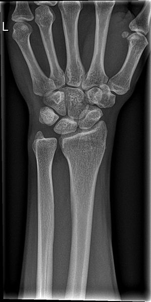
Physical examination revealed wasting of the intrinsic muscles of the right hand, most pronounced in the first dorsal interosseous muscles with weak intrinsic movements when comparison to the left side. Following initial examination a series of investigation and imaging was conducted:

It is not uncommon for golfers to fracture the hook of hamate based on the type of grip and dynamics of the golf swing. Furthermore, they can develop stress fractures of the hook of the hamate, which subsequently do not unite. [11,12]

Whilst this may have been the mechanism for the development of injury, an alternative explanation implicates a congenital anomaly where the primary ossification center the hamate fails to unite with the hook of the hamate giving rise to a bipartite bone (os hamulus proprius). [3]

Findings

This patient had a well-established long-standing asymptomatic non-union of the hamate or an os hamuli proprius, which subsequently became symptomatic following a motor vehicle accident in January 2005 resulting in an acute eight-week history of fine motor control deficit in the right hand.

Surgical intervention

A mobile segment of the hook of the hamate was identified.  Pressure over the mobile segment of the hook of the hamate compressed the motor branch of the median nerve as it traversed around the ulnar and distal hook of the hook of the hamate. The motor branch of the median nerve was swollen proximal to the point where the mobile segment of the hook of the hamate dynamically impacted on the nerve. This had the appearance of a ‘neuroma in continuity’ commonly seen from failure of regenerating nerve growth cone to reach peripheral targets.

The ulnar nerve was released in Guyon’s Canal. The motor branch of the ulnar nerve was identified and dissected as it coursed around the hook of the hamate. The hook of the hamate was very mobile and unstable. Manipulation of the mobile hook of the hamate demonstrated how it impacted and compressed the motor branch of the median nerve distal to the swollen segment of the motor branch of the median nerve. This was surgically excised.

The patient noticed a marked improvement of symptoms within two days post-operatively commenting on a return of ‘power and movement’. Following rehabilitation through daily grip strength exercises; this was further demonstrated on clinical examination at eighteen days confirming a return of intrinsic muscle power in the right hand.

The following five images describe the surgical repair of Os Hamulus Proprius as performed in this case.

Discussion

The hook of the hamate is an important structure providing mechanical stability on the ulnar aspect and protecting the motor branch of the ulnar nerve as it traverses deep into the hand from Guyon’s canal. It is also an important structure for insertion of the flexor retinaculum and as a result the muscles on the ulnar side of the hand. [16]

It is very likely that this abnormality of the hook of the hamate was present prior to his injury. The most likely explanation is that it is a secondary ossification center of the hook of the hamate (os hamulus proprius) which went on to unite. However, it is not possible to completely rule out that this represents a long standing non-union of the hook of the hamate and at some stage in the past he may have sustained a stress fracture which resulted in a non-union. [1,3,6,8,11,17]

Clinical examination plays a crucial role in isolating cases of os hamulus proprius. Patients will often present with clinical signs suggesting ulnar neuropathy such as intrinsic muscle weakness and altered sensation of the hand. In differentiating a case of bipartite hamulus, there will also be marked local tenderness over the hook of hamate with symptomatic pain due to dynamic compression such as when performing a power grip. Further hand and upper limb evaluation can compliment the diagnosis by quantifying and comparing loss of strength in the hand. [17]

The patient had marked motor (intrinsic hand muscles) weakness and some minor impairment of sensation in the ulnar distribution, which is consistent with the electrophysiological abnormalities in the hand. Surgery to remove the mobile segment of hamulus resulted in major improvement – particularly in terms of the level of his symptoms and restoration of normal power to the intrinsic muscles of the hand. Excision of the mobile os hamulus proprius has restored control and sensation of his left hand and enabled him to resume his career as a professional golfer.

Ulnar nerve compression in the hand could be due to a multitude of factors, including a tumour, a ganglion cyst, a fracture of either the pisiform or the hamate, compression in Guyon’s Canal, and an aneurysm of the ulnar artery. [18] To discriminate between a congenital bipartite hamulus or a non union of the hook of the hamate five criteria [17] have been described:

  • Bilaterally similar bipartite hamulus
  • Absence of history or signs of previous trauma
  • Equal size and uniform signal intensity of each part on imaging
  • Absence of progressive degenerative changes between the two components of the hamate or elsewhere in the wrist
  • Smooth well corticated and rounded margins of the hamate and mobile separate hook

Treatment

There are a limited number of options to treat a mobile hamulus segment causing ulnar nerve compression. [8] Initial splinting of the hand can be trialed to prevent dynamic compression of the nerve in the hope that pain and weakness resolve. [5] Furthermore, avoidance of sports relying on grip strength may provide symptomatic relief. If these interventions do not result in the resolution of symptoms, then there is the option of surgically excising the accessory ossification center on the tip of the hook of the hamate with subsequent decompression and release of the ulnar nerve such as presented in this case.

Consent declaration

Informed consent was obtained from the patient for publication of this case report and accompanying figures. IMAGE ONE is taken from http://upload.wikimedia.org/wikipedia/commons/3/31/Gray422.png. This image is in the public domain because its copyright has expired. This applies worldwide.

Acknowledgments

This paper was written under the supervision of Jeff Ecker from the Western Orthopaedic Clinic in Perth, WA.

Conflict of interest

None declared.

Correspondence

S Moniz: monizsheldon@gmail.com

References

[1] Andress M, Peckar V. Fracture of the hook of the hamate. British Journal of Radiology. 1970;43(506):141-143.

[2] Blum AG, Zabel J-P, Kohlmann R, Batch T, Barbara K, Zhu X, et al. Pathologic Conditions of the Hypothenar Eminence: Evaluation with Multidetector CT and MR Imaging1. Radiographics. 2006;26(4):1021-1044.

[3] Bianchi S, Abdelwahab I, Federici E. Unilateral os hamuli proprium simulating a fracture of the hook of the hamate: a case report. Bulletin of the Hospital for Joint Diseases Orthopaedic Institute. 1990;50(2):205.

[4] Grave K, Brown T. Skeletal ossification and the adolescent growth spurt. American journal of orthodontics. 1976;69(6):611-619.

[5] Chow JC, Weiss MA, Gu Y. Anatomic variations of the hook of hamate and the relationship to carpal tunnel syndrome. The Journal of hand surgery. 2005;30(6):1242-1247.

[6] Greene M, Hadied A. Bipartite hamulus with ulnar tunnel syndrome–case report and literature review. The Journal of hand surgery. 1981;6(6):605.

[7] O’Driscoll SW, Horii E, Carmichael SW, Morrey BF. The cubital tunnel and ulnar neuropathy. Journal of Bone & Joint Surgery, British Volume. 1991;73(4):613-617.

[8] Pierre-Jerome C, Roug I. MRI of bilateral bipartite hamulus: a case report. Surgical and Radiologic Anatomy. 1998;20(4):299-302.

[9] Garzón-Alvarado D, García-Aznar J, Doblaré M. Appearance and location of secondary ossification centres may be explained by a reaction–diffusion mechanism. Computers in biology and medicine. 2009;39(6):554-561.

[10] Freyschmidt J, Brossmann J. Koehler/Zimmer’s Borderlands of Normal and Early Pathological Findings in Skeletal Radiography. TIS; 2003.

[11] Koskinen SK, Mattila KT, Alanen AM, Aro HT. Stress fracture of the ulnar diaphysis in a recreational golfer. Clinical Journal of Sport Medicine. 1997;7(1):63.

[12] Torisu T. Fracture of the hook of the hamate by a golfswing. Clinical orthopaedics and related research. 1972;83:91-94.

[13] Viegas SF, Patterson RM, Hokanson JA, Davis J. Wrist anatomy: incidence, distribution, and correlation of anatomic variations, tears, and arthrosis. The Journal of hand surgery. 1993;18(3):463-475.

[14] Berger R, Garcia-Elias M. General anatomy of the wrist. In: Biomechanics of the wrist joint: Springer; 1991.

[15] SHEA JD, McCLAIN EJ. Ulnar-nerve compression syndromes at and below the wrist. The Journal of Bone & Joint Surgery. 1969;51(6):1095-1103.

[16] LaStayo P, Michlovitz S, Lee M. Wrist and hand. Physical Therapies in Sport and Exercise. 2007:338.

[17] Evans MW, Gilbert ML, Norton S. Case report of right hamate hook fracture in a patient with previous fracture history of left hamate hook: is it hamate bipartite? Chiropractic & osteopathy. 2006;14(1):1-7.

[18] Zeiss J, Jakab E, Khimji T, Imbriglia J. The ulnar tunnel at the wrist (Guyon’s canal): normal MR anatomy and variants. AJR. American journal of roentgenology. 1992;158(5):1081-1085.

Categories
Case Reports Articles

Adult Onset Still’s Disease – a diagnostic dilemma

Introduction

ASOD is characterised by fever, an evanescent skin rash, polyarthralgia, hepatosplenomegaly, leucocytosis, liver enzyme elevation and a high serum ferritin level. [1,2,3] It is a difficult diagnosis to make, as there is no pathognomonic test for the disease and it is a great mimicker of other conditions, such as autoimmune disorders and haematological malignancies.

Despite being a separate clinical entity to JIA and rheumatoid arthritis, there is evidence to suggest that AOSD as well as JIA are triggered by viral infections. [2,3,4] The following case demonstrates a young man who was diagnosed with AOSD following an infection with Epstein Barr Virus. This is impetus for a discussion of the interplay between AOSD and a viral aetiology, and the innate and adaptive immune responses in guiding effective therapy.

Case Presentation

In 2012, a previously healthy 20 year old male presented with a sore throat, malaise, tender cervical lymphadenopathy and fever, consistent with infectious mononucleosis. He was transferred to a secondary referral hospital where paired EBV serology was positive for an active infection despite a negative monospot test. The patient’s travel history and past medical history were unremarkable apart from regular alcohol binge drinking.

After being discharged, he began to experience intermittent fevers and night sweats. In addition to this, he had ongoing malaise and was forced to stop work as a mechanic. Weight loss of 10kg occurred during a two month period, along with a persisting microcytic anaemia, with a haemoglobin level of approximately 8.0 g/dL.

His polyarticular pain was distributed mainly to his ankles, knees, shoulders and wrists, and associated with morning stiffness and visible swelling. The pain was partially responsive to regular ibuprofen. He also complained of intermittent pleuritic chest pain. Over a course of two months, his weight stabilised and night sweats improved, but his anaemia and polyarthralgias persisted.

Approximately two months after his initial diagnosis of infectious mononucleosis, the patient represented to hospital with severe polyarthralgias and was unable to walk. During this admission, he was afebrile but had some mild tender cervical lymphadenopathy with no hepatosplenomegaly. A pleural rub was auscultated. He had a salmon-coloured non-blanching rash on the medial aspect of both legs that felt like a ‘sunburn’; this was biopsied. Although the diagnosis of ASOD had previously been considered, the patient was investigated for other causes for these symptoms. The results of these investigations are presented in Table 1. His investigations included a bone marrow and trephine biopsy, which revealed a markedly hypercellular bone marrow. The skin biopsy of the rash on his legs showed a leucocytoclastic vasculitis with perivascular neutrophilic invasion, but negative staining for complement. This finding is non-specific to the condition and can occur due to drug reaction, immune-complex deposition or be idiopathic. [5] As test results did not indicate another likely cause for his symptoms, the patient was commenced on treatment for ASOD and was referred to a rheumatologist.

Case Discussion

This case illustrates the unique clinical and laboratory picture of AOSD, with its intermitting and remitting fevers, polyarthralgias, myalgias, lymphadenopathy, transient macular rash and pleuritis. It is likely that the patient had a degree of pleuritis, as suggested clinically with a pleural rub and on CT imaging. Serositis manifesting as pleuritis, pleural effusions or pericarditis can be encountered in ASOD, but is rare. [3,6] The rash is fleeting and may only last for hours or days, and skin biopsies generally reveal a non-specific perivascular inflammation. [1] Our patient’s thrombocytosis and markedly elevated serum ferritin are reactive changes. The serum ferritin level has been suggested as a predictive marker for AOSD as it is invariably elevated and often higher than levels found in other autoimmune or inflammatory diseases, with a five-fold increase in serum ferritin being 41% specific and 80% sensitive as a diagnostic test. [9] The markedly high ferritin level in AOSD has been attributed to hyper-production by the reticuloendothelial system or hepatocyte damage, and is unrelated to iron metabolism. [8] The patient’s blood results illustrated a microcytic anaemia, although the iron studies point towards an inflammatory reaction.

The leukocyte count appears to correlate well with the activity of illness. The underlying mechanism of this is probably bone marrow granulocyte hyperplasia, as demonstrated on bone marrow biopsy in our patient. It is not uncommon to see marked reductions in red cell counts, weight loss and hypoalbuminaemia in active disease. [8]

In our patient, causes of fever of unknown origin with or without rash were considered, such as endocarditis, haematological malignancies and systemic vasculitides. The single cytopaenia, normal LDH and bone marrow biopsy excludes leukaemia, lymphoma and myelodysplasia. It is unlikely he had a protracted course of EBV due to the nature of his symptoms and degree of anaemia, in addition to the negative EBV IgM serology. Given the recent heavy rainfall, migrating polyarthritic conditions such as Ross River and Barmah Forest viruses were considered in the differentials.

The diagnosis of ASOD is made after taking into account the patient’s medical history and risk factors for other infectious agents, environment and relevant infectious diseases epidemiology. Although being a diagnosis of exclusion, there are two commonly used clinical criteria in practice, that being Yamaguchi (Table 2), which has been shown to be most sensitive (93.5%) followed by Cush’s (80.6% sensitivity). [7,8]

In regards to the aetiology of AOSD, there have been numerous case reports of AOSD following viral infection, [4,10] with one citing an older female patient diagnosed with AOSD after EBV infection. [2] Other implicated viruses include rubella, mumps, cytomegalovirus, parainfluenza, human herpes virus 6, echovirus, parvovirus B19, and bacterial infections like mycoplasma pneumoniae, chlamydia pneumonia, yersinia enterocolitica and borrelia. [2,8] Although relevant to our patient, the link between infections and AOSD has not been robustly established from an aetiological perspective, [10] and probably only forms part of the multifaceted pathogenesis, that being a dysregulated immune system combined with susceptible HLA loci. However, no consistent associations between AOSD and particular HLA loci have been elucidated, although HLA-B17, HLA-B18, HLA-B35 and HLA-DR2 have been implicated. [6]

It does appear that pathogenesis of the condition overlies autonomous activity of both innate and adaptive immune systems. Patients with AOSD often show hypercomplementaemia, and serum levels of IL-1β, IL-6, IL-18, TNFα, IFN Ɣ and macrophage-colony stimulating factor (M-CSF) have been found to be considerably higher than compared with controls. [6,7,11] These cytokines also appear to share a role in increasing the production of ferritin. [1,12] IL-18 is predominantly secreted by macrophages and has been implicated in hepatotoxicity [13] and joint disease, [7] and may be the cause of liver enzyme derangement characteristic of AOSD. Serum IL-18 levels also appear to correlate significantly with serum ferritin levels. [8] Furthermore, IL-18 may be seen as the part of the bridge between activation of the innate and adaptive immune systems in AOSD, as it facilitates the Th1 response and induces other cytokines like IL-1 β, TNFα and IFNƔ. [6] Pro-inflammatory cytokines such as IL-6, TNFα and IFN Ɣ also increase the expression of Toll-like receptors (TLR), and high circulating levels of cytokines leads to a higher sensitivity of TLR to anti-microbial or viral peptides, thus creating a self-perpetuating cycle of inflammatory response and augmentation. [14]

On the adaptive immunity side of the pathogenesis, the role of T cells in pathogenesis has been well documented. [11,14] Dysregulated production of a particular subset of T helper cells, called Th17 cells, that secrete IL-17 have been implicated in the development of autoimmune diseases. [15] Significantly higher levels of Th17 cells and serum IL-17 levels were found in both AOSD and SLE patients, and there was a parallel decrease with clinical remission. [10] IL-17 stimulates monocytes to produce IL-6 and IL-1β, which are also principle cytokines involved in the differentiation of CD4+ T cells into Th17 cells. [6] These therefore augment and maintain the inflammatory cascade. [16]

Non-steroidal anti-inflammatory drugs (NSAIDS) had previously been the first line medication for ASOD, despite only being effective monotherapy in less than 15% of patients. [10] The benefits of corticosteroids are higher when patients have more pronounced joint disease, with a response rate of two thirds of the patient population. [10]

Highlighting the implicated cytokines, namely IL-1β, IL-6 and TNFα, [17,18] will guide the use of targeted therapies such as the disease modifying anti-rheumatic drugs (DMARDS). There have been favourable results with corticosteroids, and more than two thirds of patients require corticosteroids after NSAIDs are attempted as symptom relief. [6] The use of DMARDS are indicated where the condition is refractory to corticosteroids without signs of remission, or in combination as corticosteroid-sparing agents. This includes methotrexate, which has indirect actions on TNFα and IL-6. Although there is a lack of robust evidence regarding TNF in the pathogenesis of ASOD compared to rheumatoid arthritis, the use of etanercept and infliximab have shown significant improvement in disease in several case series. [10] Of particular note, there is increasing evidence to suggest that anakinra, an IL-1 receptor antagonist, is well tolerated, and several case series have yielded positive results in ameliorating the disease at a haematological, biochemical and cytokine level. An excess of IL-1β inducing factor has been demonstrated in JIA, a condition that also shares similar pathogenesis to that of AOSD. [6,19]

The clinical course of AOSD is heterogeneous, with patients falling into one of three clinical patterns. The first group which affects about 60% of patients [8] is a monocyclic systemic group with only one episode of systemic manifestations, with complete remission within one year of the onset of symptoms. The second group is polycyclic systemic, whom experience more than one episode which is followed by partial or total remission. The third group is a chronic articular group, with persistent polyarthritis lasting longer than 6 months. [6] In the chronic group, the average duration of disease is 10 years, the symptoms appear to be less permanent than other rheumatological diseases and the disease shows less propensity to interfere with social functioning or time off from work despite disability and the need for long-term medication. [20]

Patient outcome

The patient improved satisfactorily with regular ibuprofen and prednisolone 20 mg daily and was discharged after day 7 with a tapering steroid dose.

He was able to resume work, but continued to experience mild intermittent polyarthralgias with no other significant systemic symptoms. Six months post-admission, deterioration in arthritic symptoms prompted the addition of methotrexate.

Key points

  • Adult Onset Stills Disease is a rare systemic inflammatory disorder that mainly affects people aged 16-35 years old.
  • It is a difficult diagnosis to make, and one that must be questioned continually, as it is a mimicker of other disorders, including other causes of fever of unknown origin, infectious diseases and malignancy.
  • It is characterised by both clinical and laboratory manifestations like fever, evanescent rash, polyarthritis and polymyalgias, microcytic anaemia, leucocytosis, thrombocytosis and marked hyperferritinaemia.
  • Treatment is based on clinical course and is similar to that of rheumatoid arthritis. A more targeted biological disease modifying therapy should be chosen with consideration of likely pathogenic pro-inflammatory cytokines.

Consent declaration

Consent from the patient was gained for the writing and distribution of this article for education purposes.

Acknowledgements

Thank you to Dr. Hedley Griffiths, Consultant Rheumatologist.

Conflict of interest

None declared.

Correspondence

S Ooi: soo@deakin.edu.au

References

[1] Mehrpoor G, Owlia M. Adult – onset Still’s disease: A review. Indian J Med Sci. 2009;63(5):207-21.

[2] Schifter T, Lewinski U. Adult onset Still’s disease associated with Epstein-Barr virus infection in a 66-year-old woman. Scand J Rheum. 1998;27(6):458-60.

[3] Mert A, Ozaras R, Tabak F, Bilir M, Ozturk R, Ozdogan H, et al. Fever of unknown origin: a review of 20 patients with adult-onset Still’s disease. Clin Rheum. 2003;22(2):89-93.

[4] Wouters J, van der Veen J, van de Putte L, de Rooij D. Adult onset Still’s disease and viral infections. Ann Rheum Dis. 1988;47(9):764-7.

[5] Koutkia P, Mylonakis E, Rounds S, Erickson A. Leucocytoclastic vasculitis: an update for the clinician. Scand J Rheum. 2001;30(6):315-22.

[6] Kontzias A, Efthimiou P. Adult-onset Still’s disease: pathogenesis, clinical manifestations and therapeutic advances. Drugs. 2008;68(3):319-37.

[7] Bagnari V, Colina M, Ciancio G, Govoni M, Trotta F. Adult-onset Still’s disease. Rheumatol Int. 2010;30(7):855-62.

[8] Kádár J, Petrovicz E. Adult-onset Still’s disease. Best Pract Res Cl Rh. 2004;18(5):663-76.

[9] Lian F, Wang Y, Yang X, Xu H, Liang L. Clinical features and hyperferritinemia diagnostic cutoff points for AOSD based on ROC curve: a Chinese experience. Rheumatol Int. 2012;32(1):189-92.

[10] Mavragani C, Spyridakis E, Koutsilieris M. Adult-Onset Still’s Disease: From Pathophysiology to Targeted Therapies. Int J Inflamm. 2012;2012:1-10.

[11] Efthimiou P, Georgy S. Pathogenesis and management of adult-onset Still’s disease. Semin Arthritis Rheu. 2006;36(3):144-52.

[12] Mehrpoor G, Owlia MB, Soleimani H, Ayatollahi J. Adult-onset Still’s disease: a report of 28 cases and review of the literature. Mod Rheumatol. 2008;18(5):480-5.

[13] Kasper D, Braunwald E, Fauci A, Hauser S, Longo D, Jameson J, Loscalzo J. Harrison’s principles of internal medicine (17th ed.). New York: McGraw-Hill Medical Publishing Division; 2008.

[14] Kramer M, Joosten L, Figdor C, van den Berg W, Radstake T, Adema GJ. Closing in on Toll-like receptors and NOD-LRR proteins in inflammatory disorders. Future Rheumatol. 2006;1(4):465-79.

[15] Lichtman M, Kipps T, Seligsohn U, Kaushansky K, Prchal J. Williams Hematology. 8th ed. USA: McGraw-Hill Companies; 2010.

[16] Chen D, Chen Y, Lan J, Lin C, Chen H, Hsieh C. Potential role of Th17 cells in the pathogenesis of adult-onset Still’s disease. Rheumatology. 2010;49(12):2305-12.

[17] Nordstrom D, Knight A, Luukkainen R, van Vollenhoven R, Rantalaiho V, Kajalainen A, et al. Beneficial Effect of Interleukin 1 Inhibition with Anakinra in Adult-onset Still’s Disease. An Open, Randomized, Multicenter Study. J Rheumatol. 2012 October 1, 2012;39(10):2008-11.

[18] Efthimiou P, Kontzias A, Ward C, Ogden N. Adult-onset Still’s disease: can recent advances in our understanding of its pathogenesis lead to targeted therapy? Nat Clin Prac Rheumatol. 2007;3(6):328-35.

[19] Allantaz F, Stichweh D, Pascual V. Interleukin-1 as a therapeutic target in systemic-onset juvenile idiopathic arthritis. Future Rheumatol. 2007;2(3):305-12.

[20] Sampalis J, Esdaile J, Medsger Jr T, Partridge A, Yeadon C, Senécal J, et al. A controlled study of the long-term prognosis of adult still’s disease. Am J Med. 1995;98(4):384-8.


Categories
Case Reports Articles

Acute viral bronchiolitis in the setting of extensive family history of asthma

This case report describes a previously healthy eleven-month old ex-preterm female with a severe presentation of acute viral bronchiolitis with an extensive family history of asthma. The link between viral bronchiolitis and asthma has always been controversial despite extensive research. Several studies have linked respiratory syncytial virus (RSV) bronchiolitis to the development of persistent wheezing or asthma later in childhood, even suggesting that a dose-response relationship may exist between the two entities. Some studies have also demonstrated that severe lower respiratory infections in the first year of life are important contributors to asthma, particular in those sensitized during infancy. On the other hand, it has also been studied as to whether an individual at risk of asthma has any impact on the severity of bronchiolitis. Despite numerous studies, results have largely been inconclusive, and the question of whether it is RSV that directly results in asthma, or if the susceptibility to RSV is conferred due to predisposing pulmonary pathology, still remains unknown.

Case

Sally was an eleven-month old ex-preterm (35 weeks) female who presented to the Emergency Department (ED) with symptoms of fever, coryzal symptoms and a wheeze, subsequently diagnosed as viral bronchiolitis.

History

Sally had become acutely febrile two nights prior to presentation, developing coryzal symptoms the following morning. Sally was initially treated for an upper respiratory tract infection (URTI) and acute otitis media with amoxicillin by her general practitioner, but began to worsen over the subsequent 24 hours, with laborious and wheezy breathing, coupled with a persistent fever of 38.4oC. Despite two doses of salbutamol, her breathing continued to deteriorate, leading to her presentation at the ED. There were no apnoeic or cyanotic episodes, rigors or any associated inspiratory stridor. During this period, Sally was anorexic, with subsequently fewer nappy changes, and was reported by her parents to be far less active than usual. She was previously well, with no known sick contacts, and her vaccinations were up-to-date.

Sally had a similar episode of bronchiolitis in at eight months of age but was treated then as an outpatient. There was an extensive family history of asthma (Figure 1), and both the patient’s siblings had bronchiolitis as infants. At eleven months, Sally was meeting all the developmental parameters for her age. There were no known drug allergies and other than salbutamol PRN, the patient was not on any other medication. There was no remarkable social history.

Examination

Sally appeared lethargic and was in respiratory distress, with tachypnoea, an audible wheeze and classical signs of increased breathing effort (nasal flaring, tracheal tug and subcostal recession). She had a respiratory rate of 78 and was saturating at 100% on 8L supplementary oxygen, which decreased to 90% on room air, and was febrile at 38.5oC. She was tachycardic at a heart rate of 170. There were no clinical signs of dehydration. On auscultation, air entry was reduced bilaterally, with an expiratory wheeze and diffuse crackles present. There was no evidence of stridor, increased vocal resonance, or dullness to percussion. An ear, nose and throat examination revealed an erythematous pharynx but was otherwise unremarkable. All other examination findings were unremarkable.

Workup and Progress

The presenting symptoms suggested a diagnosis of acute viral bronchiolitis. However, with the extensive family history of asthma, Sally’s presentation could be her first virus- triggered asthma attack. While the clinical presentation suggested otherwise, there were concerns over the possibility of pneumonia, which had to be ruled out in the workup. In consideration of the severity of her initial presentation and the likely further deterioration until its peak at day 2-3, basic investigations were ordered. These included a full blood count, urea, electrolytes and creatinine parameters, a chest X-ray and a nasopharyngeal aspirate. There were no abnormal findings.

Sally was commenced on immediate supportive therapy. An IV line was inserted and she was commenced on 75% maintenance fluids as per guidelines to avoid Syndrome of Inappropriate Antidiuretic Syndrome. [1] She was also commenced initially on 2L oxygen as per guidelines for respiratory distress, [2] but failed to saturate appropriately until given 8L of humidified oxygen via nasal prongs, where she maintained 100% O2 saturation. While the efficacy of short-acting beta agonists (SABAs) in the acute management of bronchiolitis has been inconclusive despite extensive research, current guidelines recommended a trial of bronchodilators in infants >6 months. [3] Sally was administered six puffs of Salbutamol MDI (100mcg/ puff) via spacer [1] but with no results, hence the regime was discontinued.

The Royal Children’s Hospital (RCH) provides further guidance regarding management based on clinical signs and symptoms. Admission to the intensive care unit (ICU) was indicated for Sally to allow continuous cardiorespiratory monitoring and supportive management. Observations were performed hourly and with only supportive management, her oxygen requirements were weaned down to 2L over 24 hours.

Discussion

Bronchiolitis during infancy and asthma in childhood – is there a causal link? Should infants at high risk of asthma receive Palivizumab immunization?

Several studies have linked respiratory syncytial virus (RSV) bronchiolitis to the development of persistent wheezing or asthma later in childhood. In a long-term prospective cohort study, there was a relative risk of 2.8 of developing wheezing at 5.5 years in children who had had bronchiolitis. [4] Sigurs et al. (2005) also reported a ten-fold excess of asthma in a similar study. [5]

It has also been suggested that a dose-response relationship exists between bronchiolitis and asthma. In a study involving 90,341 children, Carroll et al. (2009) demonstrated that the odds ratios (OR) for asthma as a child were 1.86 (95% CI, 1.7-2.0), 2.41 (95% CI, 2.2-2.6) and 2.8 (95% CI, 2.6-3.0) in the outpatient, ED, and hospitalization groups, respectively, compared to children without bronchiolitis. [6] Henderson et al. (2005) also noted an OR of 2.5 (95% CI 1.4-4.3) of developing asthma with hospitalization for RSV bronchiolitis, [7] as did two prospective studies, which showed a 30-40% likelihood of subsequent asthma. [8]

RSV bronchiolitis as a direct cause of asthma

In an extensive seven-year REBEL prospective cohort study, Bacharier et al. (2012) reported that increased Chemokine (C-C motif) Ligand 5 (CCL5) expression in nasal epithelial cells during RSV infection carried an OR of 3.8 (95% CI, 1.2-2.4) for developing asthma. [9] This is in concordance with studies demonstrating increased CCL5 levels in subjects with asthma, [10] as well as in vitro studies demonstrating increased expression and transcription by RSV. [11] Unfortunately, the study failed to measure CCL5 levels prior to infection and thus the causal relationship has not been established. Hence, whether it was RSV that directly resulted in asthma, or if the susceptibility to RSV was conferred due to predisposing pulmonary pathology, [12] still remains unknown.

A five-year cohort study on children at high risk of atopy by Kusel et al. (2007) demonstrated that severe lower respiratory infections in the first year of life are important contributors to asthma, particular in those sensitized during infancy. [13] These findings suggest that protecting high-risk individuals from infection during infancy may be considered for long-term asthma prevention.

Effect of family history of asthma or atopy on severity of bronchiolitis

It has also been studied whether an individual at risk of asthma has any impact on the severity of bronchiolitis. This was particularly relevant in the Sally’s case, with her significant family history of asthma. Results in this field have been conflicting, with most studies not eliciting any significant association. However, Gurwitz et al. (1981) demonstrated that hospitalized cases were associated with a higher incidence of first-degree relatives with bronchial hyper-responsiveness. [14] A study by Trefny et al. (2000) also demonstrated similar results. [15]

Should high-risk atopic individuals receive Palivizumab immunization during RSV season for prevention of asthma?

Passive immunization with Palivizumab is currently recommended only for high-risk infants to prevent serious complications arising from RSV infections. [16] However, a recent double-blinded RCT in the Netherlands has begun examining its preventive effect on recurrent wheeze in healthy preterm children 33-35 weeks gestational age (MAKI trial), based on a non-randomized trial suggesting a prevention of wheeze in 50% of preterm children. [17] Such a study would complement this case study’s patient profile, and would be especially relevant in the context of her rich family history of asthma which puts her at high risk, and the abovementioned association but inconclusive causation between bronchiolitis and asthma.

From an economic standpoint, studies assessed the cost-effectiveness of Palivizumab, albeit in the context of high-risk premature infants (32-35 weeks). Unfortunately, the predisposition of these infants to a higher disease burden and costlier hospitalizations constitutes a higher cost per QALY [18]      compared to this case study’s patient, but even then there is still considerable controversy over its cost-effectiveness, especially across various healthcare systems.

Conclusion

In summary, this was a case of severe viral bronchiolitis warranting ICU admission for supportive management, on a background of an extensive family history of asthma. While studies have shown a clear association of bronchiolitis with asthma, causation has not been conclusively established, with family history of atopy possibly interacting in the development of asthma. Current research is lacking in the area of Palivizumab prophylaxis in the interest of asthma prevention in healthy children, but the evidence would suggest that it is likely to be cost-ineffective.

Conflict of interest

None declared.

Correspondence

G Yong: glenn.yong.kw@gmail.com

References

[1] Royal Children’s Hospital. Bronchiolitis- Ongoing Management. http://www.rch.org.au/rchcpg/hospital_clinical_guideline_index/Bronchiolitis_Ongoing_Management/#Fluid_requirements (accessed 11 March 2013)

[2] Royal Children’s Hospital. Oxygen Delivery. http://www.rch.org.au/rchcpg/hospital_clinical_guideline_index/Oxygen_delivery/ (accessed 11 March 2013)

[3] [Guideline] Diagnosis and management of bronchiolitis. Pediatrics. 2006 Oct;118(4):1774-93.

[4] Murray M, Webb MS, O’Callaghan C, Swarbrick AS, Milner AD. Respiratory status and allergy after bronchiolitis. Arch Dis Child. 1992 April;67(4): 482-7.

[5] Sigurs N, Bjarnason R, Sigurbergsson F, Kjellman B. Respiratory syncytial virus bronchiolitis in infancy is an important risk factor for asthma and allergy at age 7. Am J Respir Crit Care Med. 2000;161:1501–7.

[6] Carrol KN, Wu P, Gebretsadik T, Griffin MR, Dupont WD, Mitchel EF, Hartert TV. The severity- dependent relationship of infant bronchiolitis on the risk and morbidity of early childhood asthma. Journal of Allergy & Clinical Immunology 2009;123:1055-61.

[7] Henderson J, Hilliard TN, Sherriff A, Stalker D, Al Shammari N, Thomas HM. Hospitalization for RSV bronchiolitis before 12 months of age and subsequent asthma, atopy and wheeze: a longitudinal birth cohort study. Pediatric Allergy Immunology. 2005;16:386–92.

[8] Sign AM, Moore PE, Gern JE, Lemanske RF, Hartert TV. Bronchiolitis to asthma A review and call for studies of gene-virus interactions in asthma causation. Am J. Respir. Crit Care Med. January15 2007;175(2):108-19.

[9] Bacharier LB, Cohen R, Schweiger T, Yin-Declue H, Christie C, Zheng J et al. Determinants of asthma after severe respiratory syncytial virus bronchiolitis. Journal of Allergy & Clinical Immunology 2012;130:91-100.

[10] Humbert M, Ying S, Corrigan C, Menz G, Barkans J, Pfister R et al. Bronchial mucosal expression of the genes encoding chemokines RANTES and MCP-3 in symptomatic atopic and nonatopic asthmatics: relationship to the eosinophilactive cytokines interleukin (IL)-5, granulocyte macrophage-colony-stimulating factor, and IL-3. Am J Respir Cell Mol Biol 1997;16:1-8.

[11] Koga S, Novick AC, Toma H, Fairchild RL. CD81T cells produce RANTES during acute rejection of murine allogeneic skin grafts. Transplantation 1999;67:854-64.

[12] Adamko DJ, Friesen M. Why does respiratory syncytial virus appear to cause asthma? Journal of Allergy & Clinical Immunology 2012 Jul;130(1):101-2.

[13] Kusel MM,  de Klerk NH, Kebadze T Vohma V, Holt PG, Johnston SL et al. Early-life respiratory viral infections, atopic sensitization, and risk of subsequent development of persistent asthma. Journal of Allergy & Clinical Immunology 2007;119:1105-10.

[14] Gurwitz D, Mindorff C, Levison H. Increased incidence of bronchial reactivity in children with a history of bronchiolitis. J Pediatr 1981;98:551–5.

[15] Trefny P, Stricker T, Baerlocher C, Sennhauser FH. Family history of atopy and clinical course of RSV infection in ambulatory and hospitalized infants. Pediatric Pulmonology 2000;30:302–6.

[16] Wang D, Byliss S, Meads C. Palivizumab for immunoprophylaxis of respiratory syncytial virus (RSV) bronchiolitis in high-risk infants and young children: a systematic review and additional economic modelling of subgroup analyses. Health Technol Assess 2011 Jan;15(5):1-124.

[17] Broughton S, Bhat R, Roberts A, Zuckerman M, Rafferty G, Greenough A. Diminished lung function, RSV infection, and respiratory morbidity in prematurely born infants. Arch Dis Child Jan 2006;91(1):26-30.

[18] Smart KA, Paes BA, Lanctot KL. Changing costs and the impact of RSV prophylaxis. Journal of Medical Economics. 2010;13(4):705-8.


Categories
Case Reports Articles

A case of solid pericardial metastases causing constrictive pericarditis in a patient with non small cell lung cancer

Introduction: Cardiac metastases are rarely diagnosed in patients with cancer despite being a common finding at autopsy. We report on a case of pericardial metastases in a patient with non small cell lung cancer (NSCLC) in the setting of coexisting direct tumour invasion into the superior vena cava (SVC) and right atrium. Case: We present a case of a 61-year old gentleman with metastatic adenocarcinoma of the lung. He presented with 2 – 3 days of progressive dyspnoea associated with pre-syncope. His physical examination was significant for bilateral elevated jugular venous pressure (JVP), bilateral arm oedema and hypotension. These clinical signs were explained by solid pericardial metastases causing constrictive pericarditis in the absence of significant pericardial effusion, and direct invasion of tumour into the SVC and right atrium producing SVC obstruction. His admission was complicated by an episode of supraventricular tachycardia (SVT), presumably caused by compromise of the electrical conduction system within the right atrium. Discussion: Cardiac metastasis is a common occurrence in advanced neoplastic disease, but is often clinically silent. The mechanism of cardiac metastases is believed to be primarily through lymphatic channels and metastasis is most commonly located in the pericardium. Symptoms of cardiac metastasis can be understood with respect to their anatomical position and are best diagnosed using transoesophageal echocardiogram (TOE). The most concerning complication of metastasis to the heart is pericardial effusion leading to life-threatening cardiac tamponade. This is an oncological emergency and is treated with pericardiocentesis and follow up preventative measures. Ultimately, cardiac metastasis signals advanced disease and poor prognosis.

Introduction

Cardiac metastases occur in 20-30% of patients with non small cell lung cancer (NSCLC) [1] but are clinically silent in the majority of cases. [2] We report on a case of constrictive pericarditis caused by solid pericardial metastases concurrent with direct invasion of tumour into the superior vena cava (SVC) and right atrium. The clinical picture was complicated by SVC obstruction and right atrial compromise causing supraventricular tachycardia (SVT).

The case

The patient was a 61-year old store worker with a previous 60 pack-year smoking history.  He was admitted with progressive dyspnoea over 2-3 days, associated with pre-syncope, on a background of metastatic NSCLC (T4N0M1b).

Past Medical History

The patient initially presented with a six month history of pain when abducting his right arm, which was associated with dyspnoea and a productive cough. A chest computed tomography (CT) identified a large right hilar lesion causing right middle lobe bronchus occlusion and collapse of the right lung. The tumour had invaded the mediastinum, and was attenuating the SVC and compromising the right atrium. Metastases to the liver, the right sternoclavicular joint and the 4th, 9th and 10th ribs were also identified.

Pathology from the bronchoscopy biopsy demonstrated that the tumour was p63 negative and Thyroid Transcription Factor 1 (TTF-1) positive via immunohistochemical staining, consistent with adenocarcinoma of the lung. Epidermal Growth Factor Receptor (EGFR) screening was negative, hence the tumour was not sensitive to treatment to EGFR tyrosine kinase inhibitors. His management plan, following multidisciplinary team discussion, was radiotherapy to his chest and clavicle for pain management, and palliative chemotherapy (carboplatin/paclitaxel).

Three months post initial diagnosis, restaging CT of the chest, abdomen, pelvis and lumbar spine was organised after completion of radiotherapy and two cycles of chemotherapy. CT scans of the chest, abdomen and pelvis showed disease progression with new right adrenal metastases and new pericardial metastases (Figures 1 and 2). CT of his lumbar spine revealed disease in L1-2 and S1. In light of his latest CT results, radiotherapy to the lumbar spine and pericardium was planned.

Medications and Allergies

At time of admission the patient’s medications included oxycodone/naloxone, omeprazole and dexamethasone. He was allergic to penicillin.

Inpatient Admission

The patient was admitted after worsening dyspnoea was noticed during radiotherapy to his lumbar spine. Initial physical examination revealed mild bilateral pitting oedema at the ankle and no other signs. His initial investigations included full blood examination (FBE), urea, electrolytes and creatinine (UEC), liver function tests (LFTs), and calcium, magnesium and phosphate (CMP). All were normal except for a decrease in haemoglobin (Hb: 83). A provisional diagnosis of anaemia secondary to chemotherapy or neoplastic disease was made based on his haemoglobin, and he was given two units of packed red blood cells.

The following morning, the patient’s dyspnoea had not improved with transfusion.  On examination, his jugular venous pressure (JVP) was elevated bilaterally at four centimetres, his arms were swollen bilaterally and his blood pressure (BP) was 95/55. He was not tachycardic. An urgent electrocardiogram (ECG) and transthoracic echocardiogram (TOE) was performed to exclude cardiac tamponade. ECG was normal and a pericardial effusion, though noted on TOE, was trivial and deemed insufficient to cause cardiac tamponade. However, the echocardiogram demonstrated pericardial metastases overlying the left and right ventricular apex as well as the lateral left ventricular wall and inferior right ventricular wall. Significant echocardiography findings consistent with constrictive pericarditis included abnormal septal motion with marked septal bounce, annulus reversus on tissue Doppler, and left diastolic dysfunction with shortened deceleration time.

On day three of his admission, the patient had an episode of supraventricular tachycardia. His tachycardia was asymptomatic and he was treated conservatively with fluid hydration. Fluid resuscitation was unsuccessful and he remained tachycardic and hypotensive. Amiodarone 200mg was delivered and his heart rate and BP normalised gradually over eight hours. Electrolyte replacement was also initiated after UEC and CMP results revealed mildly decreased potassium and magnesium.

Outcome

The patient remained as an inpatient for a further two weeks, amiodarone was gradually reduced from 200mg three times daily to 200mg daily and he had no further episodes of SVT. He completed radiotherapy to his lumbar spine and pericardium. Chemotherapy was ceased due to disease progression and functional decline.

Before discharge, the patient enquired about his prognosis which was carefully explained to him and communicated to his family. He was discharged to a palliative care unit, where he died six days later.

Discussion

Cardiac metastases are rare clinical ante-mortem diagnoses, as they are silent in more than 90% of patients. [2] Most cases of cardiac metastases are diagnosed post-mortem and, as a result, most epidemiological data regarding cardiac metastases are from autopsy results. The reported incidence according to the literature up to 15% [3] in oncology patients.  An increased incidence has been reported due to modern diagnostic tools and improved survival of cancer patients, secondary to improved treatment and change in the natural cancer history. [2]

In theory, any primary malignancy has the potential to spread to the heart. The rates of metastasis in different tumour types were reported by Bussani et al. in 2007 [4] in a review of post-mortem studies performed at the University of Trieste, Italy, where over 80% of in-hospital deaths are examined by autopsy. They reviewed data from 1994 to 2003 and their reported rates of cardiac metastasis in different tumour types is summarised in the table below (Table 1). Currently, the only tumours which have not been demonstrated to metastasise to the heart are central nervous system tumours. [5]

Tumours can spread to the heart via one of four routes: 1) direct invasion, 2) haematogenous spread, 3) lymphatic spread and 4) intracavitary diffusion. [4] Tumours which originate near the heart, such as bronchial and oesophageal tumours, can directly invade the heart. Lymphatic channels facilitate pericardial metastases, whereas haematogenous routes seed myocardial metastases. [5] Endocardial metastases arise from a combination of haematogenous and intracavitary diffusion through other layers of the heart. [4]

The most common site of cardiac metastases is the pericardium, followed by the myocardium and endocardium. [4-7] With a preference for lymphatic spread, lung and breast carcinomas commonly spread to the pericardium, whereas lymphomas, leukaemias, sarcomas and malignant melanomas spread haematogenously and seed in the myocardium. [5] Only in isolated cases has there been tumour spread to the valves. [4,7]

Clinical evidence of cardiac metastases is variable ante-mortem. However, common presenting symptoms of cardiac involvement include dyspnoea, cough, palpitations, syncope and chest pain. [2] Presentations of cardiac metastases may be obscured by symptoms of advancing primary malignancy, but they can also present as life-threatening emergencies, such as cardiac tamponade, myocardial rupture, ventricular arrhythmia and, rarely, acute myocardial infarction. [2] In some cases, the rise of symptoms from cardiac involvement may be the only indication of an underlying malignancy. [8,9]

In our case, cardiac metastases presented with dyspnoea and elevated JVP. Relevant differentials for dyspnoea associated with elevated JVP included intracardiac SVC obstruction, cardiac tamponade, constrictive pericarditis, radiation pericarditis and restrictive cardiomyopathy. CT chest demonstrated SVC obstruction, and echocardiography findings were suggestive of constrictive pericarditis caused by solid pericardial metastases. As such, the patient’s dyspnoea and elevated JVP were likely to have been caused by a combination of tumour compression of the SVC reducing venous return to the right atrium and impaired diastolic filling due to an inelastic pericardium in constrictive pericarditis.

Presentations of cardiac metastases can be explained by the anatomical position of the metastases. Pericardial lesions cause pericarditis, which lead to serosanguineous or haemorrhagic pericardial effusions and, in most cases, cardiac tamponade. [5] Replacement of the myocardium and endocardium with tumour can cause systolic or diastolic heart failure, particularly if the ventricles are involved. [4] Myocardial infarctions occur when a neoplasm-induced embolus occludes the coronary circulation, or when coronary arteries are directly compressed or invaded by tumour or pericardial effusion. [4] Arrhythmias are common in the setting of any neoplastic involvement of the heart. [10]

The investigation of choice in detecting cardiac metastases is transoesophageal echocardiography. [5] Pericardial involvement is strongly indicated by a thickened pericardium, or in some cases, as a cauliflower-like projection into the pericardial fluid space. [11] Pericardial effusions can be detected with high sensitivity, and pericardiocentesis can be immediately performed under ultrasound guidance, quickly verifying the diagnosis of metastatic disease. Other imaging modalities such as MRI and CT can determine the size and extension of the tumour more precisely, and provide information on the characteristics of the lesion. [5,12] As such, myocardial metastases are better demarcated by CT and MRI over ultrasonography.

ECG findings in cardiac metastases are non-specific, although more than two thirds of patients with cardiac metastases show some degree of abnormality. [2] Similarly, chest radiography has limited use, but may reveal an increased cardiac silhouette from pericardial effusion or pericardial tumour. Chest radiography may demonstrate a primary lung tumour or pleural effusion resulting from heart failure. [5] There have been rare cases of osteogenic sarcoma metastases to the heart which contained bone and were visualised on the chest radiography.  [13]

Biopsy of cardiac metastases is rarely indicated, as less invasive imaging techniques are usually adequate to suggest tumour type and determine if surgery is feasible. However, biopsies of the heart can be done using fluoroscopy- or ultrasound-guided techniques, or through open surgery. [5] Coronary angiogram studies have value in surgical planning. However, the above techniques are rarely utilised in secondary cardiac tumours, and are more significant in the evaluation of likely primary tumours. [5,14]

Surgical treatment of cardiac tumours is uncommon and reserved for those with good long-term prognosis. Radiotherapy is commonly used to relieve local symptoms, provide local control and obtain haemodynamic stability. [2] Chemotherapy is also employed if the tumour is chemo-sensitive, as in the case of lymphomas, leukaemias and germ cell tumours. The life expectancy of pericardial metastases without treatment is reported to be 1.75 weeks. [15] With treatment, namely radiotherapy and periocentesis when necessary, life expectancy was extended to 22.5 weeks. [15]

Immediate treatment is required in patients presenting with pericardial effusions leading to life-threatening cardiac tamponade. Drainage of the pericardial fluid by pericardiocentesis is required, but effusions return in up to 60% of cases. [16] Thus, treatment of the initial effusion is combined with prevention of recurrence, which can be achieved with prolonged catheter drainage, obliteration of the pericardial space or creation of a permanent pericardial window which drains into the pleural or peritoneal cavity. The utilisation of sclerosing agents and instillation of chemotherapeutic agents in the pericardium have also been shown to prevent effusions. [2]

Conclusion

This case demonstrates salient features of cardiac involvement in metastatic lung cancer, including primary invasion into the SVC and right atrium as well as metastatic involvement of the pericardium. While most cardiac metastases are silent, or obscured by advanced disease, this case has highlighted clinical complications of cardiac involvement, including SVC obstruction, SVT and constrictive pericarditis.  Suspicion of cardiac metastases should always be high in oncology, as it allows prompt treatment and optimal comfort of the patient.

Consent declaration

Informed consent was obtained from the patient for publication of this case report and accompanying figures.

Acknowledgements

The authors would like to thank Dr. Vishal Boolell and Dr. Peter Briggs for their advice and supervision.

Conflict of interest

None declared.

Correspondence

J Ng: jwng9@student.monash.edu

References

[1] Tamura A, Matsubara O, Yoshimura N, Kasuga T, Akagawa S, Aoki N. Cardiac metastasis of lung cancer. A study of metastatic pathways and clinical manifestations. Cancer 1992;70(2):437-42.

[2] Al-Mamgani A, Baartman L, Baaijens M, de Pree I, Incrocci L, Levendag PC. Cardiac metastases. Int J Clin Oncol 2008;13(4):369-72.

[3] Mukai T, Shinka T, Tominaga K, Shimosata Y. The incidence of secondary tumours of the heart and pericardium: a 10-year study. Jpn J Clin Oncol 1988;18(3):195–201.

[4] Bussani R, De-Giorgio F, Abbate A, Silvestri F. Cardiac metastases. J Clin Pathol 2007;60(1):27-34.

[5] Reynen K, Kockeritz U, Strasser RH. Metastases to the heart. Ann Oncol 2004;15(3): 375-81.

[6] Klatt EC, Heitz DR. Cardiac Metastases. Cancer 1990;65(6):1456-9.

[7] Malaret GE, Aliaga P. Metastatic disease to the heart. Cancer 1968;22(2):457-66.

[8] Imazio M, Demichelis B, Parrini I, Favro E, Beqaraj F, Cecchi E, Pomari F, Demarie D, Ghisio A, Belli R, Bobbio M, Trinchero R. Relation of acute pericardial disease to malignancy. Am J Cardiol. 2005;95(11):1393-4.

[9] Sosinska-Mielcarek K, Sosvinska-Mielcarek K, Senkus-Konefka E, Jassem J, Kulczycka J, Jendrzejewski J, Jaskiewicz K. Cardiac involvement at presentation of non-small-cell lung cancer.J Clin Oncol. 2008;26(6):1010-1.

[10] Nakamura A, Suchi T, Mizuno Y. The effect of malignant neoplasms on the heart: a study on the electrographic abnormalities and the anatomical findings in cases with and without cardiac involvement. Jpn Circ J. 1975;39(5):531-42.

[11] Chandraratna PA, Aronow WS. Detection of pericardial metastases by cross-sectional echocardiography. Circulation 1981;63(1):197–199.

[12] Chiles C, Woodard PK, Gutierrez FR, Link KM. Metastatic involvement of the heart and pericardium: CT and MR imaging. Radiographics  2001;21(2):439-49.

[13] Seibert KA, Rettenmier CW, Waller BF, Battle WE, Levine AS, Roberts WC. Osteogenic sarcoma metastatic to the heart. Am J Med 1982; 73(1):136–141.

[14] Vander Salm TJ. Unusual primary tumours of the heart. Semin Thorac Cardiovasc Surg 2000;12(2):89-100.

[15] Quaraishi MA, Constanzi JJ, Honkanson J. The natural history of lung cancer with pericardial metastases. Cancer 1983;51(4):740-2.

[16] Tsang TS, Seward JB, Barnes ME, Bailey KR, Sinak LJ, Urban LH, Hayes SN. Outcomes of primary and secondary treatment of pericardial effusion in patients with malignancy. Mayo Clin Proc 2000;75(3):248-53.

Categories
Articles Case Reports

Blood culture negative endocarditis – a suggested diagnostic approach

This case report describes a previously healthy male patient with a subacute presentation of severe constitutional symptoms, progressing to acute pulmonary oedema, and a subsequent diagnosis of blood culture negative endocarditis with severe aortic regurgitation. Blood culture negative endocarditis represents an epidemiologically varying subset of endocarditis patients, as well as a unique diagnostic dilemma. The cornerstones of diagnosis lay in careful clinical assessment and exposure history, as well as knowledge of common aetiologies and appropriate investigations. The issues of clinically informed judgement and having a systematic approach to the diagnosis of these patients, especially within an Australian context, are discussed. Aetiological diagnosis of these patients modifies and directs treatment, which is fundamental in minimising the high morbidity and mortality associated with endocarditis.

Case

Mr NP was a previously healthy, 47 year old Caucasian male who presented to a small metropolitan emergency department with two days of severe, progressive dyspnoea which was subsequently diagnosed as acute pulmonary oedema (APO). This occurred on a three month background of dry cough, malaise, lethargy and an unintentional weight loss of 10 kilograms.

History

Apart from the aforementioned, Mr NP’s history of the presenting complaint was unremarkable. In the preceding three months Mr NP was previously treated in the community for pertussis and atypical pneumonia, resulting in no significant improvement. Notably, this therapy included two courses of antibiotics (the specifics unable to be remembered by the patient), with the latest course completed the week prior to admission. He had no relevant past medical or family history, specifically denying a history of tuberculosis, malignancy, and heart and lung disease. There were no current medications or known allergies; he denied intravenous or other recreational drug use, reported minimal alcohol use, and had never smoked.

Mr NP lived in suburban Melbourne with his wife and children. He kept two healthy dogs at home. There had been no sick contacts and no obvious animal or occupational exposures, although he noted that he occasionally stopped cattle trucks on the highway as part of his occupation, but had no direct contact with the cattle. He recently travelled to Auckland, New Zealand for two weeks, two months prior. There were no stopovers, notable exposures or travel throughout the country.

During the initial assessment of Mr NP’s acute pulmonary oedema, blood cultures were drawn with a note made of oral antibiotics during the preceding week. A transthoracic echocardiogram (TTE) found moderate aortic regurgitation with left ventricular dilatation. A subsequent transoesophageal echocardiogram (TOE) noted severe aortic regurgitation, a one centimetre vegetation on the aortic valve with destruction of the coronary leaflet, LV dilation with preserved ejection fraction greater than 50%. Blood cultures, held for 21 days, revealed no growth.

Empirical antibiotics were started and Mr NP was transferred to a large quaternary hospital for further assessment and aortic valve replacement surgery.

Table 1. A suggested schema for assessing exposures to infectious diseases during the clinical history, illustrated using the commonly used CHOCOLATES mnemonic.

Exposure Assessment Schemata: CHOCOLATES mnemonic
Country of origin

Household environment

Occupation

Contacts

Other: Immunisations, intravenous drug user, immunosuppression,

splenectomy, etc.

Leisure activities/hobbies

Animal exposures

Travel and prophylaxis prior

Eating and drinking

Sexual contact

AVR – Aortic valve replacement; ANA – Antinuclear antibodies; ENA – Extractable nuclear antigens

Examination

Examination of Mr NP, after transfer and admission, showed an alert man, pale but with warm extremities, with no signs of shock or sepsis. Vital signs revealed a temperature of 36.2°C, heart rate of 88 beats per minute, blood pressure of 152/50 mmHg (wide pulse pressure of 102 mmHg) and respiratory rate of 18 breaths per minute, saturating at 99% on room air.

No peripheral stigmata of endocarditis were noted, and there was no lymphadenopathy. Examination of the heart and lungs noted a loud diastolic murmur through the entire precordium, which increased with full expiration, but was otherwise normal with no signs of pulmonary oedema. His abdomen was soft and non-tender with no organomegaly noted.

Workup and Progress

Table 2 shows relevant investigations and results from Mr NP.

Table 2. Table outlining the relevant investigation results for Mr NP performed for further assessment of blood culture negative endocarditis.


Investigation Result
Blood Cultures Repeat Blood Cultures x 3 (on antibiotics) No growth until date; held for 21 days
Autoimmune Rheumatoid Factor Weak Positive – 16 [N <11]
ANA Negative
ENA Negative
Serology Q Fever Phase I Negative Phase II Negative
Bartonella Negative
Atypical Organisms; (Legionella, Mycoplasma) Negative
Valve Tissue (post AVR) Histopathology Non-specific chronic inflammation and fibrosis
Tissue Microscopy and Culture Gram positive cocci seen. No growth until date.
16S rRNA Streptococcus mitis
18S rRNA Negative

Empirical antibiotics for culture negative endocarditis were initiated during the initial presentation and were continued after transfer and admission:

Benzylpenicillin for streptococci and enterococci

Doxycycline for atypical organisms and zoonoses

Ceftriaxone for HACEK organisms

Vancomycin for staphylococcus and resistant gram positive bacteria.

During his admission, doxycycline was ceased after negative serology testing and microscopy identifying gram positive cocci. Benzylpenicillin was changed to ampicillin after a possible allergic rash. Ceftriaxone, ampicillin and vancomycin were continued until the final 16S rRNA result from valvular tissue identifying Streptococcus mitis, a viridians group Streptococci.

The patient underwent a successful aortic valve replacement (AVR) and was routinely admitted to the intensive care unit (ICU) post cardiac surgery. He developed acute renal failure, most likely due to acute tubular necrosis from a combination of bacteraemia, angiogram contrast, vancomycin, and the stresses of surgery and bypass. Renal functional gradually returned after resolution of contributing factors without the need for removal of vancomycin, and Mr NP was discharged to the ward on day six ICU.

Clinical improvement was seen in Mr NP, as well as through a declining white cell count and a return to normal renal function. He was discharged successfully with Hospital in the Home for continued outpatient IV vancomycin for a combined total duration of four weeks and for follow up review in clinic.

Discussion

There is an old medical adage, that “persistent bacteraemia is the sine qua non of endovascular infection.” The corollary is that persistently positive blood cultures is a sign of an infection within the vascular system. In most clinical situations this is either primary bacteraemia or infective endocarditis, although other interesting, but less common differentials, exist (e.g. septic thrombophlebitis/Lemierre’s Syndrome, septic aneurysms, aortitis, etc.). Consequently, blood culture negative endocarditis (BCNE) becomes both an oxymoron, and a unique clinical scenario.

BCNE can be strictly defined as endocarditis (as per Duke criteria) without known aetiology after three separate blood cultures with no growth after at least seven days, [1] although less rigid definitions have been used throughout the literature. The incidence is approximately 2-7% of endocarditis cases, although it can be as much as 31%, due to multiple factors such as regional epidemiology, the administration of prior antibiotics and the definition of BCNE used. [1-3] Importantly, the morbidity and mortality associated with endocarditis remains high despite multiple advances, and early diagnosis and treatment remains fundamental. [1,4,5]

The most common reason for BCNE is prior antibiotic treatment before blood culture collection, [1-3] as was the case with Mr NP. Additional associated factors for BCNE include exposure to zoonotic agents, underlying valvular disease, right-sided endocarditis and presence of a pacemaker. [1,3]

Figure 1 shows the aetiology of BCNE; Table 3 lists clinical associations and epidemiology of common organisms which may be identified during assessment. Notably, there is a high prevalence of zoonotic infections, as well as a large portion remaining unidentified. [2] Additionally, the incidence of normal endocarditis organisms is comparatively high, which in most cases have been suppressed through prior antibiotic use. [2]

Table 3. Common aetiologies in BCNE and associated clinical features and epidemiology. [1,2,5-9]

Aetiology Clinical Associations and Epidemiology
Q Fever (Coxiella burnetii) Zoonosis: contact with farm animals (commonly cattle, sheep, and goats). Farmers, abattoir workers, veterinarians, etc. Check for vaccination in aforementioned high risk groups.
Bartonella spp. Zoonosis: contact with cats (B henselae); transmitted by lice, poor hygiene, homelessness (B quintana).
Mycoplasma spp. Ubiquitous. Droplet spread from person to person, increased with crowding. Usually causes asymptomatic or respiratory illness, rarely endocarditis.
Legionella spp. Usually L pneumophila; L longbeachae common in Australia. Environmental exposures through drinking/inhalation. Colonises warm water, and soil sediments. Cooling towers, air conditioners, etc. help aerosolise bacteria. Urinary antigen only for L pneumophila serogroup 1. Usually respiratory illness, rarely endocarditis.
Tropheryma whipplei Associations with soil, animal and sewerage exposures. Wide spectrum of clinical manifestations. Causative organism of Whipple’s Disease (malabsorptive diarrhoeal illness).
Fungi Usually with Candida spp. Normal GIT flora. Associated with candidaemia, HIV/immunosuppression, intravascular device infections, IVDU, prosthetic valves, ICU admission, parenteral feeding, broad spectrum antibiotic use. Associated with larger valvular vegetations.
HACEK organisms* Haemophilus, Actinobacillus, Cardiobacterium, Eikenella, and Kingella spp. Fastidious Gram negative rods. Normal flora of mouth and upper GI. Associated with poor dentition and dental work. Associated with larger valvular vegetations.
Streptococcus viridans group* Umbrella term for alpha haemolytic streptococci commonly found as mouth flora. Associated with poor dentition and dental work.
Streptococcus bovis* Associated with breaches of colonic mucosa: colorectal carcinoma, inflammatory bowel disease and colonoscopies.
Staphylococcus aureus* Normal skin flora. IVDU, intravascular device infections, post-operative valve infections.

IVDU – Intravenous drug user; GIT – Gastrointestinal tract.

* Traditional IE organisms. Most BCNE cases with usual IE bacteria isolated where antibiotics given before culture. [1-3]


The HACEK organisms (Haemophilus, Actinobacillus, Cardiobacterium, Eikenella, and Kingella) are fastidious (i.e. difficult to grow), gram negative oral flora. Consequently (and as a general principle for other fastidious organisms), these slow growing organisms tend to produce both more subacute presentations as well as larger vegetations at presentation. They have been traditionally associated with causing culture negative endocarditis, but advancements in microbiological techniques have resulted in the majority of these organisms being able to be cultured within five days, and now have a low incidence in true BCNE. [1]

Q Fever is of particular importance as it is both the most common identified aetiology of BCNE, as well as an important offender in Australia, given the large presence of primary industry and the consequent potential for exposure. [1-3,6] Q Fever is caused by the Gram negative obligate intracellular bacteria Coxiella burnetii, (named after Australian Nobel laureate Sir Frank Macfarlane Burnet), and is associated in particular with various farm animal exposures (see Table 4). The manifestations of this condition are variable and nonspecific, and the key to diagnosis often lies in an appropriate index of suspicion and an exposure history. [6] In addition, Q fever is a very uncommon cause of BCNE in Northern Europe and UK, and patient exposures in this region may be less significant. [1,2,6]

The clinical syndrome is separated into acute and chronic Q Fever. This differentiation is important to note for two reasons: firstly, Q fever endocarditis is a manifestation of chronic, not acute, Q fever, and secondly because of the implication on serological testing. [6] Q fever serology is the most common diagnostic method used, and is separated into Phase II (Acute Q Fever) and Phase I (Chronic Q Fever) serologies. Accordingly, to investigate Q fever endocarditis, Phase I serology must be performed. [6]

Given the large incidence of zoonotic aetiologies, the modified Duke criteria suggests that positive blood culture or serology for Q fever be classed as a major criterion for diagnosis of endocarditis. [10] However, Lamas and Eykyn [3] found that even with the modifications to the traditional Duke criteria this is still a poor predictor for BCNE, identifying only 32% of their pathologically proven endocarditis patients. Consequently, they suggest the addition of minor criteria to improve sensitivity, making particular note of rapid onset splenomegaly or clubbing which can occur especially in patients with zoonotic BCNE. [3]

Figure 2 outlines the suggested diagnostic approach, modified from the original detailed by Fournier et al. [2] The initial steps are aimed at high incidence aetiologies and to rule out non-infectious causes, with stepwise progression to less common causes. Additionally, testing of valvular tissue plays a valuable role in aiding diagnosis in situations where this is available. [1,2,11,12]

16S ribosomal RNA (rRNA) gene sequence analysis and 18S rRNA gene sequence analysis are broad range PCR tests, which can be used to amplify genetic material that may be present inside a sample. Specifically, it identifies sections of rRNA which are highly preserved against mutation, and are specific to a species of organism. When a genetic sequence has been identified, it is compared against a library of known genetic codes to identify the organism if listed. 16S identify prokaryotic bacteria, and 18S is the eukaryotic fungal equivalent. These tests can play a fundamental role in the identification of aetiology where cultures are unsuccessful, although they must be interpreted with caution and clinical judgement, as they are highly susceptible to contamination and false positives due to their high sensitivity. [11-13] Importantly, antibiotic sensitivity testing is unable to be performed on these results, as there is no living microorganism isolated. This may necessitate broader spectrum antibiotics to allow for potential unknown resistance – as was demonstrated by the choice of vancomycin in the case of Mr NP.

The best use of 16S and 18S rRNA testing in the diagnosis of BCNE is upon valvular tissue; testing of blood is not very effective and not widely performed. [2,11,13] Notwithstanding, 18S rRNA testing on blood may be appropriate in certain situations where first line BCNE investigations are negative, and fungal aetiologies become much more likely. [2] This can be prudent given that most empirical treatment regimes do not include fungal cover.

Fournier et al. [2] suggested the use of a Septifast© multiplex PCR (F Hoffmann-La Roche Ltd, Switzerland) – a PCR kit designed to identify 25 common bacteria often implicated in sepsis – in patients who have had prior antibiotic administration. Although studies have shown its usefulness in this context, it has been excluded from Figure 2 because, to the best of the author’s knowledge, this is not a commonly used test in Australia. The original diagnostic approach from Fournier et al. [2] identified aetiology in 64.6% of cases, with the remainder being of unknown aetiology.

Conclusion

BCNE represents a unique and interesting, although uncommon, clinical scenario. Knowledge of the common aetiologies and appropriate testing underpins the timely and effective diagnosis of this condition, which in turn modifies and directs treatment. This is especially important due to the high morbidity and mortality rate of endocarditis and the unique spectrum of aetiological organisms which may not be covered by empirical treatment.

Acknowledgements

The author would like to thank Dr Adam Jenney and Dr Iain Abbott for their advice regarding this case.

Consent declaration

Informed consent was obtained from the patient for publication of this case report and accompanying figures.

Conflict of interest

None declared.

Correspondence

S Khan: sadid.khan@gmail.com

References

[1] Raoult D, Sexton DJ. Culture negative endocarditis. In: UpToDate, Basow, DS (Ed), UpToDate, Waltham, MA, 2012.
[2] Fournier PE, Thuny F, Richet H, Lepidi H, Casalta JP, Arzouni JP, Maurin M, Célard M, Mainardi JL, Caus T, Collart F, Habib G, Raoult D. Comprehensive diagnostic strategy for blood culture negative endocarditis: a prospective study of 819 new cases. CID. 2010; 51(2):131-40.
[3] Lamas CC, Eykyn SJ. Blood culture negative endocarditis: analysis of 63 cases presenting over 25 years. Heart. 2003;89:258-62.
[4] Wallace SM, Walton BI, Kharbanda RK, Hardy R, Wilson AP, Swanton RH. Mortality from infective endocarditis: clinical predictors of outcome.
[5] Sexton DJ. Epidemiology, risk factors & microbiology of infective endocarditis. In: UpToDate, Basow, DS (Ed), UpToDate, Waltham, MA, 2012.
[6] Fournier PE, Marrie TJ, Raoult D. Diagnosis of Q Fever. J. Clin. Microbiol. 1998, 36(7):1823.
[7] Apstein MD, Schneider T. Whipple’s Disease. In: UpToDate, Basow, DS (Ed), UpToDate, Waltham, MA, 2012.
[8] Baum SG. Mycoplasma pneumonia infection in adults. In: UpToDate, Basow, DS (Ed), UpToDate, Waltham, MA, 2012.
[9] Pedro-Botet ML, Stout JE, Yu VL. Epidemiology and pathogenesis of Legionella infection. In: UpToDate, Basow, DS (Ed), UpToDate, Waltham, MA, 2012.
[10] Li JS, Sexton DJ, Mick N, Nettles R, Fowler VG Jr, Ryan T, Bashore T, Corey GR. Proposed modifications to the Duke criteria for the diagnosis of infective endocarditis. CID. 2000; 30:633-38.
[11] Vondracek M, Sartipy U, Aufwerber E, Julander I, Lindblom D, Westling K. 16S rDNA sequencing of valve tissue improves microbiological diagnosis in surgically treated patients with infective endocarditis. J Infect. 2011; 62(6):472-78
[12] Houpikian P & Raoult D. Diagnostic methods: Current best practices and guidelines for identification of difficult-to-culture pathogens in infective endocarditis. Infect Dis Clin N Am. 2002; 16:377–92
[13] Muñoz P, Bouza E, Marín M, Alcalá L, Créixems MR, Valerio M, Pinto A. Heart Valves Should Not Be Routinely Cultured. J Clin Microbiol. 2008; 46(9):2897.

Categories
Case Reports Articles

Metastatic melanoma: a series of novel therapeutic approaches

The following report documents the case of a 63 year old male with metastatic melanoma following a primary cutaneous lesion. Investigation into the molecular basis of melanoma has identified crucial regulators in melanoma cell proliferation and survival, leading to the inception of targeted treatment and a shift toward personalised cancer therapy. Recently, the human monoclonal antibody ipilimumab and the targeted BRAF inhibitor vemurafenib have demonstrated promising results in improving both progression-free and overall survival.

Introduction

A diagnosis of metastatic melanoma confers a poor prognosis, with a median overall survival of six to ten months. [1-3] This aggressive disease process is of particular relevance in Australia, owing to a range of adverse risk factors including a predominantly fair-skinned Caucasian population and high levels of ultra-violet radiation. [4-6] While improved awareness and detection have helped to stabilise melanoma incidence rates, Australia and New Zealand continue to display the highest incidence of melanoma worldwide. [4-7] Clinical trials have led to two breakthroughs in the treatment of melanoma: ipilimumab, a fully human monoclonal antibody, and vemurafenib, a targeted inhibitor of BRAF V600E.

Case Presentation

The patient, a 63 year old male, initially presented to his general practitioner ten years ago with an enlarging pigmented lesion in the centre of his back. Subsequent biopsy revealed a grade IV cutaneous melanoma with a Breslow thickness of 5mm. A wide local excision was performed, with primary closure of the wound site. Sentinel node biopsy was not carried out, and a follow-up scan six months later found no evidence of melanoma metastasis.

In mid-2010, the patient noticed a large swelling in his left axilla. A CT/ PET scan demonstrated increased fluorodeoxyglucose avidity in this area, and an axillary dissection was performed to remove a tennis ball- sized mass that was histopathologically identified wholly as melanoma. A four week course of radiotherapy was commenced, followed by six weeks of interferon therapy. However, treatment was discontinued when he developed acute abdominal pain caused by pancreatitis.

CT/PET scans were implemented every three months; in early 2011 pancreatic metastases were detected.

The tumour was tested for a mutation in BRAF, a protein in the mitogen activating protein kinase (MAPK) signaling pathway. BRAF mutations are found in approximately half of all cutaneous melanoma, and this is a target for a recently developed inhibitor, vemurafenib. [8-11] The patient’s test was negative, and he was commenced on a clinical trial of nanoparticle albumin bound (nab) paclitaxel. He completed a nine month course of nab-paclitaxel, and experienced many adverse side effects including extreme fatigue, nausea, and arthralgia. A CT/PET scan demonstrated almost complete remission of his pancreatic lesions. Despite this progress, three months after completing treatment, a follow-up CT/PET scan revealed liver metastases that were confirmed by biopsy.

In 2012 he was commenced on the novel immunotherapy agent ipilimumab, which involved a series of four infusions of 10mg/kg three weeks apart. One week after his second dose, he was admitted to hospital with a two day history of maintained high fevers reaching above 40oC, rigors, sweats, and diffuse abdominal pain. These symptoms were preceded by a week long mild coryzal illness. On investigation he had elevated liver enzymes, more than double the reference range, and his blood cultures were negative. His symptoms settled within eight days, and he was discharged after an admission of two weeks in total.

The patient remains hopeful about his future, and is optimistic about the ‘fighting chance’ that this novel therapy has presented.

Discussion

The complexity of the melanoma pathogenome poses a major obstacle in developing efficacious treatments; however, the identification of novel signaling pathways and oncogenic mutations is challenging this paradigm. [12,13] The resultant development of targeted treatment strategies has clinical importance, with a number of new molecules targeting melanoma mutations and anomalies specifically. The promise of targeted treatments is evident for a number of other cancers, with agents such as trastuzumab in HER-2 positive breast cancer and imatinib in chronic myelogenous leukaemia now successfully employed as first-line options. [14,15]

This patient’s initial treatment with interferon alpha aimed to eradicate remaining micro-metastatic disease following tumour resection. While interferon-alpha has shown disease-free survival benefit, studies have failed to consistently demonstrate significant improvement in overall survival. [16-18]

Favourable outcomes in progression-free and median survival have been indicated for the taxane-based chemotherapy nab-paclitaxel that he next received; however, it has also been associated with concerning toxicity and side effect profiles. [19]

Ipilimumab is a promising development in immunotherapy for metastatic melanoma, with significant improvement in overall survival reported in two recent phase III randomised clinical trials. [20,21] This novel monoclonal antibody modulates the immune response by blocking cytotoxic T lymphocyte-associated antigen 4 (CTLA-4), which competitively binds with B7 on antigen presenting cells to prevent secondary signaling. When ipilimumab occupies CTLA-4, the immune response is upregulated and host versus tumour activity is improved. Native and tumour-specific immune response modification has led to a profile of adverse events associated with ipilimumab that is different from those seen with conventional chemotherapy. Immune- related dermatologic, gastrointestinal, and endocrine side effects have been observed, with the most common immune specific adverse events being diarrhoea, rash, and pruritis (see Table 1). [20,21] The resulting patterns of clinical response to ipilimumab also differ from conventional therapy. Clinical improvement and favourable outcomes may manifest as disease progression prior to response, durable stable disease, development of new lesions while the original tumours abate, or a reduction of baseline tumour burden without new lesions. [22]

Recently discovered clinical markers may offer predictive insight into ipilimumab benefit and toxicity, and are a key goal in the development of personalised medicine. Pharmacodynamic effects on gene expression have been demonstrated, with baseline and post-treatment alterations in CD4+ and CD8+ T cells implicated in both likelihood of relapse and occurrence of adverse events. [23] Novel biomarkers that may be associated with a positive clinical response include immune- related tumour biomarkers at baseline and a post-therapy increase in tumour-infiltrating lymphocytes. [24]

Overall survival was reported as 10 and 11.2 months for the two phase III studies compared with 6.4 and 9.1 months in the control arms. [20,21] Furthermore, recently published data on the durability of response to ipilimumab has indicated five year survival rates of 13%, 23%, and 25% for three separate earlier trials. [25]

Somatic genetic alterations in the MAPK signaling cascade have been identified as key oncogenic mutations in melanoma, and research into independent BRAF driver mutations has resulted in the development of highly selective molecules such as vemurafenib. Vemurafenib inhibits constitutive activation of mutated BRAF V600E, thereby preventing upregulated downstream effects that lead to melanoma proliferation and survival. [26,27] A multicentre phase II trial demonstrated a median overall survival of 15.9 months, and a subsequent phase III randomised clinical trial was ended prematurely after pre-specified statistical significance criteria was attained at interim analysis. [8,9] Crossover from the control arm to vemurafenib was recommended by an independent board for all surviving patients. [8] Conversely, in patients with mutated upstream RAS and wild-type BRAF mutation status, the use of vemurafenib is unadvisable on the basis of preclinical models. For these mutations, BRAF inhibition may lead to paradoxical gain-of-function mutations within the MAPK pathway, and drive tumourigenesis rather than promoting downregulation. [13] The complexity of BRAF signaling and reactivation of the MAPK pathway is highly relevant in the development of intrinsic and acquired drug resistance to vemurafenib. Although the presence of the V600E mutation generally predicts response, acquisition of secondary mutations has resulted in short-lived treatment duration. [28]

Ipilimumab and vemurafenib, when used individually, clearly demonstrate improvements in overall survival. Following the success of these two agents, a study examining combination therapy in patients testing positive to the BRAF V600E mutation is currently underway. [29]

With the availability of new treatments for melanoma, the associated health care economics of niche market therapies need to be acknowledged. It is likely that the cost of these drugs will be high, making it difficult to subsidise in countries such as Australia where public pharmaceutical subsidies exist. Decisions about public subsidy of drugs are often made on cost-benefit analyses, which may be inadequate in expressing the real life benefits of prolonging a patient’s lifespan in the face of a disease with a dismal prognosis. Non-subsidy may lead to the availability of these medicines to only those who can afford it, and it is concerning when treatment becomes a commodity stratified by individual wealth rather than need. This problem surrounding novel treatments is only expected to increase across many fields of medicine with the torrent of medical advances to come.

Conclusion

This case illustrates the shift in cancer therapy for melanoma towards a model of personalised medicine, where results of genomic investigations influence treatment choices by potentially targeting specific oncogenes driving the cancer.

Conflict of interest

None declared.

Consent declaration

Informed consent was obtained from the patient for publication of this case report.

Correspondence

J Read: jazlyn.read@griffithuni.edu.au