Categories
Original Research Articles

Comparison study of two methods of identifying the adrenal glands on Computed Tomography (CT)

Background: The adrenal glands (AG) are common sites for metastases in cancer patients. Identification of the AG on computed tomography (CT) is complicated by surrounding anatomical structures of similar radiological density, and may be difficult for non-radiologists.
Aim: This study compared the accuracy of two landmarks commonly used to identify the AG on CT.
Methods: 1,112 consecutive patients attending a comprehensive cancer centre received CT scans of their abdomen or chest over a one-month period. Scans were retrospectively analysed on a PACS workstation by a radiologist. The distance between the AG and two easily identifiable CT landmarks were measured. The landmarks included the upper pole of the kidneys (UP) and the coeliac axis (CA).
Results: CT scans were analysed to find the distance (using axial slices) between the AG and the landmarks. When they occurred on the same slice, the distance was given a value of zero. The CA and the right AG occurred at the same level in 65% of patients, and the CA and the left AG in 88% of patients. For the right UP, the right AG was at the same level in 42% of patients; and the left UP, at the same level as the left AG in 58% (p<0.001). The mean distance of the CA from the right and left AG was 4.45±7.14 mm and 1.15±3.59 mm respectively. The right and left UP were at a mean distance of 8.05±9.42 mm and -4.30±5.94 mm respectively from the AG. Paired t-test showed a significant correlational difference between the CA and UP as landmarks for the AG (p<0.0001). Conclusion: This study showed that the CA was closer to the AG than the UP. The CA may provide an accurate landmark for identification of the AG on CT scans.

Introduction

Comparison study of two methods of identifying the adrenal glands on computed tomographyThe adrenal glands (AG), locoregional lymph nodes, and the liver are common sites of metastases in cancer patients, with more than half of adult malignancies from renal cell carcinomas affecting the AG. [1-3] The majority of AG metastases are identified on computed tomography (CT) imaging. [1,4-8] Occasionally, one may encounter primary adrenal tumours such as adrenal adenomas and rarely, adrenal carcinomas. [3,6] It is important for clinicians to be able to accurately identify the AG in cancer patients to allow early identification and subsequent management of metastases. [9-12] As there are often delays in the time for CT scans to be assessed by a radiologist, early diagnosis of adrenal metastases by physicians may be of benefit in terms of earlier treatment and cost savings. [4,13] Furthermore, physicians who are able to quickly identify adrenal lesions on CT and provide detailed information to their patients in clinic may aid their patients in the understanding of their illness. This may potentially improve treatment outcomes, especially for patients with metastatic cancer. [13-16]
Identification of the AG on CT can be difficult for a number of reasons. Firstly, surrounding anatomical structures can complicate the diagnosis of adrenal lesions by superimposing upon the AG, or mimicking an adrenal mass (pseudotumour). [1-4,7] Secondly, the AG may cover the upper pole of the kidneys (UP) or be pressed up against the crus of the diaphragm. [17] Thirdly, the AG may move with respiration, making them more difficult to identify without a stable landmark. [9,18,19] Finally, a paucity of intra-abdominal fat can hinder identification of the AG. Generally, the right AG is easier to identify than the left AG owing to its different anatomy and surrounding structures. [20]
There is limited research available to compare the different methods for identifying the AG. [7,12] Known methods use different landmarks: most commonly the UP and possibly the coeliac axis (CA). [18,21] These landmarks are used to aid in the identification of the AG, and are especially useful for medical professionals not trained in cross-sectional radiography. [1,12] While the UP are frequently used landmarks, [18,21] the CA, a major branch of the abdominal aorta that gives rise to the hepatic and gastric arteries, is suggested to be an accurate landmark as it is present in most patients. [22,23] Although the hepatic and gastric branches move with respiration, the origin of the CA (which comes off the aorta) demonstrates minimal movement with respiration. It is currently unclear whether the CA is an accurate landmark for identification of the AG. Accordingly, we conducted a cross-sectional study with the aim of comparing the distances of the CA and UP from the AG on CT scans.

Methods

Patient Population

Consecutive patients (n=1,112) attending a comprehensive cancer centre over a one-month period underwent CT scans of their abdomen or chest as part of their routine medical management. CT images and clinical data were reviewed retrospectively in accordance with Institutional Review Board (IRB) and ethics approval. Patients were excluded if they had medical conditions that were thought to significantly alter the location of the AG (n=75). This included patients with surgery adjacent to the AG (n=9), splenomegaly (n=1), renal lesions (n=27), massive adrenal nodules (n=7) and massive adrenal metastases (n=31). Patients who had not received IV contrast (n=74) were not excluded from the study.

CT acquisition and analysis

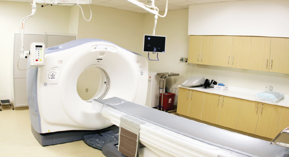
Patients were scanned on a LightSpeed © 64-Slice VCT CT system, manufactured by GE Healthcare (Little Chalfont, Buckinghamshire, UK). Contiguous axial 0.6 mm slices were obtained and the images were reconstructed at 5 mm intervals for viewing on a Carestream PACS v10.2 workstation (Carestream Health, Rochester, NY, USA). All images were reviewed by a radiologist with over 20 years of experience in cross-sectional imaging.
Two methods were studied to determine the more effective method of land marking and identifying the AG. The first and more commonly known method involved the identification of the UP, following it slice by slice superiorly until reaching the mid-level of the AG. The second method employed the CA, as the landmark (Figure 1.A). [22] For this study, the mid-level of the AG was identified, and its position was noted in relation to the upper margin of the respective UP and to the CA. Distances between the AG and landmarks were calculated by counting the number of axial slices between the structures and converting this to millimetres (craniocaudal distance). Each CT DICOM had a slice thickness of 5 mm (Table 1). A distance of 0 mm indicated that the landmark and AG were present on the same slice. Positive values indicated the AG were superior to the landmark and negative values indicated that the AG were inferior to the landmark.

Figure 1.a. A CT scan reveals the coeliac axis (CA) on the same slice (and level) to the right (R.AG) and left adrenal gland (L.AG).
Figure 1.b. A CT scan that demonstrates the tail of the pancreas (PT) interfering with the left adrenal gland (L.AG) and the right adrenal gland is absent from the image.

Statistical analysis

Results were analysed using IBM SPSS ® version 20 (Armonk, NY, USA). The distance between the AG and the defined landmarks were compared using paired t-tests. A p-value of <0.05 was considered to be statistically significant. Results

In total there were 1,037 patients with a median age of 60 years (Inter-quartile range: 52-68 years) and male to female ratio of 527:510. Twelve (1.2%) of the patients in the study had non-massive adrenal lesions and 74 (7.1%) of the patients had not received IV contrast. The images without IV contrast were scrutinised in greater depth because it was more difficult to distinguish the CA on non-contrast CT, but it did not markedly affect the identification of normal AG. [12,21,24] The results below provide an overview of the relational distance between AG and landmarks, calculated by slice levels that were numbered and converted to millimetres (Table 1). Graphical representation of the mean distances of the AG to the landmarks are shown in Figure 2. Statistical assessment of differences between landmarks are shown in Table 2 and Figure 3.

Table 1. A comparison of the distance between the adrenal glands and their respective landmarks (coeliac axis and upper poles of the kidney). IQR, inter-quartile range; CA, coeliac axis; UP, upper pole; AG, adrenal gland. * The number of cases in which the AG were on the same slice (0 mm, level=0) as the respective landmarks, with the valid percentage included. † Both the right and left AG and their joint relationship with the landmarks, CA and UP.
Table 1. A comparison of the distance between the adrenal glands and their respective landmarks (coeliac axis and upper poles of the kidney).
IQR, inter-quartile range; CA, coeliac axis; UP, upper pole; AG, adrenal gland.
* The number of cases in which the AG were on the same slice (0 mm, level=0) as the respective landmarks, with the valid percentage included.
† Both the right and left AG and their joint relationship with the landmarks, CA and UP.

Figure 2. A comparison of the mean absolute distance of the adrenal glands from the respective landmarks. The AG to CA (blue), and AG to UP (red).
Figure 2. A comparison of the mean absolute distance of the adrenal glands from the respective landmarks.
The AG to CA (blue), and AG to UP (red).

Table 2. Paired t-tests samples show a significant decrease in distance within individual patients when using the coeliac axis as a landmark.

Pair 1 is the comparison of the mean distance between the right adrenal gland and its landmarks, the coeliac axis and the upper pole of the right kidney. Pair 2 is the comparison of the mean distance between the left adrenal gland and its landmarks, the coeliac axis and the upper pole of the left kidney. Differences are expressed as mean and standard deviation.

Figure 3. Paired t-test analysis demonstrates significant difference between the adrenal glands and landmark, the means and 95% CI, 95% Confidence Interval of the Difference. The measurements for CA – Right UP (blue box) and CA – Left UP (red box) are shown in Table 2.

The CA and the right AG occurred at the same level in 65% of patients, and the CA and the left AG in 88% of patients. For the right UP, the right AG was at the same level in 42% of patients; and the left UP, at the same level as the left AG in 58% (p-value<0.001). The median distance for the CA to the right AG (0 mm) was less than the median distance between the right UP and right AG (5 mm). The left AG had the same median distance from the CA and the left UP (0 mm). The CA also had smaller interquartile ranges, 0 – 10 for the right AG and 0 – 0 for the left AG, compared to the UP with interquartile ranges, 0 – 15 for the right AG and 0 – 10 for the left AG. Pair 1 compared the right UP with the CA, and Pair 2 compared the left UP with the CA. Both Pair 1 (the distance of the right AG from the CA and right UP) and Pair 2 (the distance of the left AG from the CA and left UP) indicated that the CA distance to AG was closer to 0 mm than the UP distances to AG (p<0.0001; Table 2). Discussion

The main finding of this study was that the AG could be more accurately identified on CT images when the CA was used as the reference landmark as opposed to the conventional method of using the UP as a landmark. The distance between the AG and the CA was significantly shorter than the distance between the AG and the UP. Furthermore, the CA was more likely to be located on the same axial slice as the left and right AG than the UP. This study also showed that the CA was a less variable landmark than the UP, with smaller interquartile ranges. These findings support the belief that the CA does not move with respiration, providing one explanation why the CA appeared to be a more accurate landmark.

Anatomical landmarks play an important role in medical imaging. Identifying a landmark proximal to the target organ can reduce distance-related errors that may occur when analysing CT images. [25] As Gunderman [26] said, “By far the most important role of imaging in the evaluation of the AG lies in the detection, characterization, and staging of adrenal tumours.” There are a limited number of studies available that explore different methods of identifying the AG, however, many studies have been undertaken assessing other radiological landmarks in the body. [7,12,27-31] For a landmark to be accurate, it must include the following general characteristics: Firstly, having a close measurable distance relationship to the target structure (preferably less than 10 mm). Secondly, the stability of the landmark should allow clinicians to accurately estimate the location of the target structure, and avoid the over- or underestimation of the target structure’s location from the landmark. [25,28-30] Thirdly, pattern approach CT imaging interpretation requires the more accurate landmark to be easily recognizable to the viewer. [28-32]

This study has several limitations. First, data was gathered from a sample of cancer patients, which is a subset of all patients that receive CT scans of the abdomen. Consequently, the results of this study may not be a true representation of the general population. Second, IV contrast was not administered to all 1,037 patients included and enhanced images may have allowed for easier identification of the CA. [12,21,24] Finally, CT images were analysed by an experienced academic radiologist. The findings may have been different had they been analysed by junior doctors or other non-radiologist physicians. This study could be improved by including a number of non-radiology clinicians as the primary readers. The data described here was from a cancer centre in Southeast Asia only. This study could be further improved with an international patient pool allowing comparison of different ethnic groups. Patients with massive renal and adrenal pathology, such as adrenal metastases, were excluded from the study. A larger study is required to investigate the impact of such pathology on the identification of AG on CT. Further research could be done to determine the possibility of reducing the time taken to identify and diagnose the status of the AG, when using the CA as a landmark. The technique of using the CA as a landmark may also be applied to the identification of primary or secondary carcinoma, and further study could aid with the locating and identification of abnormal AG.

Conclusion

The CA is an easily identifiable landmark that is closer to the adrenal gland than the UP. This landmark may be applied by clinicians to facilitate timely detection of the AG on CT scans.

Conflicts of Interest

None declared.

Acknowledgements

Professor John Morley and Associate Professor Noel Young for their assistance in proof-reading the abstract. Eric Martin and Diane Premnath for their patience and kindness in reviewing the paper. Jeremiah Schmidt for his aid with the statistics. Michelle Ng, Sophie Kobuch, Chatwin Lee, Lisa Leow, Corinne Fulford, Simon Chow, and Karlene Zhu for their patience in proof-reading the paper.

References

[ 1] Chabner BA, Thomas J. Lynch J, Longo DL. Harrison’s Manual of Oncology. 16th ed. Shanahan J, Davis KJ, editors. New York: McGraw-Hill Companies, Inc.; 2008. p. 345-356.

[ 2] Gokan T, Ohgiya Y, Nobusawa H, Munechika H. Commonly encountered adrenal pseudotumours on CT. Br J Radiol. 2005;78:170-4.

[ 3] Ma G, Liu SW, Zhao ZM, Lin XT, Lou L, Li ZP, et al. Sectional anatomy of the adrenal gland in the coronal plane. Surg Radiol Anat. 2008;30:271-80.

[ 4] Benitah N, Yeh BM, Qayyum A, Williams G, Breiman RS, Coakley FV. Minor morphologic abnormalities of adrenal glands at CT: prognostic importance in patients with lung cancer. Radiology. 2005;235(2):517-22.

[ 5] Boland G, Blake MA. Adrenal Imaging. Boston: Humana Press; 2009.

[ 6] Higham CE, Coen JJ, Boland GWL, Trainer PJ. The Adrenals in Oncology. In: Blake MA, Boland GWL, editors. Adrenal Imaging. Totowa: Humana Press Springer Science+Business Media; 2009. p. 65-76.

[ 7] Al-Hawary MM, Francis IR, Korobkin M. Adrenal Imaging Using Computed Tomography: Differentiation of Adenomas and Metastasis. In: Blake MA, Boland GWL, editors. Adrenal Imaging. Totowa: Humana Press, Springer Science+Business Media; 2009. p. 127-39.

[ 8] Grumbach MM, Biller BM, Braunstein GD, Campbell KK, Carney JA, Godley PA, et al. Management of the clinically inapparent adrenal mass (“incidentaloma”). Ann Intern Med. 2003;138(5):424-9.

[ 9] Ellis H, Logan BM, Dixon AK. Human Sectional Anatomy: Atlas of Body Sections, CT and MRI Images. Oxford: Butterworth Heinemann; 1999. p. 134-163.

[ 10] Johnson PT, Horton KM, Fishman EK. Adrenal mass imaging with multidetector CT: pathologic conditions, pearls, and pitfalls. Radiographics. 2009;29(5):1333-51.

[ 11] Arnold DT, Reed JB, Burt K. Evaluation and management of the incidental adrenal mass. Proc (Bayl Univ Med Cent). 2003;16:7-12.

[ 12] Ilias I, Sahdev A, Reznek RH, Grossman AB, Pacak K. The optimal imaging of adrenal tumours: a comparison of different methods. Endocr Relat Cancer. 2007;14(3):587-99.

[ 13] Abujudeh HH, Kaewlai R, McMahon PM, Binder W, Novelline RA, Gazelle GS, et al. Abdominopelvic CT increases diagnostic certainty and guides management decisions: A Prospective investigation of 584 patients in a large academic medical center. AJR Am J Roentgenol. 2011;196:238-43.

[ 14] Brundage MD, Feldman-Stewart D, Cosby R, Gregg R, Dixon P, Youssef Y, et al. Cancer patients’ attitudes toward treatment options for advanced non-small cell lung cancer: implications for patient education and decision support. Patient Educ Couns. 2001;45(2):149-57.

[ 15] Leydon GM, Boulton M, Moynihan C, Jones A, Mossman J, Boudioni M, et al. Cancer patients’ information needs and information seeking behaviour: in depth interview study. BMJ. 2000;320(7239):909-13.

[ 16] Silvestri G, Pritchard R, Welch HG. Preferences for chemotherapy in patients with advanced non-small cell lung cancer: descriptive study based on scripted interviews. BMJ. 1998;317(7161):771-5.

[ 17] Ellis H, Mahadevan V. Clinical Anatomy: Applied Anatomy for Students and Junior Doctors. Tweleth Edition ed. Chichester: Wiley-Blackwell; 2010. p. 61-168.

[ 18] Johnson PT, Horton KM, Fishman EK. Adrenal imaging with multidetector CT: evidence-based protocol optimization and interpretative practice. Radiographics. 2009;29(5):1319-31.

[ 19] Mayo-Smith WW, Boland GW, Noto RB, Lee MJ. State-of-the-art adrenal imaging. Radiographics. 2001;21(4):995-1012.

[ 20] Welch TJ, Sheedy SP, II PFS. Adrenal Glands. In: Haaga JR, Dogra VS, Forsting M, Gilkeson RC, Ha HK, Sundaram M, editors. CT and MRI of the Whole Body. 5 ed. Philadelphia: Mosby Elseiver; 2009. p. 1813-62.

[ 21] Kawashima A, Sandler CM, Fishman EK, Charnsangavej C, Yasumori K, Honda H, et al. Spectrum of CT findings in nonmalignant disease of the adrenal gland. Radiographics. 1998;18(2):393-412.

[ 22] Henry G. Gray’s Anatomy: The Anatomical Basis of Medicine and Surgery. 38th ed. Williams PL, editor. New York: Churchill Livingstone; 1995.

[ 23] Özbülbül NI. CT angiography of the celiac trunk: anatomy, variants and pathologic findings. Diagn Interv Radiol. 2011;17:150-7.

[ 24] Boland GW, Blake MA, Hahn PF, Mayo-Smith WW. Incidental adrenal lesions: principles, techniques, and algorithms for imaging characterization. Radiology. 2008;249(3):756-75.

[ 25] Nemoto M, Masutani Y, Hanaoka S, Nomura Y, Miki S, Yoshikawa T, et al., editors. Coarse-to-fine localization of anatomical landmarks in CT images based on multi-scale local appearance and rotation-invariant spatial landmark distribution model. Proc SPIE Medical Imaging: International Society for Optics and Photonics 8669; 2013.

[ 26] Gunderman RB. Essential Radiology. New York: Thieme Medical Publishers; 1998. p. 261-264

[ 27] Boland GW. Adrenal imaging: why, when, what, and how? Part 1. Why and when to image? AJR Am J Roentgenol. 2010;195(6):377-81.

[ 28] Aronson D, Kier R. CT pelvimetry: the foveae are not an accurate landmark for the level of the ischial spines. AJR Am J Roentgenol. 1991;156(3):527-30.

[ 29] Connor SE, Arscott T, Berry J, Greene L, O’Gorman R. Precision and accuracy of low-dose CT protocols in the evaluation of skull landmarks. Dentomaxillofac Radiol. 2007;36(5):270-6.

[ 30] Lou L, Lagravere MO, Compton S, Major PW, Flores-Mir C. Accuracy of measurements and reliability of landmark identification with computed tomography (CT) techniques in the maxillofacial area: a systematic review. Oral Surg Oral Med Oral Pathol. 2007;104(3):402-11.

[ 31] Karthikeyan D, Chegu D. CT Abdomen: A Pattern Approach: Jaypee Brothers Medical Publishers; 2007. p. 222-231

[ 32] Macari M, Megibow AJ, Balthazar EJ. A Pattern Approach to the Abnormal Small Bowel: Observations at MDCT and CT Enterography. AJR Am J Roentgenol. 2007;188(5):1344-55.

Categories
Review Articles Articles

Is plasmapheresis the optimal treatment option for acute pancreatitis secondary to hypertriglyceridemia? A systematic review

Background: Hypertriglyceridemia is an uncommon cause of acute pancreatitis, which is a life-threatening illness. Conventional management involves fasting, lipid-lowering medication, insulin and heparin. Plasmapheresis is an approach which is used occasionally to achieve rapid lowering of triglyceride levels in patients where conventional management is unsuccessful. It is currently unclear whether plasmapheresis improves outcome in patients with hypertriglyceridaemia-induced pancreatitis. Aim: A literature review and critical analysis was conducted to assess the effectiveness of plasmapheresis in improving patient outcomes in patients with acute pancreatitis secondary to hypertriglyceridemia Methods: The PICO model (Population, Intervention, Comparator, Outcomes) was used to synthesise a research question. Thereafter, a search was conducted through the Scopus database (includes complete MEDLINE coverage) applying the terms ‘plasmapheresis’ OR ‘plasma exchange’ OR ‘lipid apheresis’ AND ‘pancreatitis’ AND ‘hypertriglyceridemia’ OR ‘hyperlipidaemia’ OR ‘hyperlipidemia’. Article titles and/or abstracts were screened for relevance to the topic. Original research articles assessing the efficacy of plasmapheresis in hypertriglyceridaemia-induced pancreatitis were included. Results: To date, no randomised controlled trials have been published assessing the efficacy of plasmapheresis in this population. Two retrospective primary research studies were identified. Both studies demonstrated a rapid reduction in triglyceride levels following plasmapheresis in the magnitude of 65.8-80%. The studies showed no significant clinical benefit in terms of mortality and morbidity, but were limited by small sample size and study design. Conclusion: Current evidence demonstrates that plasmapheresis in the setting of hypertriglyceridemia-induced pancreatitis reduces triglyceride levels by 46-80%. [1] However there is insufficient data to suggest a beneficial effect on clinical outcomes. Well-designed prospective studies with adequate follow-up are required to elucidate whether plasmapheresis is associated with reduced morbidity and mortality in this population.

Introduction

Hypertriglyceridemia is an uncommon cause of acute pancreatitis, accounting for 1.3-3.8 % of cases with an incidence of 18/100,000 per year in the United States of America. [1,2] Primary (genetic) and secondary causes, such as uncontrolled diabetes mellitus, hypothyroidism, alcohol, obesity, certain medications, and pregnancy, are associated with hypertriglyceridemia-induced pancreatitis. [1,3] The mechanism for severe hypertriglyceridemia-inducing pancreatitis remains unclear, [3] although triglyceride levels exceeding 10 mmol/l (1000 mg/dl) can trigger a bout of pancreatitis. [3,4] One postulated theory involves the idea that pancreatic lipase hydrolyses excess triglycerides to produce free fatty acids around the pancreas. These free fatty acids can damage the pancreatic acinar cells and pancreatic vascular endothelium, resulting in ischaemia and inflammation. The acidic environment can further amplify the free fatty acid toxicity in a vicious cycle. [3,4]

Hypertriglyceridemic pancreatitis is a life-threatening illness with a mortality rate of 7-30%. [5] Complications include sepsis, pancreatic necrosis, abscess formation and renal insufficiency; which account for the high mortality seen in this disease. [3] Optimal management of hypertriglyceridemia-induced pancreatitis is essential to reduce morbidity and mortality. Current management includes fasting, lipid-lowering medication (such as fenofibrate), and insulin and heparin, used to ‘accelerate lipoprotein lipase activity’. [2,4] These interventions have shown limited efficacy in reducing inflammation and life-threatening complications associated with severe acute pancreatitis. Novel therapies are needed to improve patient outcomes. [5]

Plasmapheresis is defined as ‘removing the plasma and replacing it with donor plasma or a plasma substitute’. [6] The term plasmapheresis is used interchangeably with the term ‘therapeutic plasma exchange’. The use of plasmapheresis in patients with hypertriglyceridemia can be traced back to a case report in 1978. [3] However, it is not widely utilised in this patient population at present. Given that plasmapheresis provides rapid removal of the triglycerides responsible for underlying inflammation in hypertriglyceridemia-induced pancreatitis, it is expected that this intervention may prove highly effective in reversing this sub-type of acute pancreatitis. The aim of this review was to assess the effectiveness of plasmapheresis in achieving positive patient outcomes (as per Table 1) in patients with acute pancreatitis secondary to hypertriglyceridemia.

Methods

The PICO model was used to synthesise the research question. [7]

Search Methodology

The literature search was conducted on Scopus, which is one of the largest databases of abstracts and citations of research literature, and includes complete coverage of the MEDLINE database. [8] The following search terms were used: (“plasmapheresis” OR “plasma exchange”) AND “pancreatitis” AND (“hypertriglyceridemia” OR “hyperlipidaemia” OR “hyperlipidemia”). The initial database search yielded a total of 139 documents, of which 110 were written in English. After further limiting the search query to include only ‘articles’ or ‘reviews’, a total of 86 documents were found. The documents were then sorted by relevance, and titles and abstracts were screened to identify articles that reported on the use of plasmapheresis in the management of hypertriglyceridemia-induced acute pancreatitis. A total of 28 relevant articles were identified: one primary research study, one review paper, one guideline (the 2010 American Society for Apheresis (ASFA) guideline), eleven case-series (62 patients), and fourteen individual case studies (Figure 1). Review of the references and citations of these studies yielded an additional two articles: one original retrospective study and one review. Focused searches revealed no additional relevant articles. The inclusion criterion for this review was limited to primary research studies only.

Results

Two cohort studies meeting the pre-specified inclusion and exclusion criteria were included. No randomised-controlled trials were identified, and are probably not feasible given the low incidence of hypertriglyceridemia-induced pancreatitis.

Chen et al. [9] conducted a retrospective cohort study to compare the mortality and morbidity in patients with hyperlipidaemic pancreatitis before and after the introduction of plasmapheresis in Shin kong Wu-Ho-Su Memorial Hospital, Taiwan, in August 1999. This study separated the patients into two cohorts: group I (pre-August 1999) and group II (post-August 1999). There were 34 patients in group I and 60 patients in group II, of which twenty patients received plasmapheresis. The cohort was recruited appropriately with all patients fitting the time-frame criteria (pre- or post-August 1999). There was no statistical difference between the demographics of group I and group II, including mean age, gender distribution, initial mean triglyceride level, diabetes mellitus, alcohol consumption, Ranson’s score ≥ 3 and Balthazar grade D or E. [9]

Furthermore, patients with severe hypertriglyceridemic pancreatitis (defined by Ranson’s score ≥ 3) were analysed separately, comparing Group A (those who received plasmapheresis) and Group B (those who did not receive plasmapheresis). There were ten patients in Group A and nineteen patients in Group B. Morbidity was defined in terms of systemic complications, including acute renal failure, upper gastrointestinal bleeding, shock and acute respiratory distress syndrome; and local complications, in particular abscess and pseudocyst formation.

The results were analysed using a t-test and chi-square test. There was no statistical significance (defined as p<0.05) between the mortality and complications of patients with severe pancreatitis who received plasmapheresis, compared to those who received conventional therapy. Similarly, there was no significant difference (defined as p<0.05) between the clinical outcomes of pre-August 1999 and post-August 1999 samples. Interestingly, Chen et al. found that the mean serum concentration of triglycerides and lipase were markedly reduced after plasmapheresis, with a 65.8% reduction of triglyceride levels and 88.8% reduction of lipase levels. [9]

Gubensek et al. [4] also carried out a retrospective cohort study. They looked at two sets of patients. The first sample consisted of 50 patients who were treated with plasmapheresis between 1992 and 2008 at a tertiary-care hospital (University Medical Center Ljubljana, Slovenia), and the triglyceride and total cholesterol levels before and after plasmapheresis were compared. The demographic characteristics of these patients revealed a gender bias, with 92% of the sample being male. The second set of patients included 40 patients treated between 2003 and 2008 with plasmapheresis. The Acute Physiology and Chronic Health Evaluation II (APACHE II) score was used as a prognostic tool, with a score of < 8 indicating mild pancreatitis and a score of ≥ 8 indicating severe pancreatitis. A comparison of mortality rates was made between these two groups. The mortality rate was 4% and 42% respectively, with statistically significant differences between the two groups (p<0.001).

The results of the first cohort in the study by Gubensek et al. [4] showed a statistically significant (defined as p<0.001) reduction in triglyceride and cholesterol levels after plasmapheresis within 24 hours. On average, a reduction in serum triglyceride levels of approximately 80% was achieved by plasmapheresis. The analysis of the second cohort showed a significantly higher mortality in patients who had an APACHE II score of ≥ 8 (42% vs. 4%). The overall mortality was 15%, which the authors acknowledged as ‘considerable’.

Discussion

The efficacy of plasmapheresis in hypertriglyceridemia-induced pancreatitis has been unclear. In an attempt to clarify this, we conducted a systematic review of published studies and guidelines investigating the benefits of this therapy. Studies by Chen et al. and Gubensek et al. reported on different populations; however both highlighted that a significant reduction in serum triglyceride levels can be achieved by plasmapheresis in patients with hypertriglyceridemic pancreatitis. Chen et al. [9] showed a reduction in triglyceride levels by 65.8%, and Gubensek et al. [4] demonstrated an average reduction of 80% in triglyceride levels. It should be highlighted that a rapid reduction in triglyceride and cholesterol levels does not necessarily imply a clinical benefit to the patient. The exact pathophysiology of hypertriglyceridemia-induced pancreatitis remains unclear. During a prolonged episode of acute pancreatitis cellular injury can occur which may remain irreversible regardless of lowering of triglyceride levels, although further pancreatic destruction and recurrent episodes may be reduced. [5,10-11] Admission triglyceride levels have not been associated with severity, complication rates or clinical course. [5] Therefore more research is required to look at the effect of the rapid lowering of triglyceride levels on patient outcomes.

The weaknesses of both studies are the small sample size and sub-optimal study design. Chen et al. only looked at a small number of patients from a single hospital, and no definitive conclusions could be reached based on the limited statistical power of the study. Moreover, Chen et al. [9] compared group I and group II outcomes, despite group II comprising of 40 patients who had not received plasmapheresis in addition to the twenty patients who had received plasmapheresis. Therefore, since group II comprised of a mixed sample of patients receiving plasmapheresis and those receiving conventional treatment, the outcomes do not give a true picture of the effect of plasmapheresis only. A statistical comparison between group I and group II only accentuates that there was no benefit in terms of mortality and morbidity between patients presenting with acute pancreatitis secondary to hypertriglyceridemia before August 1999 or after August 1999 at that specific hospital. Thus, any conclusion on the effectiveness of plasmapheresis at reducing patient morbidity and mortality using these statistical results is invalid.

On the other hand, Chen et al. [9] should be commended on their detailed explanation of the apheresis procedure performed, which was well controlled. However, replacement fluid was either fresh frozen plasma (N=8) or isovolumetric 5% albumin solution (N=12). The authors did not adjust their results for this potentially confounding variable. The mean serum concentration of triglycerides and lipase were markedly reduced after plasmapheresis, with a 65.8% reduction of triglyceride levels and 88.8% reduction of lipase levels. However, the time period in which these reductions occurred post-procedure is unclear, and no comparisons were made with the reduction in serum lipase and triglycerides in patients presenting pre-August 1999. The authors did not follow the patients over a considerable time period and were unable to assess recurrence of acute pancreatitis or mortality between patient samples.

The research study by Chen et al. [9] is essentially the only study comparing the use of plasmapheresis and conventional treatment in acute pancreatitis due to hypertriglyceridemia. This study found no differences in mortality and morbidity between conventional therapy and plasmapheresis, but the results need to be carefully evaluated, as mentioned above. A review of this study by the 2010 ASFA Guidelines highlight that ‘adequate information was not provided to ascertain the comparability of the two groups’. [1] Chen et al. stated that earlier intervention might provide positive outcomes for plasmapheresis, given that the median time for starting plasma exchange was three days for their patients. [9] This is a possible explanation for the results, and further studies are needed to evaluate the relationship between the time of initiating plasmapheresis and patient outcomes. A review by Tsuang et al. reported that ‘early initiation of treatment for hypertriglyceridemic pancreatitis is likely to be beneficial’, [3] based on findings from the retrospective case series by Kyriakidis et al., who reported positive patient outcomes in eight out of nine patients treated with plasmapheresis within 48 hours of diagnosis. [2]

The study by Gubensek et al. is also limited, in that it did not compare the effect of plasmapheresis with conventional treatment options.  No comparison was made with the mortality rate of patients who were treated conventionally. The study makes a strong point on the effectiveness of plasmapheresis in acutely reducing serum triglyceride and cholesterol levels, but does not determine whether there is any clinical benefit of this to the patient.

The indications and criteria for applying plasmapheresis was not consistent across the literature. Plasmapheresis is commonly used in settings where there is inadequate outcomes achieved using conventional management. [5] However, the patients treated with plasmapheresis in Chen et al. [9] and Gubensek et al. [4] include those with mild to moderate pancreatitis as well as severe pancreatitis (evaluated using Ranson’s scores or APACHE II score), but no information was given about any conventional treatment prior to the initiation of plasmapheresis. Therefore, it is unclear whether any prior conventional treatment had an effect on patient outcomes.

Neither study reviewed the potential symptomatic relief from plasmapheresis. A possible reason could be due to the subjectivity in measuring this outcome and the retrospective nature of the studies. Evaluation of potential symptomatic relief from plasmapheresis may be useful in minimising the severe pain associated with hypertriglyceridaemic pancreatitis.

ASFA [1] conducted a literature review to evaluate the rationale for plasmapheresis in patients with hypertriglyceridemic pancreatitis. This review is consistent with the findings of studies by Chen et al. and Gubensek et al., and reported no randomised controlled trial evaluating the effectiveness of plasmapheresis in these patients.  Their search on PubMed yielded twelve case-series and 28 case reports, with sample sizes ranging from 100-300. The ASFA have given a Category III indication to the use of plasmapheresis in patients with hypertriglyceridemic pancreatitis, which implies that the optimum role of apheresis therapy in these patients is not established. ASFA highlight the role of clinicians in making individualised decisions due to the weak evidence in this area. [1]

There is clearly a need for stronger evidence to determine the effectiveness of plasmapheresis in patients with hypertriglyceridemic pancreatitis. However, the low incidence of this disease means that randomised controlled trials may not be feasible. Gubensek et al. [4] argue that it would be questionable to perform a randomised controlled trial, given the large reduction in triglyceride levels by plasmapheresis. The ethical issues and feasibility of performing a randomised controlled trial on a small sample are barriers in answering this question. Nevertheless, large cohort studies with sufficient follow-up and appropriate adjustments for population stratification should provide immense support either in favour or against the use of plasmapheresis. Furthermore, appropriate studies assessing the effectiveness of rapidly lowering triglyceride levels on patient outcomes should be conducted.

Conclusion

There is insufficient evidence to confirm that plasmapheresis is a beneficial treatment option for patients with acute pancreatitis secondary to hypertriglyceridemia. Current literature shows that plasmapheresis promotes a rapid reduction in triglyceride levels of 46-80% [1], however its effect on patient outcomes remains unclear. Adequately powered prospective studies with long term follow-up are recommended to elucidate whether plasmapheresis is associated with reduced morbidity and mortality in this population.

Acknowledgements

Thank you to Dr Channa Perera, Endocrinologist at Campbelltown Hospital, for the inspiration to research and write up on this topic.

Conflict of interest

None declared.

Correspondence

M Rehmanjan: mrehmanjan@gmail.com

References

[1] Szczepiorkowski ZM, Winters JL, Bandarenko N, Kim HC, Linenberfer ML, Marques MB, et al. Guidelines on the use of therapeutic apheresis in clinical practice. Journal of Clinical Apheresis 2010; 25:83-177.

[2] Kyriakidis AV, Raitsiou B, Sakagianni A, Harisopoulou V, Pyrgioti M, Panagopoulou A, et al. Management of Acute Severe Hyperlipidemic Pancreatitis. Digestion 2006; 73(4):259-64.

[3] Tsuang W, Navaneethan U, Ruiz L, Palascak JB, Gelrud A. Hypertriglyceridemic Pancreatitis: Presentation and Management. The American Journal of Gastroenterology 2009;104:984-91

[4] Gubensek J, Buturovic-Ponikvar J, Marn-Pernat A, Kovac J, Knap B, Premru V, et al. Treatment of hyperlipidemic acute pancreatitis with plasma exchange: A single-center experience. Therapeutic Apheresis and Dialysis 2009;13(4):314–17.

[5] Lebenson J, Oliver T. Acute pancreatitis [Internet]. Rijeka, Croatia: InTech; 2012. Chapter 16, Hypertriglyceride Induced Acute Pancreatitis [cited 2013 Aug 25]. Available from: http://www.intechopen.com/books/acute-pancreatitis/hypertriglyceride-induced-acute-pancreatitis

[6] Brooker C. Medical Dictionary. Philadelphia. Churchill Livingstone Elsevier; 2008.

[7] University of Warwick Library. The Pico Method [Internet]. 2010 Sep 1 [cited 2012 Aug 6]. Available from: http://www2.warwick.ac.uk/services/library/tealea/sciences/medicine/evidence/pico/

[8] Elsevier. Scopus Database [Internet]. 2012 [cited 2012 Aug 6]. Available from: http://www.scopus.com/home.url?null

[9] Chen JH, Yeh JH, Lai HW, Liao CS. Therapeutic plasma exchange in patients with hyperlipidemic pancreatitis. World Journal of Gastroenterology 2004; 10(15):2272-74.

[10] Banks PA, Conwell DL, Toskes PP. The Management of Acute and Chronic Pancreatitis. Gastroenterol Hepatol 2010; 6(2 Suppl 5):1–16.

[11] Piolot A, Nadler F, Cavallero E, Coquard JL, Jacotot B. Prevention of recurrent acute pancreatitis in patients with severe hypertriglyceridemia: value of regular plasmapheresis. Pancreas 1996; 13(1):96-9.

Categories
Feature Articles Articles

The history of breast cancer surgery: Halsted’s radical mastectomy and beyond

Breast cancer is common. One in eight Australian women will be diagnosed by the time they turn 85, and it has been estimated that this year in Australia approximately 14,600 women will receive the diagnosis, around 40 women each day. [1] A significant proportion will undergo surgery, mostly as the first means of treatment. Over the past forty years many advances have been made in the surgical approach to breast cancer. [2] New techniques and approaches have been developed and efforts for further improvements are ongoing. This article will explore the journey that breast surgery has undergone and what we can learn from its evolution.

William Halsted (born 1852) was an American surgeon whose contributions have influenced surgical principles to this day. [3] He is considered one of the ‘Big Four’ founding physicians of John Hopkins Hospital. [4] He pioneered the use of the hospital chart, advocated careful handling of tissue during surgery and stressed the importance of haemostasis. [3-6] His name is also synonymous with the radical mastectomy that he introduced in 1882. [3] At that time attempts at breast surgery had resulted in poor long term results and prognosis. [6,7] This new surgical approach was revolutionary in the treatment of breast cancer. The radical mastectomy was implemented for breast cancer no matter the size of the tumour, type, or the patient’s age. [8] It typically involves removal of all breast tissue, axillary lymph nodes and both pectoralis muscles. [6] It often results not only in severe disfigurement of the patient but also weakened arm function and disabling lymphoedema. [9] Whilst revolutionary at the time it was pioneered by Halsted, it was still widely used in the 1970s with a ‘one size fits all’ approach. [8] That approach is very different to the one taken today. [2,10,11]

While there had been some exploration of modifications to the procedure, such as sparing of the pectoralis muscles, as well as further dissection with removal of the internal mammary nodes, the surgical approach to breast cancer remained relatively static for more than eighty years. [8] Although there are many potential reasons this state of inactivity is surely multifactorial. Feminist authors have claimed that the mastectomy was not altered by male surgeons because of the power and control it gave them; that they had no understanding of the importance of a woman’s breast to a woman and treated patients in response to this view. [12] Others have suggested that Halsted was held in such high regard that no one dared alter his procedure, with surgeons ‘indoctrinated’ into his way of thinking. [12,13] Perhaps it is also that the nature of the disease affected how it was approached, with surgeons hesitant to make changes to a procedure they believed could save the lives of countless women. This seemed to be the case for Halsted himself who suggested, “After all, disability, ever so great, is a matter of very little importance as compared with the life of the patient.” [6] It must be acknowledged that in Halsted’s time there was no method of grading or staging cancers as there is now, a problem he recognised stating, “the importance of such a classification, if it were to any extent possible, is so evident that it is unnecessary to emphasize it.” [7] Had he had such information available to him his approach to individual cases may have varied greatly.

Alterations to the mastectomy were taken cautiously. There were forays into and case reports of the super-radical mastectomy, simple mastectomy and use of radiation therapy, as well as some use of simple excision; however no clear evidence as to the differences was available. [8] In 1969 the World Health Organisation approved a randomised control trial comparing radical mastectomy to the ‘quadrantectomy’. [8] Recruitment began in 1973 of patients staged with T1N0 disease who were aged less than 70 years. The quandrantectomy was combined with complete axillary dissection and postoperative radiotherapy. Early data demonstrated no difference in regards to survival rates, and the similarities in the long term survival rates were confirmed in data released in 2002. [8] In 1971 Fisher et al commenced a randomised trial comparing the radical mastectomy with total mastectomy with or without radiotherapy. [14] Studies such as these heralded the advent of breast conserving surgery and the acknowledgement that routine radical mastectomy may not always be the most appropriate surgical management.

Halsted proposed that although breast cancer begins as a local disease, it spreads in a contiguous manner away from the primary site through the lymphatic system. [6,15] This proposal led to his emphasis on aggressive locoregional treatment to prevent further spread. [6,7,12,15] This principle, however (known as the ‘Halsted Theory’), was also critical in introducing the concept of a sentinel node in relation to breast cancer. [15] Research into the sentinel node led to the use of the sentinel node biopsy which has dramatically influenced surgical management and outcomes for patients. One of the first studies demonstrating the benefits of lymphatic mapping for breast cancer was published by Guiliano et al in 1994. [16] Since that time the evidence, understanding and surgical skills in this area have grown rapidly.

Modern day surgery for breast cancer has changed significantly compared to that performed in the 1970s. The combination of breast conserving surgery alongside a sentinel biopsy allows patients to be left with good cosmetic results. [17,18] Oncoplastic techniques such as remodelling mammoplasty are also being utilised to improve cosmetic outcomes without compromising adequate tumour removal. [19,20] There is still however an appropriate place for the mastectomy. [20] Breast conservation is desirable, but needs to be acceptable cosmetically and not result in compromise to local control of the disease or survival benefit. [17,19,20] If local recurrence does occur following breast conservation, then salvage mastectomy is considered the standard approach, with salvage breast-conserving surgery only currently appropriate for consideration in select patients. [21]

Reconstructive breast surgery is an important part of management utilised by surgeons today. It is significant in improving the psychological morbidity associated with breast cancer surgery, particularly following mastectomy. [2,22] Once again Halsted had an influence in this area of breast surgery. He believed reconstruction was a “violation of the local control of the disease”. [10] Although there were a few early attempts at breast reconstruction by the likes of Czerny, Tanzini and Ombredanne, [23-25] the opinion put forward by Halsted and the view that local recurrence may not be detected if reconstruction occurred caused it to be avoided. [2,10] It seems, however, that surgical exploration of reconstructive procedures during this period was considered more than the possibility of breast conservation was. [8,10] This supports the view that surgeons at the time believed the mastectomy was crucial to life saving treatment and it was this belief that prevented progress to other initial surgical approaches. [13]

Following the introduction of the mastectomy in 1882 there were surgeons willing to attempt reconstructions to improve the quality of life of their patients. [2,10,22] In 1963 these efforts were bolstered by the silicone gel breast implant introduced by Cronin and Gerow. [26] In 1971 Snyderman and Guthrie placed an implant under the chest wall immediately following a mastectomy, as opposed to the delayed technique that had been used, and this was then accepted as the new technique. [27] In 1982 Radovan introduced the concept of skin expanders for those with significant skin deficits so that these patients too could be eligible for reconstructive surgery. [28] Following this skin-sparing mastectomies were introduced, with results demonstrating similar rates of local recurrence. [29] There have also been advances with the use of flaps as a method of reconstruction. [2,10] In regards to reconstruction of the nipple-areola complex, tattooing is now commonplace as initially suggested by Becker in 1986. [30] Today a woman who thirty years ago would have been left with almost no chance of reconstruction can have a relatively symmetrical result. Decisions relating to the method of reconstruction depend on many variables, however it can be seen that important progress has been made in this area of breast surgery. [2,10] This can improve patient perceptions towards treatment and significantly improve their quality of life. [2,22]

The evolution of breast cancer surgery demonstrates important principles when evaluating any surgical procedure. No matter what procedure is undertaken, the most appropriate management needs to be carefully considered with clear clinical reasoning and evidence where available. Despite this there are also many elements which need to be considered when deciding what is most suitable, not all of which are clear without a thorough understanding of both the patient being treated and the disease. The disease cannot be treated in isolation but must be regarded in consideration of the patient’s wishes, and often in regards to other health issues. This is especially true in oncological surgery. Treatment decisions cannot often be made simply or alone. They are best made by the patient and the surgeon as part of a multidisciplinary team. [2,22] With regard to breast cancer the tumour itself plays a critical role – its type, size, determination of its spread to lymph nodes or metastatic sites, and whether it is hormonally responsive. [2,22] The size of the tumour is crucial in determining operability, especially alongside a consideration of the size of the breast itself. Tumour size, breast size and the location are important when assessing for the likelihood of future local recurrence as well as the impact on cosmetic outcomes. [2,11,17]

For the appropriate management to be undertaken the surgeon must obtain as much information as possible. The patient’s family history and potential for a recognisable genetic factor requires thought. [11] Genetics play an increasingly important role in the management of patients with breast cancer. Along with the well-known BRCA1 and BRCA2 mutations, there are other unidentified genes which lead to strong familial associations. [31] Knowledge of these factors impacts on decisions regarding the surgical management of a particular patient as well as other forms of treatment.

Cost and availability of treatment options are also important in the surgical management of breast cancer. Cost is not often seen to impact on the treatment patients receive in Australia, however it can be overlooked, as can the availability of services and travel required to undergo particular surgical options. [2] These factors are much more pronounced in other parts of the world where only the exceptionally wealthy may be eligible for surgery. [32] It is important to consider the impact of a particular surgical treatment on the need for ongoing follow up and the level to which this will be required. [2,11] No matter which surgical approach is taken it is vital that realistic expectations of the prognostic as well as cosmetic results are discussed with each patient prior to surgery.

Surgical excision deals with local, known disease and comes alongside radiotherapy, chemotherapy, hormonal therapy and biological agents where appropriate. [11,22] These other treatment modalities impact on both prognosis and cosmetic results. [2,11,17,18,22] The role of surgery must be considered in relation to these other factors. Management options for breast cancer will continue to expand in coming years as current therapies improve and new ones emerge, requiring ongoing collaboration. [33]

At a multidisciplinary team (MDT) meeting the many stakeholders involved in a patient’s treatment come together. [11,22] No matter which path is chosen for each area of management, it is done in consultation with other experts, with each responsible for justifying their position. [33] Communication barriers are broken down and the full clinical picture is able to be understood by all involved. Although there is variation worldwide as to how MDTs are run, it is perceived that they improve clinical decision making, treatment quality and the practice of evidence based medicine. [33] Interestingly, Halsted himself viewed it as important that the surgeon had an intimate awareness of the pathology which he excised, writing

“There is a gap between the surgeon and pathologist which can be filled only by the surgeon. The pathologist seldom has the opportunity to see diseased conditions as the surgeon sees them. A tumor on a plate and a tumor in the breast of a patient, how different!” [7]

Halsted seemed to advocate that the surgeon’s interest and understanding of the pathology was more significant than that of the pathologist. The recognition of multi-disciplinary meetings is that all parties have significant “incentive” (as Halsted put it), [7] and by working together the gaps between the theatres, the laboratory and the chemotherapy centre can be closed.

The radical mastectomy is now famous for its brutality. [12] Despite its poor reputation today, by bringing it into existence Halsted caused many women’s lives to be saved. We should respect those who have gone before us and learn from their work, whilst at the same time be willing to question and improve upon it. We need to guard ourselves from repeating mindlessly that which we have been taught, without seeking to develop it. Much has changed since Halsted boldly stated, “Tumours should never be harpooned, nor should pieces ever be excised from malignant tumors for diagnostic purposes.” [7] This may seem strange considering how we now utilise biopsies, although in years to come the flaws in our own thoughts and practices will be exposed.

Halsted was innovative, bringing discoveries to his time, and he will always hold an important role in surgical history. The French philosopher Gaston Bachelard displayed wisdom in saying “the characteristic of scientific progress is our knowing that we did not know”. It should be added that scientific progress comes through our knowing there is much we still do not know, and it is up to us to seek the answers.

Conflict of interest

None declared.

Correspondence

R Young: rebeccayoung07@gmail.com

References

[1] Cancer Australia. Report to the nation – breast cancer 2012. Surry Hills, New South Wales: Cancer Australia; 2012.

[2] Penninton D. Breast reconstruction after mastectomy: current state of the art [Review]. ANZ J Surg. 2005;75(6):454-8.

[3] Rankin J. William Stewart Halsted: a lecture by Dr Peter D. Olch. Ann Surg. 2006;243(3): 418-25.

[4] Roberts C. H.L Mencken and the four doctors: Osler, Halsted, Welch, and Kelly. Proc Bayl Univ Med Cent. 2010;23(4):377-88.

[5] Lathan S. Dr Halsted at Hopkins and at High Hampton. Proc Bayl Univ Med Cent. 2010;23(1):33-7.

[6] Halsted W. The results of operations for the care of cancer of the breast performed at the Johns Hopkins hospital from June, 1889, to January, 1894. Ann Surg. 1894;20(5):497-555.

[7] Halsted W. I. A clinical and histological study of certain adenocarcinomata of the breast: and a brief consideration of the supraclavicular operation and of the results of operations for cancer of the breast from 1889 to 1898 at the Johns Hopkins Hospital. Ann Surg. 1898;28(5):557-76.

[8] Veronesi M, Cascinelli N, Mariani L, Greco M, Saccozzi R, Lunini A et al. Twenty-year follow-up of a randomized study comparing breast-conserving surgery with radical mastectomy for early breast cancer. N Engl J Med. 2002;347:1227-32.

[9] Feigenberg Z, Zer M, Dintsman M. Comparison of postoperative complications following radical and modified radical mastectomy. World J Surg. 1977; 1(2): 207-10.

[10] Uroskie T, Colen L. History of breast reconstruction. Semin Plast Surg. 2004;18(2):65-9.

[11] Association of Breast Surgery at BASO BAPRAS and the Training Interface Group in Breast Surgery. Oncoplastic breast surgery – a guide to good practice. EJSO. 2007;33:S1-23.

[12] Bland C. The Halsted mastectomy: present illness and past history. West J Med. 1981;134(6):549-55.

[13] Veronesi U. Rationale and indications for limited surgery in breast cancer: current data. 1987;11(4):493-8.

[14] Fisher B, Jeong J, Anderson S, Bryant J, Fisher E, Wolmar N. Twenty-five-year follw-up of a randomized trial comparing radical mastectomy, total mastectomy, and total mastectomy followed by irradiation. N Engl J Med. 2002;346:567-75.

[15] Tanis P, Nieweg O, Olmos R, Rutgers E, Kroon B. History of sentinel node and validation of the technique. Breast Cancer Res. 2001;3(2):109-12.

[16] Giuliano A, Kirgan D, Guenther J, Morton D. Lymphatic mapping and sentinel lymphadenectomy for breast cancer. Ann Surg. 1994;220:391-8.

[17] Taylor M, Perez C, Halverson K, Kuske R, Philpott G, Garcia D et al. Factors influencing cosmetic results after conservation therapy for breast cancer. Int J Radiat Oncol Biol Phys. 1995;31(4):753-64.

[18] Rose M, Olivotto I, Cady B, et al. Conservative surgery and radiation therapy for early breast cancer: long-term cosmetic results. Arch Surg. 1989;124(2):153-7.

[19] Clough K, Lewis J, Couturand B, Fitoussi A, Nos C, Falcou M. Oncoplastic techniques allow extensive resections for breast-conserving therapy of breast carcinomas. Ann Surg. 2003;237(1):26-34.

[20] Clough K, Kaufman G, Nos C, Buccimazza I, Sarfati I. Improving breast cancer surgery: a classification and quadrant per quadrant atlas for oncoplastic surgery. Ann Surg. 2010;17(5):1375-91.

[21] Suarez J, Arthur D, Woodward W, Kuerer H. Breast preservation in patients with local recurrence after breast-conserving therapy. Curr Breast Cancer Rep. 2011;3(2):88-96.

[22] Rozen W, Ashton M, Taylor G. Defining the role for autologous breast reconstruction after mastectomy: social and oncologic implications. Clin Breast Cancer. 2008;8(2):134-42.

[23] Czerny V. Plastic replacement of the breast with a lipoma. Chir Kong Verhandl. 1895;2:216.

[24] Tanzini I. Spora il mio nuova processo di amputazione della mammella. Riforma Medica. 1906;22:757.

[25] Teimourian B, Adham M. Louis Ombredanne and the origin of muscle flap use for immediate breast mount reconstruction. Plast Recontr Surg. 1983;72:907-10.

[26] Cronin T, Gerow F. Augmentation mammoplasty: a new “natural feel” prosthesis. Transections of the third international congress of plastic surgery, Amsterdam. Excerpta Medica. 1964;66:41-9.

[27] Snyderman R, Guthrie R. Reconstruction of the female breast following radical mastectomy. Plast Reconstr Surg. 1971;47:565-7.

[28] Radovan C. Breast reconstruction after mastectomy using the temporary expander. Plast Reconstr Surg. 1982:69:195-208.

[29] Lanitis S, Tekkis P, Sgourakis G, Dimopoulos N, Al Mufti R, Hadjiminas D. Comparison of skin-sparing mastectomy versus non-skin-sparing mastectomy for breast cancer: a meta-analysis of observational studies. Ann Surg. 2010;251(4):632-9.

[30] Becker H. Breast reconstruction using an inflatable breast implant with detachable reservoir. Plast Reconst Surg. 1984;73:678-83.

[31] Sotiriou C, Pusztai L. Gene-expression signatures in breast cancer. N Engl J Med. 2009;360:790-800.

[32] Agarwal G, Ramakant P, Forgach E, Rendón J, Chaparro J, Basurto C et al. Breast cancer care in developing countries. World J Surg. 2009;33(10):2069-76.

[33] Saini K, Taylor C, Ramirez A, Palmieri C, Gunnarsson U, Schmoll H et al. Role of the multidisciplinary team in breast cancer management: results from a large international survey involving 39 countries. Ann Onc. 2012;23:853-9.