Categories
Review Articles Articles

The future of personalised cancer therapy, today

With the human genome sequenced a decade ago and the concurrent development of genomics, pharmacogenetics and proteomics, the field of personalised cancer treatment appears to be a maturing reality. It is recognised that the days of ‘one-sizefi ts-all’ and ‘trial and error’ cancer treatment are numbered, and such conventional approaches will be refined. The rationale behind personalised treatment is to target the genomic aberrations driving tumour development while reducing drug toxicity due to altered drug metabolism encoded by the patients’ genome. That said, a number of key challenges, both scientific and non-scientific, must be overcome if we are to fully exploit knowledge of cancer genomics to develop targeted therapeutics and informative biomarkers. The progress of research has yet to be translated to substantial clinical benefits, with the exception of a handful of drugs (tamoxifen, imatinib, trastuzumab). It is only recently that new targeted drugs have been integrated into the clinical armamentarium. So the question remains: Will there be a day when doctors no longer make treatment choices based on population-based statistics but rather on the specific characteristics of individuals and their tumours?

Introduction

In excess of 100,000 new cases of cancer were diagnosed in Australia in 2010, and the impact of cancer care on patients, their carers, and the Australian society is hard to ignore. Cancer care itself consumes $3.8 billion per year in Australia, constituting close to one-tenth of the annual health budget. [1] As such, alterations to our approach to cancer care will have wide-spread impacts on the health of individuals as well as on our economy. The first ‘golden era’ of cancer treatment began in the 1940s, with the discovery of the effectiveness of the alkylating agent, nitrogen mustard, against non-Hodgkin’s lymphoma. [2] Yet the landmark paper that demonstrated cancer development required more than one gene mutation was published only 25 years ago. [3] With the discovery of the human genome sequence, [4] numerous genes have been implicated in the development of cancer. Data from The Cancer Genome Atlas (TCGA) [5] and the International Cancer Genome Consortium (ICGC) [6] reveal that even within a cancer subtype, the mutations driving oncogenesis are diverse.

The more we learn about the molecular basis of carcinogenesis, the more the traditional paradigm of chemotherapy ‘cocktails’ classified by histomorphological features appears inadequate. In many instances, this classification system correlates poorly with treatment response, prognosis and clinical outcome. Patients within a given diagnostic category receive the same treatment despite biological heterogeneity, meaning that some with aggressive disease may be undertreated, and some with indolent disease may be overtreated. In addition, these generalised cytotoxic drugs have many side eff ects, a low specifi city, low concentration being delivered to tumours, and the development of resistance, which is an almost universal feature of cancer cells.

In theory, personalised treatment involves targeting the genomic aberrations driving tumour development while reducing drug toxicity due to altered drug metabolism encoded by the patient’s genome. The outgrowth of innovations in cancer biotechnology and computational science has enabled the interrogation of the cancer genome and examination of variation in germline DNA. Yet there remain many unanswered questions about the efficacy of personalised treatment and its applicability in clinical practice, which this review will address. The transition from morphology-based to a genetics-based taxonomy of cancer is an alluring revolution, but not without its challenges.

This article aims to outline the current methods in molecular profiling, explore the range of biomarkers available, examine the application of biomarkers in cancers common to Australia, such as melanoma and lung cancer, and to investigate the implications and limitations of personalised medicine in a 21st century context.

Genetic profiling of the cancer genome

We now know that individual tumour heterogeneity results from the gradual acquirement of genetic mutations and epigenetic alterations (changes in DNA expression that occur without alterations in DNA sequence). [7,8] Chromosomal deletions, rearrangements, and gene mutations are selected out during tumour development. These defects, known as ‘driver’ mutations, ultimately modify protein signalling networks and create a survival advantage for the tumour cell. [8-10] As such, pathway components vary widely among individuals leading to a variety of genetic defects between individuals with the same type of cancer.

Such heterogeneity necessitates the push for a complete catalogue of genetic perturbations involved in cancer. This need for a large-scale analysis of gene expression has been realised by current high throughput technologies such as DNA array technology. [11,12] Typically, a DNA array is comprised of multiple rows of complementary DNA (cDNA) samples lined up in dots on a small silicon chip. Today, arrays for gene expression profiling can accommodate over 30,000 cDNA samples. [13] Pattern recognition software and clustering algorithms promote the classification of tumour tissue specimens with similar repertoires of expressed genes. This has led to an explosion of genome-wide association studies (GWAS) which have identified new chromosomal regions and DNA variants. This information has been used to develop multiplexed tests that hunt for a range of possible mutations in an individual’s cancer, to assist clinical decision-making. The HapMap aims to identify the millions of single nucleotide polymorphisms (SNPs), which are single nucleotide differences in the DNA sequence, which may confer individual differences in susceptibility to disease. The HapMap has identified low-risk genes for breast, prostate and colon cancers. [14] TCGA and ICGC have begun cataloguing significant mutation events in common cancers. [5,6] OncoMap provides such an example, where alterations in multiple genes are screened by mass spectrometry. [15]

The reproduction and accuracy of microarray data needs to be addressed cautiously. ‘Noise’ from analysing thousands of genes can lead to false predictions and, as such, it is difficult to compare results across microarray studies. In addition, cancer cells alter their gene expression when extrapolated from their environment, potentially yielding misleading results. The clinical utility of microarrays is difficult to determine, given the variability of the assays themselves as well as the variability between patients and between the laboratories performing the analyses.

Types of cancer biomarkers

This shif offerrom entirely empirical cancer treatment to stratified and eventually personalised approaches requires the discovery of biomarkers and the development of assays to detect them (Table 1). With recent technological advances in molecular biology, the range of cancer biomarkers has expanded, which will aid the implementation of effective therapies into the clinical armamentarium (Figure 1). However, during the past two decades, fewer than twelve biomarker assays have been approved by the US Food and Drug Administration (FDA) for monitoring response, surveillance or the recurrence of cancer. [16]

Early detection biomarkers

Most current methods of early cancer detection, such as mammography or cervical cytology, are based on anatomic changes in tissues or morphologic changes in cells. Various molecular markers, such as protein or genetic changes, have been proposed for early cancer detection. For example, PSA is secreted by prostate tissue and has been approved for the clinical management of prostate cancer. [17] CA-125 is recognised as an ovarian cancer-specific protein. [18]

Diagnostic biomarkers

Examples of commercial biomarker tests include the Oncotype DX biomarker test and MammaPrint test for breast cancer. Oncotype DX is designed for women newly diagnosed with oestrogen-receptor (ER) positive breast cancer which has not spread to lymph nodes. The test calculates a ‘recurrence score’ based on the expression of 21 genes. Not covered by Medicare, it will cost US$4,075 for each woman. One study found that this test persuaded oncologists to alter their treatment recommendations for 30% of their patients. [19]

Prognostic biomarkers

The tumour, node, metastasis (TNM)-staging system is the standard for prediction of survival in most solid tumours based on clinical, gross and pathologic criteria. Additional information can be provided with prognostic biomarkers, which indicate the likelihood that the tumour will return in the absence of any further treatment. For example, for patients with metastatic nonseminomatous germ cell tumours, serum-based biomarkers include α-fetoprotein, human chorionic gonadotropin, and lactate dehydrogenase.

Predictive biomarkers

Biomarkers can also prospectively predict response (or lack of response) to specific therapies. The widespread clinical usage of ER and progesterone receptors (PR) for treatment with tamoxifen, and human epidermal growth factor receptor-2 (HER-2) for treatment with trastuzumab, is evidence of the usefulness of predictive biomarkers. Epidermal growth factor receptor (EGFR) is overexpressed in multiple cancer types. EGFR mutation is a strong predictor of a favourable outcome if treated with EGFR tyrosine kinase inhibitors such as gefitinib in non-small cell lung carcinoma (NSCLC) and anti-EGFR monoclonal antibodies such as cetuximab or panitumumab in colorectal cancer. [20] Conversely, the same cancers with KRAS mutations are associated with primary resistance to EGFR tyrosine kinase inhibitors. [21,22] This demonstrates that biomarkers, such as KRAS mutation status, can predict which patient may or may not benefit from anti-EGFR therapy (Figure 2).

Pharmacodynamic biomarkers

Determining the correct dosage for the majority of traditional chemotherapeutic agents presents a challenge because most drugs have a narrow therapeutic index. Pharmacodynamic biomarkers, in theory, can be used to guide dose selection. The magnitude of BCR–ABL kinase activity inhibition was found to correlate with clinical outcome, possibly justifying the personalised selection of drug dose. [23]

The role of biomarkers in common cancers

Biomarkers currently have a role in the prediction or diagnosis of a number of common cancers (Table 2).

Breast Cancer

Breast cancer can be used to illustrate the contribution of molecular diagnostics to personalised treatment. Discovered in the 1970s, tamoxifen was the first targeted cancer therapy against the oestrogen signalling pathway. [8] Approximately three quarters of breast cancer tumours express hormone receptors for oestrogen and/or progesterone. Modulating either the hormone ligand or the receptor has been shown to be effective in treating hormone receptorposi tive breast cancer for over a century. Although quite effective for a subset of patients, this strategy has adverse partial oestrogenic eff ects in the uterus and vascular system, resulting in an increased risk of endometrial cancer and thromboembolism. [9,10] Alternative approaches to target the ligand production instead of the ER itself was hypothesised to be more effective with fewer side effects. Recent data suggest that the use of specific aromatase inhibitors (anastrozole, letrozole and exemestane), which block the formation of endogenous oestrogen, may be superior in both the adjuvant [24] and advanced disease settings. [25]

Lung Cancer

Lung cancer is the most common cause of cancer-related mortality affecting both genders in Australia. [26] Many investigators are using panels of serum biomarkers in an attempt to increase sensitivity of prediction. Numerous potential DNA biomarkers such as the overactivation of oncogenes, including K-ras, myc, EGFR, and Met, or the inactivation of tumour suppressor genes, including p53 and Rb, are being investigated. Gefitinib was found to be superior to carboplatin– paclitaxel in EGFR-mutant non-small cell lung cancer cases [20] and to improve progression-free survival, with acceptable toxicity, when compared with standard chemotherapy. [27]

Melanoma

Australia has the highest skin cancer incidence in the world. [28] Approximately two in three Australians will be diagnosed with skin cancer before the age of 70. [29] Currently, the diagnosis and prognosis of primary melanoma is based on histopathologic and clinical factors. In the genomic age, the number of modalities for identifying and subclassifying melanoma is rapidly increasing. These include immunohistochemistry of tissue sections and tissue microarrays and molecular analysis using RT-PCR, which can detect relevant multidrug resistance-associated protein (MRP) gene expression and characterisation of germ-line mutations. [30] It is now known that most malignant melanomas have a V600E BRAF mutation. [31] Treatment of metastatic melanoma with PLX4032 resulted in complete or partial tumour regression in the majority of patients. Responses were observed at all sites of disease, including the bone, liver, and small bowel. [32]

Leukaemia

Leukaemia has progressed from being seen merely as a disease of the blood to one that consists of 38 different subtypes. [33] Historically a fatal disease, chronic myeloid leukaemia (CML) has been redefined by the presence of the Philadelphia chromosome. [34] In 1998, imatinib was marketed as a tyrosine kinase inhibitor. This drug has proven to be so effective that patients with CML now have mortality rates comparable to those of the general population. [35]

Colon Cancer

Cetuximab was the first anti-EGFR monoclonal antibody approved in the US for the treatment of colorectal cancer, and the first agent with proven clinical efficacy in overcoming topoisomerase I resistance. [22] In 2004, bevacizumab was approved for use in the first-line treatment of metastatic colorectal cancer in combination with 5-fluorouracil-based chemotherapy. Extensive investigation since that time has sought to define bevacizumab’s role in different chemotherapy combinations and in early stage disease. [36]

Lymphoma

Another monoclonal antibody, rituximab, is an anti-human CD20 antibody. Rituximab alone has been used as the first-line therapy in patients with indolent lymphoma, with overall response rates of approximately 70% and complete response rates of over 30%. [37,38] Monoclonal antibodies directed against other B-cell-associated antigens and new anti-CD20 monoclonal antibodies and anti-CD80 monoclonal antibodies (such as galiximab) are being investigated in follicular lymphoma. [39]

Implication and considerations of personalised cancer treatment

Scientific considerations

Increasing information has revealed the incredible complexity of the cancer tumourigenesis puzzle; there are not only point mutations, such as nucleotide insertions, deletions and SNPs, but also genomic rearrangements and copy number changes. [40-42] These studies have documented a pervasive variability of these somatic mutations, [7,43] so that thousands of human genomes and cancer genomes need to be completely sequenced to have a com¬plete landscape of causal mutations. And what about epigenetic and non-genomic changes? While there is a lot of intense research being conducted on the sorts of molecular biology techniques discussed, none have been prospectively validated in clinical trials. In clinical practice, what use is a ‘gene signature’ if it provides no more discriminatory value than performance status or TNM-staging?

Much research has so far been focused on primary cancers; what about metastatic cancers, which account for considerable mortality? The inherent complexity of genomic alterations in late-stage cancers, coupled with interactions that occur between tumour and stromal cells, means that most often we are not measuring what we are treating. If we choose therapy based on the primary tumour, but we are treating the metastasis, we are likely giving the wrong therapy. Despite our increasing knowledge about metastatic colonisation, we still hold little understanding of how metastatic tumour cells behave as solitary disseminated entities. Until we identify optimal predictors for metastases and an understanding of the establishment of micrometastases and activation from latency, personalised therapy should be used sagaciously.

In addition, from a genomic discovery, it is difficult, costly and timeconsuming to deliver to patients a new targeted therapy with suitable pharmacokinetic properties, safety and demonstrable efficacy in randomised clinical trials. The first cancer-related gene mutation was discovered nearly thirty years ago – a point mutation in the HRAS gene that causes a glycine-to-valine mutation at codon twelve. [44,45] The subsequent identification of similar mutations in the KRAS family [46- 48] ushered in a new field of cancer research activity. Yet it is only now, three decades later, that KRAS mutation status is affecting cancer patient management as a ‘resistance marker’ of tumour responsiveness to anti-EGFR therapies. [21]

Ethical and Moral Considerations

The social and ethical implications of genetic research are significant, in fact 3% of the budget for the Human Genome Project is allocated for the same reason. These worries range from “Brave New Worldesque” fears about the beginnings of “genetic determinism” to invasions of “genetic privacy”. An understandable qualm regarding predictive genetic testing is discrimination. For example, if a person is discovered to be at genetically-predisposed to developing cancer, will employers be allowed to make such individuals redundant? Will insurance companies deny claims on the same basis? In Australia, the Law Reform Commission’s report details the protection of privacy, protection against unfair discrimination and maintaining ethical standards in genetics, of which the majority was accepted by the Commonwealth. [49,50] In addition, the Investment and Financial Services Association states that no applicant will be required to undergo a predictive genetic test for life insurance. [51] Undeniably, the potentially negative psychological impact of testing needs to be balanced against the benefits of detection of low, albeit significant, genetic risk. For example, population-based early detection testing for ovarian cancer is hindered by an inappropriately low positive predictive power of existing testing regimes.

As personalised medicine moves closer to becoming a reality, it raises important questions about health equality. Such discoveries are magnifying the disparity in the accessibility of cancer care for minority groups and the elderly, evidenced by their higher incidence rates and lower rates of cancer survival. This is particularly relevant in Australia, given the pre-existing pitfalls of access to medical care for Indigenous Australians. Even when calibrating for later presentations and remoteness, there have still been significant survival disparities between the Indigenous and non-Indigenous populations. [52] Therefore, a number of questions remain. Will personalised treatment serve only to exacerbate the health disparities between the developing and developed world? Even if effective personalised therapies are proven through clinical trials, how will disadvantaged populations access this care given their difficulties in accessing the services that are currently available?

Economic Considerations

The next question that arises is: Who will pay? At first glance, stratifying patients may seem unappealing to the pharmaceutical industry, as it may mean trading the “blockbuster” drug offered to the widest possible market for a diagnostic/therapeutic drug that is highly effective but only in a specific patient cohort. Instead of drugs developed for mass use (and mass profi t), drugs designed through pharmacogenomics for a niche genetic market will be exceedingly expensive. Who will cover this prohibitive cost – the patient, their private health insurer or the Government?

Training Considerations

The limiting factor in personalised medicine could be the treating doctor’s familiarity with utilising genetic information. This can be addressed by enhancing genetic ‘literacy’ amongst doctors. The role of genetics and genetic counselling is becoming increasingly recognised, and is now a subspecialty within the Royal Australian College of Physicians. If personalised treatment improves morbidity and mortality, the proportion of cancer survivors requiring follow-up and management will also rise, and delivery of this service will fall on oncologists and general practitioners, as well as other healthcare professionals. To customise medical decisions for a cancer patient meaningfully and responsibly on the basis of the complete profile of his or her tumour genome, a physician needs to know which specific data points are clinically relevant and actionable. For example, the discovery of BRAF mutations in melanoma [32] have shown us the key first step in making this a reality, namely the creation of a clear and accessible reference of somatic mutations in all cancer types.

Downstream of this is the education that medical universities provide to their graduates in the clinical aspects of genetics. In order to maximise the application of personalised medicine it is imperative for current medical students to understand how genetic factors for cancer and drug response are determined, how they are altered by genegene interactions, and how to evaluate the significance of test results in the context of an individual patient with a specific medical profile. Students should acquaint themselves with the principles of genetic variation and how genome-wide studies are conducted. Importantly, we need to understand that the same principles of simple Mendelian genetics cannot be applied to the genomics of complex diseases such as cancer.

Conclusion

The importance of cancer genomics is evident in every corner of cancer research. However, its presence in the clinic is still limited. It is undeniable that much important work remains to be done in the burgeoning area of personalised therapy; from making sense of data collected from the genome-wide association studies and understanding the genetic behaviour of metastatic cancers to regulatory and economic issues. This leaves us with the parting question, are humans just a sum of their genes?

Conflicts of interest

None declared.

Correspondence

M Wong: may.wong@student.unsw.edu.au

Categories
Letters Articles

The cardiac surgeon, a dying breed?

The innovation of the cardiopulmonary bypass machine in 1951 had now allowed surgeons the ability to operate on the heart without any time constraints. It was not soon after that Russian Cardiac Surgeon, Dr. Vasilii Kolesov, had performed the first successful coronary artery bypass surgery. His success and the innovation of the heart valve prostheses had led to the rapid development of one the most glamorous specialities of medicine. Despite this dramatic rise of cardiac surgery there has only been modest technological advances within the field. Although noticeable improvements from the standard operation including arterial grafting, off-pump surgery, small incision surgery and endoscopic conduit harvesting have been made, the reluctance to tamper with original success has meant that only a niche group of surgeons have adopted such modifications. Outside the surgical realm, advances in the anatomic treatment of cardiovascular disease has been dramatic and paramount. Percutaneous transluminal coronary angioplasty (PTCA) has since progressed from primitive ineffective use of balloon angioplasty. New drug eluting stents and strong platelet inhibitors are available for the treatment of cardiovascular disease.

Coronary vascular disease is not the only cardiac entity that is amenable to catheter-based intervention. Indeed the treatment of valvular heart disease has now been attempted percutaneously with successful percutaneous aortic valve implants in patients with significant co-morbidities, unsuitable for surgical intervention, and balloon valvotomy in mitral valve stenosis. [1] Research however is evident that such percutaneous interventions (PCI) are by far inferior to the corresponding surgical approaches. Currently, percutaneous valvular interventions utilise first generation devices and one can be certain that newer devices that are more deliverable, user friendly, efficacious and safer will be available in the near future. Not too dissimilar to exponential growth of PTCA, the impact of percutaneous valvular interventions will soon be apparent. This is undeniably having numerous implications on the future of cardiac surgery. With an aging population and a preference for minimally invasive therapeutic intervention what does the future hold for cardiac surgery? In a study published by the Australian Institute of Health and Welfare, the number of PTCA operations has dramatically increased between 2000–2001 and 2007–2008 with the number of PCIs performed increasing by 57%. [2] Subsequently there has been a 19% reduction in the number of coronary artery bypass grafts performed between 2000–2001 and 2007–2008, from 16,696 to 13,612. [2]

Extrapolation of the above data shows clearly that there will be a reduction in the number of operations performed through median sternotomy. However this route is not obsolete, nor will it be so in the near future. Despite the advances in PTCA, the surgical approach is still required for those with multivessel disease and diabetic vessel disease. Coronary bypass grafting has been an effective strategy in these patients and will continue to be effective.

Treating ischaemic heart disease, has led to another problem of congestive heart failure which is on the rise with 30,000 plus new cases per year in Australia alone. [3] A large percentage of these patients have functional mitral valve regurgitation and are refractory to medical therapy requiring surgical intervention. A limited heart donor pool for transplantation has resulted in heart failure patients requiring other surgical treatments including the use of annuloplasty rings, the Dor procedure, direct remodelling, left ventricular assist and total artificial heart devices. All of which are significant advances in the area of heart failure surgery, improving patient mortality and morbidity. The surgical treatment of atrial fibrillation is another frontier that is in its infancy. The Maze procedure has been associated with conversion rates of up to 99%. This is far superior to the 50% of patients that will sustain a sinus rhythm with percutaneous catheter ablation or medical therapy. [4]

Those within the cardiac field state that there will be a shortage in qualified cardiac surgeons being able to combat high risk cases in the future due to inadequate training consequential of catheter-based intervention. Training programs already have a difficult time providing effective clinical training in many open procedures including valve repair, complex bypass grafts, off-pump surgery and homograft valve surgery. Technological advances will result in a further subspecialisation of the field and move away from the “general” cardiac surgeon. Small volume cardiac surgery hospitals will diminish with the future progressing towards a limited number of superspecialised cardiothoracic surgical institutions centred in metropolitan areas that are able to combat the high risk difficult cardiac cases.

Currently, at the Australian college of surgeon level, there is a push towards a combined vascular and cardiothoracic training program with cardiothoracic fellows already pursuing fellowships in vascular surgery and vice versa as the differing surgical skills required in the two fields will complement each other, better equipping the surgeon with skills to utilise modern technological devices and resulting in an amalgamation of both specialties. Countries outside of Australia, such as Germany, Canada and Japan have always had separate paths for training in Cardiovascular and Thoracic surgery. Perhaps one may see a shift towards these countries in the future.

For those who believe the cardiac surgeon is a dying breed, this is far from the truth and a mere myth. Interventional cardiologists have become more skilled and adventurous with the catheter-based technologies, but they are limited to that one approach. Cardiac surgery will expand as it encompasses newer technologies. The next generation cardiac surgeons will be equipped at complex bypass grafting, heart transplant and congestive heart failure treatment modalities, percutaneous mitral valvular repair and be equipped with endoluminal vascular surgical skills. A change from an individual treatment approach is also required in the field of cardiac medicine, with a multidisciplinary team comprising of both the cardiac surgeon and the cardiologist. At the end of the day, it is the patient’s interest that should be the centre of focus, eliminating conflicts between areas of expertise and allowing the practice of evidence-based medicine.

Acknowledgements

The author wishes to thank A/Prof C Juergens, A/Prof R Dignan and Mr PP Punjabi for fostering his appreciation of cardiac medicine. Thanks also to Prof PG Bannon and Dr N Jepson from the University of New South Wales

References

[1] Cribier A, Eltchaninoff H, Bash A, Borenstein N, Tron C, Bauer F, et al. Percutaneous transcatheter implantation of an aortic valve prosthesis for calcific aortic stenosis: first human case description. Circulation. 2002;106:3006–8.

[2] Australian Institute of Health and Welfare. Cardiovascular disease: Australian Facts 2011. Cardiovascular disease series. Canberra:AIHW;2011. Cat. no. CVD 53.

[3] AIHW: Field B. Heart failure…what of the future? Bulletin no. 6. Canberra:AIHW;2003. Cat. No. AUS 34.

[4] Cox JL, Ad N, Palazzo T, Fitzpatrick S, Suyderhoud JP, DeGroot KW, et al. Current status of the Maze procedure for the treatment of atrial fibrillation. Semin Thorac Cardiovasc Surg. 2000;12(1):15–9.

Categories
Letters Articles

Metformin and PCOS: Potential benefit to reduce miscarriage risk

I am writing in response to the review article by Wong (AMSJ Volume 2, Issue 2). [1] Polycystic ovary syndrome (PCOS) is associated with an increased risk of miscarriage, occurring in 30% of pregnancies. [2] Although the mechanism is unclear, several interrelated factors appear to increase the risk of spontaneous miscarriages, including higher luteinising hormone levels, obesity, hyperandrogenisation, insulin resistance and infertility treatments. [3]

The advantage of clomiphene citrate over metformin for induction of ovulation has been well published. However, successful achievement of pregnancy that results in miscarriage is perhaps more devastating to a patient than anovulation. The role of metformin is not restricted to its effects on infertility and ovulation alone and the potential for treatment of PCOS-related miscarriage should be acknowledged.

As medical students, we are taught to cherish randomised controlled trials and meta-analyses for their ability to eliminate potential retrospective and investigator bias. Meta-analyses [3,4] have examined the effect of metformin on miscarriage rates in PCOS patients, one as a primary outcome. [3] Both failed to demonstrate a statistically significant benefit of metformin administration on miscarriage rates. The statistical heterogeneity among the trials and authors’ recommendations, that further well-designed randomised trials were required, were of concern.

The seventeen trials included in the Palomba meta-analysis [3] were scrutinised. None of the trials examined miscarriage rates as a primary outcome. Nor were they sufficiently powered to detect differences in miscarriage incidence. Metformin administration in all trials was either ceased at human chorionic gonadotropin injection or diagnosis of pregnancy. Thus meta-analyses published to date can only indicate that there is unlikely to be a reduction of miscarriage rates in PCOS patients, when metformin is administered prior to conception and ceased in early pregnancy.

This is essentially consistent with preliminary evidence which suggests, continued use for the full first trimester or throughout pregnancy [5] may reduce miscarriage risk compared with earlier cessation. To our knowledge, only non-randomised studies have evaluated the effect of metformin use during pregnancy on outcomes in PCOS patients.

A pilot study suggested continuing metformin throughout pregnancy reduced first-trimester spontaneous miscarriage without teratogenicity. [6] These findings have since been repeated in other studies, with significant reductions in first-trimester spontaneous miscarriage rates. [7]

Although these results are promising, these studies were non-randomised, often retrospective and used historical miscarriage rates, contributing to potential bias. Further large, well designed randomised controlled trials examining miscarriage rate as a primary outcome in women who continue to take metformin in the first trimester are indicated.

Recurrent miscarriage with three or more consecutive early pregnancy losses affects about one percent of the population, but the prevalence of PCOS is 40% in this population; almost eight-fold higher than in the general population. [4] However, PCOS was only given a brief mention in the updated European Society of Human Reproduction and Embryology protocol for investigation and management of recurrent miscarriage and there was no mention of metformin as a possible treatment. [8]

Since this updated protocol, a case study of a PCOS patient with recurrent miscarriage demonstrated live birth after physiologic pregnancy with metformin administration before and throughout pregnancy. [9] This indicates a possible role for metformin in the setting of PCOS patients with recurrent miscarriage and supports a need for further investigation.

As for the safety of metformin administration during pregnancy, the Australian risk categorisation places metformin as a category C medication, indicating no evidence for any teratogenesis or adverse foetal effects, but lacks evidence to prove this definitively. Australian and long-term overseas research of metformin use in pregnant patients with diabetes mellitus or gestational diabetes mellitus demonstrates no evidence of teratogenesis. [10]

Of the studies with metformin administration during pregnancy in PCOS patients, there have been no reports of teratogenic effect. [6,7] Meta-analysis on limited data suggested no evidence of increased risk of major malformations when metformin is administered during the first trimester. [11]

In conclusion, metformin could still be an effective treatment of PCOS in the setting of miscarriage and recurrent miscarriage. Further large, well designed randomised controlled trials examining miscarriage rates in PCOS patients as a primary outcome are indicated. Metformin should be administered throughout the first trimester in these trials, consistent with promising preliminary evidence. Patients receiving metformin during pregnancy should be counselled of the risks, but can largely be reassured from the current safety evidence.

References

[1] Wong S. Management of infertility in the setting of polycystic ovary syndrome. Australian Medical Student Journal. 2011;2(2):45-8.

[2] Sagle M, Bishop K, Ridley N, Alexander FM, Michel M, Bonney RC, et al. Recurrent early miscarriage and polycystic ovaries. BMJ. 1988;297(6655):1027-8.

[3] Palomba S, Falbo A, Orio F Jr, Zullo F. Effect of preconceptional metformin on abortion risk in polycystic ovary syndrome: a systematic review and meta-analysis of randomized controlled trials. Fertil Steril. 2009;92(5):1646-58.

[4] Tso LO, Costello MF, Albuquerque LE, Andriolo RB, Freitas V. Metformin treatment before and during IVF or ICSI in women with polycystic ovary syndrome. Cochrane Database Syst Rev. 2009;(2):CD006105.

[5] Kattab S, Mohsen IA, Foutouh IA, Ramadan A, Moaz M, Al-Inany H. Metformin reduces abortion in pregnant women with polycystic ovary syndrome. Gynecol Endocrinol. 2006;22(12):680-4.

[6] Glueck CJ, Phillips H, Cameron D, Sieve-Smith L, Wang P. Continuing metformin throughout pregnancy in women with polycystic ovary syndrome appears to safely reduce first-trimester spontaneous abortion: a pilot study. Fertil Steril. 2001;75(1):46-52.

[7] Glueck CJ, Wang P, Goldenberg N, Sieve-Smith L. Pregnancy outcomes among women with polycystic ovary syndrome treated with metformin. Hum Reprod. 2002;17(11):2858-64.

[8] Jauniaux E, Farquharson RG, Christiansen OB, Exalto N. Evidence-based guidelines for the investigation and medical treatment of recurrent miscarriage. Hum Reprod. 2006;21(9):2216-22.

[9] Palomba S, Falbo A, Orio F Jr, Russo T, Tolino A, Zullo F. Metformin hydrochloride and recurrent miscarriage in a woman with polycystic ovary syndrome. Fertil Steril. 2006;85(5):1511.e3-5.

[10] Simmons D, Walters BNJ, Rowan JA, McIntyre HD. Metformin therapy and diabetes in pregnancy. Med J Aust. 2004;180(9):462-4.

[11] Gilbert C, Valois M, Koren G. Pregnancy outcome after first-trimester exposure to metformin: a meta-analysis. Fertil Steril. 2006;86(3):658-63.

Categories
Feature Articles

Reflections on an elective in Kenya

In Africa, you do not view death from the auditorium of life, as a spectator, but from the edge of the stage, waiting only for your cue. You feel perishable, temporary, transient. You feel mortal. Maybe that is why you seem to live more vividly in Africa. The drama of life there is amplified by its constant proximity to death.” – Peter Godwin. [1]

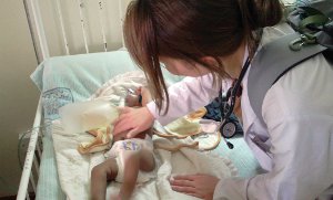
Figure 1. Baby hospitalised for suspected bacterial pneumonia.

Squeezing into our rusty mutatu (bus), we handed over the fare to the conductor, who returned to us less than expected change. In response to our indignant questioning, he defiantly stated, “You are mzungu (white person) and mzungu is money.” This was lesson one in a crash course we had inadvertently stumbled into: “Life in Kenya for the naïve tourist.” More unsettling than being scammed in day to day life, however, was the rampant corruption in the hospital and university setting.

We completed our placement at Kenyatta National Hospital, the largest referral centre in Kenya, with 1,800 beds, 50 wards and 24 operating theatres. I was based within the paediatric ward and paediatric emergency department…

Categories
Review Articles

Management of infertility in the setting of polycystic ovary syndrome

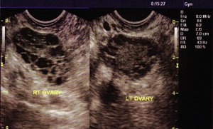
Polycystic ovary syndrome (PCOS) is a common endocrine disorder which affects a significant number of premenopausal women in Australia. PCOS has long-term clinical implications which can lead to decreased quality of life and psychological morbidity. A major contributing factor to this is the impact of PCOS on a woman’s fertility. However, there are a number of treatment modalities that may be used to treat PCOS-related infertility and with appropriate treatment, a woman’s prognosis with regards to PCOS-related infertility can be excellent.

Categories
Original Research Articles

Emergency Department management and referral of self-harm patients

Aim: To outline the socio-demographic characteristics, the means of arrival, management and referral pathways for mental health presentations to the Emergency Department (ED) where the main reason for presentation is self-harm. Methods: A retrospective study conducted in a metropolitan hospital in Sydney. Sampled data were collected from mental health presentations to the ED for the month of May in 2005, 2006 and 2007. The data collected included patient demographics as well as management, referral and follow-up outcomes. Results: There were 606 patients in the sampled data (99.3% of all mental health presentations). The gender distribution of the patient cohort was 63:37 (male n=380 and female n=226) and the average age was 36 ± 16.7 years. Two hundred and three (33.5%) patients had self-harmed and 403 (66.5%) had other mental health problems. Self-harm patients’ mode of arrival included ambulance (38.4%), self-presentations (36.5%), police (14%), and other. Self-harmers were mainly admitted to Psychiatric Emergency Care Centre (PECC) (28%) or discharged home (51.7%). More than one third (35.5%) of self-harm patients did not receive adequate follow-up. Conclusion: Important variations between self-harm patients and other mental health patients were identified in their management and referral outcomes from the ED. Clinicians need to ensure that optimal patient care is provided through appropriate follow-up of every self-harm patient post-discharge from hospital.


Categories
Feature Articles

Photograph: Tumaini


This photograph was taken during a four week elective placement at Ilula Lutheran Hospital, located in the southern highlands of rural Tanzania, East Africa. It emphasises the innocence and resilience of this country’s generous, kind people.
Ilula Lutheran Hospital is a 70-bed facility with a geographically broad service area. Patients often travel long distances to seek attention at the facility, and present most commonly with conditions such as malaria, complications of HIV, malnutrition, trauma, burns, respiratory and diarrhoeal illnesses, often in their advanced stages.
This photo was taken while visiting a village on an HIV outreach clinic. Nurses and doctors attend villages monthly to diagnose new patients, dispense anti-retroviral therapy and perform general check-ups. The rate of HIV infection in the Ilula area has not been accurately measured; however, the infection rate has been estimated at approximately 20% in the general community and 50% amongst hospital inpatients. The day this photo was taken, the nurses and doctors were helping villagers form a support group to facilitate communication between them and the hospital, to encourage new patients to seek help and to give existing patients a support network to aid with compliance. This little boy was shy as he hid behind the skirt of his HIV-positive mother. The support group was named Tumaini – hope.

This photo was the winner of the 2011 Medical Students’ Aid Project photo competition. MSAP is a not-for-profit organisation run by medical students from the University of New South Wales. MSAP’s goal is to send targeted aid to developing world hospitals visited by UNSW medical students on their elective terms. This is done through collecting donations of equipment from hospitals and doctors around the state, as well as fundraising to purchase additional equipment and arrange for delivery of these supplies. To ensure that the equipment sent is appropriate and useful, the hospitals are asked to compile a “wishlist” of required supplies. In addition, MSAP also educates medical students on issues associated with global health throughout the year. To find out more, and how you can help, visit www.msap.unsw.edu.au today!

Categories
Review Articles

Causes of death in neonatal intensive care units

Introduction

Of the approximately 130 million babies born each year, an estimated four million babies die in the neonatal period. Globally, the main causes of death are estimated to be preterm birth (28%), severe infections (26%) and asphyxia (23%). [1] New South Wales (NSW) is the most populous state in Australia with approximately 86,000 births per year. In 2006, 6,044 babies in NSW were registered to neonatal intensive care units (NICUs), representing 2.3% of total live births in that year. [2] Gestational age is highly correlated with birth outcomes including mortality. Each extra week of time spent in utero increases an infant’s chances of survival significantly, and by 27 weeks of pregnancy, over 90% of infants will survive.

There are significant differences between common causes of death in the pre-term population (less than 37 weeks gestation) and the term population (37+ weeks gestation). The most notable difference is an approximate five-fold increase in deaths caused by congenital neurological malformations in the term population compared with the pre-term population [3]. Premature infants have a considerably higher chance of dying than full-term infants. However, improved neonatal care, particularly the widespread use of surfactant replacement and antenatal steroids, has almost halved neonatal mortality in many parts of the world. [1] Between 1985 and 1991 in the United States, the overall neonatal mortality rate declined from 5.4 to 4.0 per 1,000 live births. An understanding of causes of neonatal death and changes in mortality rates is critical for prenatal counselling, decision making, quality control and further improvement in management.

In NSW, newborn infants are admitted to NICUs under the following criteria: gestational age less than 32 weeks, birth weight less than 1,500 grams, need for mechanical ventilation for four hours or more, continuous positive airways pressure for four hours or more and/or major surgery, defined as opening of the body cavity.

Newborns admitted to NICUs are cared for by a highly specialised team of medical, nursing and allied health staff. Despite the level of sickness and intensity of morbidities of NICU patients, mortality rates are relatively low. Neonatal mortality rate is defined as the number of neonatal deaths per 1,000 live births and includes all deaths of infants within 28 days after birth. Each neonate who dies in the NICU represents not only a financial cost to the community but more importantly, a significant emotional stress and grief for the involved parents and staff…

Categories
Original Research Articles

The effect of Duchenne Muscular Dystrophy on Purkinje cell number in the mdx mouse

Figure 1. Comparison of Nissl stained and Calbindin-D28k immunostained sections. (a) Calbindin immunopositive PCs clearly visible along the PC layer. (b) PCs not visible in Nissl stained section.

Background: Duchenne muscular dystrophy (DMD) is an X-linked recessive disease which causes skeletal muscle wasting in males, resulting in premature death during their early to mid 20s. Males with DMD carry defects in the gene encoding for dystrophin, a protein important in ensuring sarcolemmal stability. Dystrophin has also been implicated in disruption to Purkinje cells in the cerebellum. This disruption to cerebellar Purkinje cells has been proposed to be involved in reducing the IQ of affected boys. Aim: To compare Purkinje cell number and distribution in mutant mdx and normal mice. Methods: Cerebellar slices from both mutant (n=4) and normal (n=4) mice were prepared and stained. The number of Purkinje cells in each slice was estimated by three different cell counting techniques. Counting methods were as follows: firstly, the actual number of Purkinje cells per lobe; secondly, a randomised estimate where five random sections of the Purkinje cells layer were selected, counted and averaged; thirdly, an estimated maximum possible count, where three segments from the Purkinje cell layer with the highest density of cells were used to estimate Purkinje cell population. Results: No statistical significance in Purkinje cell numbers between the two groups was found. However, there was a trend towards a decrease in the median number of Purkinje cells in the mutant group, particularly in lobules 3, 4/5, 6 and 10. Conclusion: The study findings suggest a decrease in Purkinje cell number in mdx mice. The small sample size of this study precludes definitive statistical analysis of Purkinje cell numbers in either group. These findings demonstrate a need for larger mouse-model studies to accurately assess differences in cell numbers between the two groups. Given that the greatest difference in cell numbers was demonstrated in lobules 3 and 4/5, the authors suggest that DMD may affect the cerebellum during the maturation of these lobules. Importantly, a reduced Purkinje cell population may be implicated in the intellectual morbidity in boys with DMD.

Categories
Review Articles

Approach to the acute abdomen during pregnancy

Abstract

Many physiological changes in pregnancy may affect the presentation of abdominal pain in the pregnant patient. Rapid diagnosis and management is required to prevent dire complications for both mother and fetus. Most radiological investigations are not harmful to the developing fetus and can avoid unnecessary and potentially detrimental explorative surgery. The role of laparoscopy in the pregnant patient is increasingly being established, particularly in centres with this surgical expertise.