Categories
Book Reviews

The Emperor of All Maladies: Cancer 101

v5_i1_a23As medical students, we are experts in rare syndromes and exotic illnesses. However cancer, the second most common cause of death in the developed world, is still a total mystery. How do we explain to patients what so many of us don’t understand? Where do we begin?

Dr. Siddhartha Mukherjee is an Indian-born American oncologist, researcher and Pulitzer Prize winner. In this captivating narrative, Mukherjee explores cancer, its complexities, changing personalities, nuances, pet-peeves and habits. Indeed, Mukherjee himself states that, ‘it felt, inescapably as if I were writing not about something but someone.’ The language, therefore, unlike the mere spouting out of facts like a medical textbook, draws one in, such that the reader feels that Mukherjee has blasted open a door and allowed us to enter a landscape of discovery.

Mukherjee describes cancer’s existence thousands of years ago in Egyptian scripts, details the origins of the ongoing battle between cancer and physicians, and depicts his own clinical experiences with cancer patients, thus pouring insight, appreciation and a deeper understanding of this dreadful disease into the reader’s mind. One cannot hope to pursue a discussion about the history of cancer without first explaining what it is. His brief explanation is easy to understand and strikes a happy medium between layman’s terms and medical jargon, hence resonating perfectly with the mind of the medical student.

Cancer’s story begins in Sidney Farber’s lab in 1947, where leukemic cells were being studied. The utilisation of folate antagonists to treat leukemia can be credited to Farber’s genius: ‘If folic acid accelerated the leukemia cells in children, what if he could cut off its supply with some other drug – an antifolate?’. Thus, the idea of molecular targets and chemotherapy was born.

As chemotherapy grew in popularity, opportunities for combination therapy were explored. The author investigates the consequences of various clinical trials such as the catastrophic dips in white cell counts, the death toll rising with every turn of the page. He then leads us through the challenges of specific cancers, such as prostate and breast cancer.

Finally, Dr. Mukherjee arrives at the present day, detailing our new interests in gene therapy. Despite advancements, the true nature of cancer continues to elude us and with it, the cure consistently slips through our fingers. Dr. Mukherjee encapsulates this perfectly by advising us to, ‘focus on prolonging life rather than eliminating death’. With cancer rates increasing, its presence is approaching a level of normality and this guidebook warns us not to underestimate ‘the emperor of all maladies’, an important lesson for all future doctors.

As a medical student, The Emperor of All Maladies is a great introduction into the world of oncology. It is an easy and fun read that is a refreshing break from the traditional textbooks we pore over daily. The reader is not only educated about the intricacies of cancer but also walks away with a great deal of empathy for the patients and families whose experiences are vividly narrated. Furthermore, in the journey from historical events to present day, Mukherjee’s exciting and thrilling perspective of cancer is a useful timeline of the events of the past and what we, as medical students, can expect in the future of Oncology.

In summary, The Emperor of All Maladies is a roadmap of the places we have been, what we have done and where we still need to go. Cancer was, is, and will be the most challenging ailment that we, as future doctors will have to face. Hence, all medical students should take a note from cancer’s biography and its master storyteller.

Conflict of Interest

None declared.

Correspondence

M Ong: michelle.ong@my.jcu.edu.au

References

Mukherjee S. The Emperor of All Maladies: A biography of cancer. London: Fourth Estate; 2011.

Categories
Feature Articles

Exercising patient-centred care: A review of structured physical activity, depression and medical student engagement

Structured physical activity has a wide range of benefits that include improving mood and preventing chronic disease. Recently, there has been an explosion of research aimed at treating diseases such as depression using nothing more than exercise. This article presents an overview of research conducted into the use of exercise to treat depression. As a body of work, the literature finds it to be a practice that has significant clinical benefits; however, its implementation is not straightforward. Issues concerning exercise adherence have hampered studies and force us to ask whether prescribing exercise for sufferers of depression is indeed appropriate. Nonetheless, is there a role for medical students in encouraging physical activity as treatment? If we re-examine the use of exercise from a patient-centred perspective, medical students have an opportunity to engage with patients, promote exercise and possibly prevent depression.

Introduction

v5_i1_a19Structured physical activity has a wide range of health benefits that include the prevention of chronic diseases such as cancer, cardiovascular disease, diabetes and obesity. [1] There is also consensus that exercise has a short-term ‘feel good’ effect that improves mood and wellbeing. [2-4] Recently, converging interest in these two areas has spawned an explosion of research aimed at treating diseases such as depression using nothing more than exercise. [5-7] A 2012 meta-analysis of over 25 trials found that prescribing exercise to treat depression is on par with pharmacological and physiological interventions, and that exercise alone is moderately more effective than no therapy. [8] The study also highlights that prescribing exercise for depression is not straightforward. Poor patient attendance rates, along with issues including exercise adherence and the type, duration and intensity of exercise, all question whether prescribing exercise for sufferers of depression is indeed appropriate. The authors of the study admit the implementation is complex and that further study is required. Nonetheless, is there a role for medical students in encouraging physical activity as treatment? Can our skills in motivational interviewing and goal setting play a role? If we re-examine the use of exercise from a patient-centred perspective, medical students can promote exercise adherence and support those who are already exercising to stay exercising. In doing so, we can facilitate the prescription of exercise and possibly prevent depression. [5,7,9]

A background to depression

Depression affects a staggering 350 million people globally, with sufferers commonly reporting changes in emotional, cognitive and physical behaviour. [10] Depression also presents with high rates of comorbidity (the occurrence of more than one condition or disease). [11] More locally, a 2007 National Survey of Mental Health and Wellbeing found that almost half of the Australian population aged 18-85 (7.3 million people) had experienced a mental illness at some point in their lifetime. [3] A study by the Australian Bureau of Statistics (ABS) in the same year revealed that of those people suffering from mental illness, only 35% actually sought treatment, suggesting that within Australia there are 2.1 million potential patients going without much-needed assistance. [12]

Mental illness, while indiscriminate, has a higher incidence within certain sub-populations. Perhaps surprisingly, doctors and medical students experience higher rates of depression and stress than the general population. Medical students report increased depressive symptoms as a result of medical school while a significant number of doctors report that they are less likely to seek treatment for depression despite their awareness of the condition. [13,14] With a reported 3668 students admitted to medical schools in Australia during 2013, these statistics highlight the importance for medical students to identify and understand depression. [15]

Pathophysiology and current treatment options

Therapeutic treatment options for patients who are depressed fall into two broad categories: psychological and pharmacological. Typically, people are treated using cognitive behavioural therapy (CBT), anti-depressant medication, or a combination of both. [4,16,17] Despite being able to treat depression, a simple pathogenesis is yet to be found. Current opinion centres on depression being a result of chemical imbalances within the brain, specifically the action of monoamine neurotransmitters, including dopamine, serotonin, and norepinephrine. [17] This approach has allowed the pharmaceutical industry to develop medications including selective serotonin reuptake inhibitors (SSRIs), which target neurotransmitter reuptake to help restore their usual balance and so reduce symptoms. [18]

One promising new development in the quest to understand the pathophysiology of depression concerns the emergence of inflammation as a mediator of depression. A recent meta-analysis of 24 separate studies found that depression is accompanied by immune dysregulation and activation of the inflammatory response system (IRS). [19] Specifically, when compared to non-depressed patients, sufferers of major depression were found to have significantly higher (p < 0.001) concentrations of the pro-inflammatory cytokines tumour necrosis factor-α and interleukin-6 in their blood. Once the increased cytokine signal reaches the brain, it is able to down-regulate the synthesis, release and reuptake of the very same monoamines targeted by antidepressant medications. [20] Understanding and preventing this interaction from occurring could lead to promising new treatments for depression. Despite widespread prescription, the use of antidepressant medication is not without its drawbacks. Many patients experience unwanted side effects and stop taking their medication, while others simply do not want to take medicine that will make them feel worse. [21] Additionally, a 2012 report on Australia’s overall health revealed that some patients who experience depression suffer worse health outcomes due to the social stigma associated with taking antidepressants. [22] As a result, many patients choose to shun medication altogether, opting for alternative therapies such as acupuncture and yoga to help manage depression. [21] Viewed from the patient’s perspective, the use of antidepressants can be seen as a choice between the lesser of two evils: treatment or depression. Exercise and depression

The link between exercise and relieving depression has been a difficult one to make. A 2009 meta-analysis published by The Cochrane Collaboration evaluated the use of exercise, defined as “repetitive bodily movement done to improve or maintain … physical fitness”, in treating depression. [23] Initially, the Cochrane review pooled data from 25 trials and found a large clinical effect in the reduction of depressive symptoms when compared to a placebo or no treatment at all. However, in 2012, when the authors repeated the study, correcting for what they saw as errors and bias in the previous analysis, the data found only a moderate effect in relieving depressive symptoms. [8] Interestingly, in both studies, the data concerned with treating depression with exercise was found to be on par with the use of antidepressant medication. Nonetheless, despite the downgraded clinical effect, the authors of the study continue to find it reasonable to prescribe exercise to people with depressive symptoms; however, they caution to expect only a moderate effect at best.

Research into a preventative, rather than curative approach to depression is also being conducted. In a large-scale longitudinal study of depression, researchers hypothesised that an inverse relationship existed between the level of physical activity and depressive symptoms: those who were more active should not become depressed. [9] Researchers grouped exercise into three categories: light activity such as ‘necessary household chores’, moderate activity which included regular walks, and strenuous activity such as ‘participation in competitive sports’. The study revealed that participants in the ‘competitive sports’ category reported less depressive symptoms than those in the ‘necessary household chores’ category – an unsurprising finding given the ability of exercise to lift mood. But perhaps more revealing is the finding that participants who decreased their activity from moderate to light, and from strenuous to light, reported the greatest increase in depressive symptoms, implying that exercise may act as a buffer against depression.

Exercising patient-centred care

While scientific analysis and treatment forms the foundation of modern health care, a focus on the patient as a person should be paramount. In 2010, the Australian government endorsed patient-centred care, a framework that enshrines the values and individual needs of the patient. Since then, patient-centred care has broadened to encompass ‘an approach to the planning, delivery, and evaluation of health care that is grounded in mutually beneficial partnerships among healthcare providers, patients, and families’. [24] While relatively new, patient-centred care now shapes the delivery of health care in Australia. It has also reaffirmed the rights of the individual in developing and delivering health care.

Patient-centred care forces us to examine whether exercise as an intervention is at all appropriate for the depressed. While there may be some clinical benefit, is it really patient-centred? The Cochrane Collaboration study of exercise as a treatment for depression highlights low exercise adherence and high dropout rates amongst participants. [8] While the reasoning behind the dropouts was absent from the report, one could easily imagine the potential challenges in asking a person who is already lacking in drive and motivation to participate in an exercise program. Is it possible that in considering the prescription of exercise to the depressed we are violating key Hippocratic notions including that of non-maleficence? Are we exposing an already vulnerable individual to a situation where they are likely to fail and experience a decline in their mental health as a result? [1] It would seem that when it comes to exercise and depression, the patient-centred perspective would advise against such risks. [24]

While it is clear that exercise is good for us, it is also clear that its prescription for depression is fraught with ethical issues. As medical students, how then can we engage in such an uncertain and potentially lethal landscape when we do not possess the skills to interact with depression in any therapeutic manner? If we re-examine the issue from a patient-centred perspective, we are able to view exercise as a preventative, rather than curative, approach to depression. This opens the door to medical student engagement. For example, we can use the motivational and goal-setting skills taught as part of the preclinical curriculum to help patients achieve and maintain their exercise goals. This could involve scheduling a progress telephone call to maintain the patient’s motivation, a periodic home visit in an effort to reduce recidivism, or the discussion and identification of barriers that may prevent them from achieving their goals. Placements are the ideal environment for us to develop these mutually beneficial partnerships. Whatever the effort, promoting a healthier and more active lifestyle is a patient-centred perspective that all medical students should feel comfortable in advocating.

Conclusion
Re-examining the issue from a patient-centred perspective sees exercise as a multi-benefit, primary prevention tool that may also safeguard against developing depression. Moreover, this is wholly within our advocacy as medical students. Not only does this approach echo recommendations supported by the Australian Government to include collaborative, patient-centred care programs in undergraduate health programs, it also provides many practical opportunities for medical student engagement. For example, during placements in rural and remote areas, students often participate in community-based activities where we leverage the associative influence of our medical profession to promote the benefits of a healthier and more active lifestyle.

Despite reviews of studies inferring the protective effect of exercise against developing depressive symptoms, prescribing exercise as a treatment option requires skill and experiences beyond the scope of medical students. However, medical students do have skills in motivational interviewing and goal-setting strategies that enable us to promote exercise adherence. Therefore, if we consider exercise as a tool for disease prevention that may also safeguard against depression, patients will experience greater health outcomes and medical students can be active in its prescription.

If you or someone you care about is in crisis and you think immediate action is needed, call emergency services (triple zero – 000) or contact your doctor or local mental health crisis service, such as Lifeline (13 11 14).

Correspondence

D Lowden: danlowden@gmail.com

Acknowledgements

I’d like to thank the James Cook University Ecology of Health subject coordinators and Dominic Lopez for the valuable feedback provided in preparing this article.  

Conflict of interest declaration

None declared. 

References

[1] Allen K, Morey M. Physical activity and adherence. In: Bosworth H, editor. Improving patient treatment adherence. New York: Springer; 2010. p. 9-38.

[2] Ströhle A. Physical activity, exercise, depression and anxiety disorders. J Neural Transm. 2009;116(6):777-84.

[3] Australian Institute of Health and Welfare (AIHW). Comorbidity of mental disorders and physical conditions 2007 [Internet]. 2012 [cited 2014 May 30]. Available from: http://www.aihw.gov.au/publication-detail/?id=10737421146.

[4] Carlson VB. Mental health nursing: the nurse-patient journey. Philadelphia: W B Saunders Co.; 2000.

[5] Blake H. How effective are physical activity interventions for alleviating depressive symptoms in older people? A systematic review. Clin Rehabil. 2009;23(10):873-87.

[6] Carlson DL. The effects of exercise on depression: a review and meta-regression analysis [dissertation]. Milwaukee: University of Wisonin; 1991.

[7] Pinquart M DP, Lyness JM. Effects of psychotherapy and other behavioral interventions on clinically depressed older adults: a meta-analysis. Aging Ment Health. 2007;11(6):645-57.

[8] Rimer J DK, Lawlor DA, Greig CA, McMurdo M, Morley W, Mead GE. Exercise for depression. Cochrane Database Syst Rev. 2012;7:CD004366.

[9] Graddy JT, Neimeyer GJ. Effects of exercise on the prevention and treatment of depression. Journal of Clinical Activities, Assignments & Handouts in Psychotherapy Practice. 2002;2(3):63-76.

[10] World Health Organization. Depression: Fact sheet no. 369 [Internet]. 2012 [cited 2014 March 14]. Available from: http://www.who.int/mediacentre/factsheets/fs369/en/.

[11] World Health Organization. The world health report 2001: mental health – new understanding, new hope [Internet]. 2001 [cited 2014 May 24]. Available from: http://www.who.int/whr/2001/en/.

[12] Department of Health and Ageing. National mental health report 2010: Summary of 15 years of reform in Australia’s mental health services under the National Mental Health Strategy 1993-2008. Canberra: Commonwealth of Australia; 2010.

[13] beyondblue. Doctors’ mental health program [Internet]. 2014 [cited 2014 March 30]. Available from: http://www.beyondblue.org.au/about-us/programs/workplace-and-workforce-program/programs-resources-and-tools/doctors-mental-health-program.

[14] Rosal MC, Ockene IS, Ockene JK, Barrett SV, Ma Y, Hebert JR. A longitudinal study of students’ depression at one medical school. Acad Med. 1997;72(6):542-6.

[15] Medical Deans Australia and New Zealand Inc. Annual tables [Internet]. 2013 [30 March 2014]. Available from: http://www.medicaldeans.org.au/statistics/annualtables.

[16] Imel ZE, Malterer MB, McKay KM, Wampold BE. A meta-analysis of psychotherapy and medication in unipolar depression and dysthymia. J Affect Disord. 2008 Oct;110(3):197-206.

[17] Carlson NR. Foundations of physiological psychology. 7th ed. Boston, MA: Allyn & Bacon; 2008. p. 108-22.

[18] Nutt DJ. Relationship of neurotransmitters to the symptoms of major depressive disorder. J Clin Psychiatry. 2008;69 Suppl E1:4-7.

[19] Dowlati Y, Herrmann N, Swardfager W, Liu H, Sham L, Reim EK, et al. A meta-analysis of cytokines in major depression. Biol Psychiatry. 2010;67(5):446-57.

[20] Miller AH, Maletic V, Raison CL. Inflammation and its discontents: the role of cytokines in the pathophysiology of major depression. Biol Psychiatry. 2009;65(9):732-41.

[21] Sarris JK, Newton DJ. Depression and Exercise. J Comp Med. 2008;7(3):48-50.

[22] Australian Institute of Health and Welfare (AIHW). Australia’s health. Canberra: AIHW; 2012.

[23] Mead GE, Morley W, Campbell P, Greig CA, McMurdo M, Lawlor DA. Exercise for depression. Cochrane Database Syst Rev. 2009 Jul 8;(3):CD004366.

[24] Australian Commission on Safety and Quality in Health Care (ACSQHC). Patient-centred care: Improving quality and safety through partnerships with patients and consumers. Canberra: ACSQHC; 2011.

Categories
Review Articles

Aripiprazole as first-line treatment of late-onset schizophrenia – a case report and literature review

Introduction: Guidelines for the first-line treatment for late-onset schizophrenia (LOS) in the elderly patient have not been established. The current recommended treatment of schizophrenia in younger age groups has been extrapolated to those in the older age groups. This report considers the effects of medication specific to this demographic.

Case study: BB, a 73-year-old male, presented to the Mental Health Unit following a request and recommendation in response to concerns from family and friends, with a history of increasing paranoia and paranoid delusions. He was managed under an involuntary treatment order and was prescribed aripiprazole 10 mg once daily.

Methods: A literature review was conducted using UpToDate, Medline, PsychOnline and Ovid databases with limits set to exclude articles that were not written in English, published before the year 2000 or which were not available as full text. Articles were found using a combination of the search terms “late onset schizophrenia”; “risperidone AND mechanism of action”; “aripiprazole AND mechanism of action”; “paraphrenia”; “schizophrenia AND Australian therapeutic guidelines”; and “atypical antipsychotics pharmacology”.

Results: The literature review confirmed the efficacy and safety of aripiprazole in the elderly patient with LOS. Studies identified fewer side effects with aripiprazole, such as cerebrovascular and cardiovascular events, than with risperidone. There were no studies identified that directly addressed the question of whether aripiprazole should be used as first-line management of LOS instead of risperidone.

Conclusion: Aripiprazole should be considered as first-line management for patients with late-onset schizophrenia.

Case study

Aripiprazole as first-line treatment of late-onset schizophrenia – a case report and literature reviewBB is a 73-year-old Caucasian male who presented to the Mental Health Unit (MHU) following a request and recommendation (under the Queensland Mental Health Act 2000) in response to concerns from family and friends. BB presented from a nursing home with paranoid delusions that incorporated persecutory themes with thoughts that the nursing staff were poisoning his food in order to kill him. He also presented with auditory hallucinations complaining of hearing people through a speaker telling him they were going to cut off his toes and genitals. BB expressed suicidal ideation to escape, however, no previous attempts at suicide or self-harm had been made. When BB was further questioned about a suicidal plan he stated that he would like to do it cleanly with towels around him so there was no mess but no instrument or method was established. There was no history of substance abuse.

BB is retired and lives in a nursing home. His wife died three years prior to this presentation.

On assessment, BB was well groomed, sitting on a hospital bed, clutching his legs in a curled up manner. Mr. BB maintained a paranoid and untrusting manner throughout the presentation, with poor eye contact. Therefore, rapport with mental health staff was difficult to establish. His speech was agitated and consistent with anxiety. Affect was restricted and he appeared apprehensive towards the interviewer. His mood was difficult to establish because of his paranoid state. He had no insight into his mental illness and judgement was poor. BB was assessed to have a moderate risk of violence.

A Mini-Mental State Examination was carried out on admission to the MHU. BB scored 30/30 and combined with further assessment in the MHU dementia-related psychosis was excluded.

BB had a one-week admission to the MHU four years previously following the death of his wife. He had neurovegetative symptoms: not eating or sleeping and not carrying out activities of daily living. He required diazepam to assist with sleep and a nasogastric tube for enteral feeding. He was later discharged with family support.

BB has a medical history of hypertension, dyslipidaemia and ischaemic cardiomyopathy.

After a request and recommendation for assessment, BB was diagnosed with late-onset schizophrenia in accordance with the Diagnostic and Statistical Manual of Mental Disorders, 4th Edition, Text Revision (DSM-IV-TR) criteria and managed under the Mental Health Act (involuntary treatment order) for three weeks for this presentation.

BB was commenced on aripiprazole 5 mg once daily (OD). His auditory hallucinations and paranoid delusions persisted with no reduction in their severity. Aripiprazole was then increased by 5 mg after four days until clinical improvement was achieved at 10 mg OD.

Throughout the admission, BB’s auditory hallucinations and paranoid delusions resolved significantly and there was an improvement in his mental state. Pharmacotherapy combined with repetitive cognitive assessment resulted in a good initial prognosis.

Following aripiprazole 10 mg daily for two weeks, BB reported no auditory hallucinations or paranoid delusions. There had been no side effects of aripiprazole. BB was discharged with aripiprazole 10 mg OD and returned to Old Persons’ Mental Health Services in the community for support.

Introduction

Schizophrenia is characterised by various symptoms including delusions, hallucinations, disorganised speech, grossly disorganised or catatonic behaviour and negative symptoms (including loss of motivation, poverty of words and affective flattening). [1,2]

The average age of onset of schizophrenia is 18 in men and 25 in women. Late-onset schizophrenia (LOS) is a separate category of patients whose onset is after 45. Very late-onset schizophrenia (VLOS) occurs after 60 years of age. Australian diagnostic criteria do not separate LOS and VLOS. [2] Therefore, in the case study, Mr. BB is diagnosed with LOS.

Late-onset cases present with some differences to early-onset schizophrenia. The clinical presentation of LOS includes more persecutory delusions and hallucination, as evident in the case study, and is less likely to include disorganised behaviour and negative symptoms. [1] LOS has been demonstrated to have an increased response to pharmacotherapy (anti-psychotics) in lower doses, resulting in an improvement in symptoms compared with early-onset schizophrenia. [2]

If smaller doses can be administered the possibility of side effects is reduced.

Aim

The current recommended treatment of schizophrenia in younger age groups has been extrapolated to those in the older age groups. This article is a review of the literature, supplemented with a case study, and outlines the evidence that is available for the effectiveness of aripiprazole as first-line management of LOS. Aripiprazole is compared with risperidone, which is the current first-line management for schizophrenia in the younger population. Risperidone has been used for this review rather than olanzapine and quetiapine as it is more frequently prescribed as initial management in the elderly population. [3]

Data collection

To address the management of LOS, a literature search was conducted using the search terms “late onset schizophrenia”; “risperidone AND mechanism of action”; “aripiprazole AND mechanism of action”; “paraphrenia”; “schizophrenia AND Australian therapeutic guidelines”; and “atypical antipsychotics pharmacology”. The databases UpToDate, Medline, PsychOnline and Ovid were used with limits set to only include articles written in English and available as full-text journals. Articles published before the year 2000 were excluded from this study. Articles including meta-analyses (Level I evidence) as well as randomised controlled trials (Level II Evidence) were reviewed. No study was found comparing the use of risperidone as first-line management for LOS with the use of aripiprazole.

Current first-line management

The Royal Australian and New Zealand College of Psychiatrists (RANZCP) Clinical Practice Guidelines for the treatment of schizophrenia (inclusive of LOS) and related disorders since 2005 include psychotherapy and pharmacotherapy. [3]

Guidelines for the introduction of pharmacotherapy recommend:

  • Notification of patients and families of the benefits and risks of drug therapy. Where it is not possible to fully discuss the choice of agent as is the case in most acute episodes, oral atypical agents are used. Risperidone, olanzapine and quetiapine are regarded as the treatments of choice in the first episode of psychosis. [3]
  • Evaluation of the efficacy of treatment subjectively as well as objectively by the clinician with regular Mental State Examinations and reviews of the patient. [3,4]
  • Titration of the dose of risperidone as appropriate for the patient. [5]

Discussion

Aripiprazole is the proposed first-line management of LOS versus risperidone.

Dosing

The recommended starting dose for aripiprazole is 10 mg to 15 mg OD. [6] Intramuscular injection has a therapeutic benefit from 10 mg to 30 mg. There is no evidence showing oral medication being more effective above 15 mg OD. It is advised that dose increases should not be more frequent than twice weekly as this is the time needed to achieve a steady state. [6] In contrast, recommended initial risperidone dosing is 2 mg with titration in increments of 1–2 mg per day as tolerated by the patient to a recommended dose of 4 to 8 mg. For schizophrenia, efficacy with risperidone has been evident from 4 mg to 16 mg.

Mechanism of action

Aripiprazole acts as a partial dopamine D2 receptor agonist in the limbic system. It also acts as a partial agonist at serotonin 5-HT1A receptors but an antagonist of 5-HT2A receptors. [6,10] This is compared with risperidone, which acts as a dopamine D2 antagonist and low-affinity antagonist of serotonin type 2 receptors. [5,8]

Efficacy

In review of the Cochrane database, aripiprazole was compared with other antipsychotics for schizophrenia. One hundred and seventy four randomised control trials (RCTs) were compared involving 17 244 participants. [10] The RCTs comparing aripiprazole against risperidone demonstrated an increase in efficacy through an improvement in mental state measured by the Brief Psychiatry Rating Scale when using aripiprazole. Additionally, for patients using aripiprazole there was also a significant increase in the quality of life. Despite these demonstrations of efficacy these RCTs do not compare the use of these medications in the elderly population, limiting their applicability to LOS.

Comparison of side effects

Aripiprazole has minimal extrapyramidal side effects. [11] Sedation is a dose-related side effect (most prominent at 30 mg) which seems to decrease with time. [7,12] Monitoring is important in the elderly because of the potential for increased falls and, subsequently, possible fractures and head injuries.

In several controlled trials aripiprazole-induced extrapyramidal side effects were reported to be reduced compared to dopamine antagonists such as risperidone. [13-16] However, aripiprazole-induced akathisia was reported as higher (approximately 20%) in patients with schizophrenia. Anticholinergic agents may be used to treat parkinsonism and dystonia but are ineffective in treating akathisia. Beta-blockers and benzodiazepines are effective in reducing akathisia.

Risperidone has been reported to have increased extrapyramidal side effects including parkinsonism, akathisia, dystonia and tremor. [5,16] Cerebrovascular side effects include stroke and altered cardiac conduction, potentially causing life-threatening arrhythmias. Nausea, constipation, dyspepsia, salivary hypersecretion, abdominal discomfort, and diarrhoea were also recorded. Prolactin inhibition may result in reduced libido, caused by reduction in levels of testosterone and oestrogen. In long-term therapy low testosterone and oestrogen levels may lead to osteoporosis, which is especially relevant for older adults.

It is important to note that there is an absence of controlled studies of risperidone in the elderly, thus making assessment of the LOS side-effect profile impossible. Nevertheless, based on the known side-effect profile of risperidone, it is contraindicated for Mr. BB with a medical history of hypertension, dyslipidaemia and ischaemic cardiomyopathy.

Table 1: Comparison of anti-psychotic medications. *PBS = Pharmaceutical Benefits Scheme

Conclusion

Aripiprazole should be considered as an option for the first-line pharmacotherapeutic management of LOS. In trials comparing aripiprazole against risperidone, aripiprazole has higher efficacy in the management of the symptoms associated with schizophrenia. Additionally, aripiprazole produces fewer cardiac conduction abnormalities, gastrointestinal side effects, and extrapyramidal side effects than risperidone. Consequently, those on aripiprazole have a reduced risk of cardiovascular and cerebrovascular events, both of which are more common in older age groups.

Despite the indirect evidence for the use of aripiprazole in LOS, there is a paucity of studies directly comparing aripiprazole and risperidone in LOS. Further controlled studies (ideally double-blinded, placebo-controlled) should be performed to assess the efficacy, side-effect profile and drug interactions in older-age patients.

Consent declaration

Informed consent was obtained from the patient for the original case report.

Conflict of interest

None declared.

Correspondence

Tasciana Gordon: tasciana.gordon@my.jcu.edu.au

References

1. American Psychiatric Association. Diagnostic and statistical manual of mental disorders. 4th ed. Text revision. Arlington, VA: American Psychiatric Publishing; 2000.

2. Bernard F, Buchanan R. Schizophrenia: clinical manifestations, course, assessment and diagnosis [Internet]. UpToDate; 2014 [updated 2013 July 26; cited 2013 June 20]. Available from: http://www.uptodate.com/contents/schizophrenia-clinical-manifestations-course-assessment-and-diagnosis.

3. RANZCP CPG. Royal Australian and New Zealand College of Psychiatrists clinical practice guidelines for the treatment of schizophrenia and related disorders. Aust N Z J Psychiatry. 2005;39:1-30.

4. Howard R, Rabins P, Seeman M. Late-onset schizophrenia and very-late-onset schizophrenia-like psychosis: an international consensus. Am J Psychiatry. 2000;157(2):172-8.

5. Lexicomp. Risperidone: patient drug information [Internet]. UpToDate; 2014 [cited 2013 June 20]. Available from: http://www.uptodate.com/contents/risperidone-patient-drug-information.

6. Lexicomp. Aripiprazole: drug information [Internet]. UpToDate; 2014 [cited 2013 June 20]. Available from: http://www.uptodate.com/contents/aripiprazole-patient-drug-information.

7. Lauriellow J, Campbell A. Pharmacotherapy for schizophrenia: long-acting injectable antipsychotic drugs [Internet]. UpToDate; 2013 [cited 2013 Oct 3]. Available from: http://www.uptodate.com/contents/pharmacotherapy-for-schizophrenia-long-acting-injectable-antipsychotic-drugs.

8. Davis J, Chen N. Clinical profile of an atypical antipsychotic: risperidone. Schizophr Bull. 2002;28(1):43-61.

9. Bahman S, Ghader Z. Side effects of risperidone. Life Sci. 2012;9(3):1463-7.

10. Khanna P, Suo T, Komossa K, Ma H, Rummel-Kluge C, El-Sayeh HG, et al. Aripiprazole versus other atypical antipsychotics for schizophrenia [Internet]. Cochrane Database of Systematic Reviews. John Wiley & Sons, Ltd; 1996 [cited 2014 Jul 11]. Available from: http://onlinelibrary.wiley.com/doi/10.1002/14651858.CD006569.pub5

11. Burris KD, Molski TF, Xu C, Ryan E, Tottori K, Kikuchi T, et al. Aripiprazole, a novel antipsychotic, is a high-affinity partial agonist at human dopamine D2 receptors. J Pharmacol Exp Ther. 2002;302(1):381-9.

12. Mamo D, Graff A. Aripiprazole’s receptor pharmacology and extrapyramidal side effects. Am J Psychiatry. 2008;165(1):398.

13. Sweeney EB, Lawlor BA. Case series: extrapyramidal symptoms associated with use of aripiprazole in older adults. Int J Geriatr Psych. 2013;28:1208-10.

14. Coley K, Scipio T, Ruby C, Lenze E, Fabian T. Aripiprazole prescribing patterns and side effects in elderly psychiatric inpatients. J Psychiatr Pract. 2009;15(2):150-3.

15. Takahashi H, Oshimo T, Ishigooka J. Efficacy and tolerability of aripiprazole in first-episode drug-naïve patients with schizophrenia: an open-label trial. Clin Neuropharmacol. 2009;32(3):149-50.

16. Kim C, Chung S, Lee JN. A 12-week, naturalistic switch study of the efficacy and tolerability of aripiprazole in stable outpatients with schizophrenia or schizoaffective disorder. Int Clin Psychopharm. 2009;24(4):181-8.

Categories
Review Articles

Glibenclamide therapy as tertiary prevention of melioidosis for Type 2 diabetics.

Background: Melioidosis is a relatively high-incidence, high-mortality tropical infectious disease caused by Burkholderia pseudomallei. To date, the few prevention and management strategies in practice have failed to reduce mortality from septic shock in patients with severe melioidosis. Up to 60.9% of patients also have pre-existing type 2 diabetes mellitus (T2DM), the most significant risk factor for melioidosis. An effective tertiary prevention strategy against melioidosis for these diabetic individuals would impact significantly on the burden of disease. Glibenclamide, a drug belonging to the sulfonylurea class and commonly used as an antidiabetic, may have the potential to be one such strategy.

Aim: To examine the underlying inflammatory cause of morbidity and mortality in melioidosis and to assess the potential for glibenclamide to ameliorate these causes.

Results: Bacteraemia with B. pseudomallei leads to widespread infection. Multi-organ damage and loss of function results from both direct bacterial damage and an excessive inflammatory response, the latter of which also causes potentially fatal septic shock in 21% of cases. Massive cellular infiltration of the lungs is particularly damaging. The immunomodulatory effects of T2DM further exacerbate the deleterious immune response into a dysregulated, hyperinflammatory state. On the other hand, the several anti-inflammatory effects of glibenclamide have been demonstrated to significantly reduce mortality from septic shock in melioidosis.

Conclusion: For diabetics living in regions where melioidosis is endemic, choosing glibenclamide over other sulfonylureas and metformin for antidiabetic treatment could be a promising tertiary prevention measure against melioidosis.

Introduction

Glibenclamide therapy as tertiary prevention of melioidosis for Type 2 diabeticsMelioidosis, a tropical infectious disease, is a significant cause of death and illness in northern Australia and Southeast Asia. Its non-specific prodrome and clinical presentation makes early detection difficult.[1] Yet, timely diagnosis is critical because responsiveness to treatment declines with progression of the disease over time.[2] Morbidity and mortality remains high despite aggressive antibiotic treatment. As of yet, no vaccine is available for primary prevention and novel developments such as granulocyte colony-stimulating factor (G-CSF) and corticosteroid therapy lack evidence as good measures of tertiary prevention. Considering the significant population health threat, any effective prevention strategy would be of value. This article suggests the potential use of glibenclamide, an antidiabetic sulfonylurea, for tertiary prevention of melioidosis in diabetics. Because diabetics are approximately 13 times more likely to contract melioidosis,[3,4] this strategy may have widespread application.

One of the major and dangerous end-stage complications of melioidosis is septic shock. Especially in settings such as Northeast (NE) Thailand where melioidosis is endemic but intensive care is unavailable, morbidity and mortality from septic shock are unacceptably high. Systemically-spread B. pseudomallei stimulates massive release of cytokines into the blood, this event being instrumental in the immunopathogenesis of septic systemic vasodilation, hypotension, vascular hyperpermeability, and disseminated intravascular coagulation (DIC). Furthermore, poor glycaemic control in diabetics increases the risk and severity of these events by precipitating a compensatory, dysregulated, and hyperinflammatory response to bacterial immunogenic stimuli. Demanding particular attention is the stimulated production of cytokine IL-1β because it promotes excessive neutrophilic recruitment to the lungs and bacterial persistence inside cells. Fortunately, there is strong evidence that glibenclamide acts protectively against IL-1β-mediated damage by targeting IL-1β production directly as well as multiple upstream components in the IL-1β production pathway. The result of glibenclamide use for diabetics is a significantly reduced morbidity and mortality from melioidosis septic shock compared with diabetics not taking the drug.

Epidemiology of Melioidosis

Melioidosis is endemic in Southeast Asia, northern Australia, India, Hong Kong, southern China and Taiwan.[5,6] Isolated sporadic cases have also appeared in Central America, the Caribbean, New Caledonia, Mauritius, Africa, and the Middle East.[7] Of particular concern are the hyperendemic hotspots – NE Thailand and the Top End of the Northern Territory of Australia. A 2006 NE Thailand study that gathered results from four large hospitals reported the region experienced an estimated minimum annual incidence of 21.3 cases per 100,000 people and case fatality rate of 40.5%, melioidosis locally being the third highest infectious cause of death and second most common pathogenic cause of community-acquired bacteraemia.[8,3] From 2004 to 2009, according to data collected at Royal Darwin Hospital (RDH), the annual incidence in the Top End was 21.6 per 100,000 people and average case fatality rate plateaued at 9% having decreased from 30% since 1989. The lower fatality rate in Darwin is attributed to greater access to intensive care unit (ICU) supportive care and early sepsis management that prolongs life in the event of fatal septic shock, a complication that develops in at least 21% of cases.[9]

Type 2 diabetes mellitus (T2DM) is the largest risk factor for melioidosis. 48% and 60.9% of individuals with melioidosis were confirmed to have T2DM at RDH and in NE Thailand respectively.[10,11] The 2006 Thai study reported that the relative risk (RR) of melioidosis in adult diabetics compared with non-diabetics was 12.4.[3] In the Top End region, combining diabetes with the additional risk factor of Indigenous ethnicity raised the RR from 13.1 to 20.6. Other notable risk factors associated with a significant RR are chronic lung disease (RR 4.3), age ≥ 45 years (RR 4.0), chronic renal disease (RR 3.2), Indigenous ethnicity (RR 3.0), male sex (RR 2.4) and hazardous alcohol consumption (RR 2.1).[6]

The presence of any of these risk factors in infected individuals also dramatically increases their chance of developing septic shock (RR 4.5) and death (RR 9.2), the risk factors with highest independent association with death being age ≥ 50 years (RR 2.1) and chronic lung disease (RR 1.5). Malignancy (RR 1.9) and rheumatic heart disease and/or congestive cardiac failure (RR 1.7) are statistically insignificant risk factors for death. Clinical presentations most likely to result in death are septic shock (RR 11.2) and, out of the non-septic shock cases, the presence of neurological infection foci (RR 4.7).[9] Additionally, case fatality during the peak rainfall months of December through February is 1.6 times greater than it is during other months.[9]

Immunopathogenesis and pathophysiology

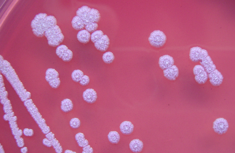
Melioidosis is caused by Gram-negative bacterium B. pseudomallei. Bacteria, found in contaminated soil, rodents, food, water, and excretions, are transmitted via inhalation, ingestion, or percutaneous inoculation – usually direct contact with open skin lesions.[12,13] Post-inoculation, B. pseudomallei can invade most human cell types. Employing Type 3 Secretion System (TTSS) clusters, bacteria enter non-phagocytic cells. If bacteria are phagocytosed, TTSS enables exit from intracellular phagocytic endosomes such that degradation is evaded. Once escaped into the cytoplasm of cells both phagocytic and non-phagocytic, bacteria replicate and self-induce polarised actin filamentation that confers motility, facilitating spread to neighbouring cells by forcing host-cell membrane protrusion and fusion.[14]

B. pseudomallei possesses highly immunogenic factors that trigger a strong host immune reaction essential to the host for early bacterial containment.[15] B. pseudomallei expresses pathogen-associated molecular patterns including lipopeptides, peptidoglycan, lipopolysaccharide, flagellin, TTSS, and DNA. They are recognised by host cell toll-like receptors (TLR) and NOD-like receptors (NLR). TLRs and NLRs are expressed by immune cells both professional – macrophages and dendritic cells – and non-professional – epithelial cells, endothelial cells, and fibroblasts.[16]

Activation of NLRC4 or NLRP3 induces binding of caspase-1, Asc (apoptosis-associated speck-like protein containing CARD) and NLRC4 or NLRP3 to form an inflammasome. The NLRC4 inflammasome in infected macrophages triggers pyroptosis, serving to limit intracellular B. pseudomallei growth and proliferation, while the NLRP3 inflammasome performs proteolytic activation of pro-IL-1β and pro-IL-18 into their mature forms for secretion.[17] IL-1β recruits neutrophils to infection site(s). However, excessive recruitment is often deleterious because neutrophils, lacking NLRC4, fail to pyroptose and instead provide a favourable intracellular environment that sustains chronic bacterial persistence. In the lungs, persistence leads to damaging pulmonary abscess formation and acute respiratory distress syndrome.[17,18] In contrast, elevated IL-18 production correlates with survival and immunoprotection because IL-18 induces IFN-γ production.[17,19] IFN-γ activates macrophages, stimulating their direct antimicrobial processes – phagolysosomal fusion and toxic reactive nitrogen species synthesis that produces nitrosative stress.[20] The cytokine also facilitates macrophage antigen processing and presentation, recruits leukocytes to infection site(s), upregulates Th1 CD4+ cell population, enhances natural killer cell function, regulates B cell anti-lipopolysaccharide antibody production and isotype switching.[21]

Glibenclamide therapy as tertiary prevention of melioidosis for Type 2 diabetics
Figure 1. NLRC4 inflammasome leads to immunoprotective effects while NLRP3 inflammasome produces both protective and deleterious responses. Glibenclamide works by inhibiting NLRP3 inflammasome formation and its deleterious effects.

TLR activation upregulates secretion of principal inflammatory mediators including type 1 interferon, chemokines, antimicrobial proteins, and pro-inflammatory cytokines.[22] Elevated expression of TLR1, TLR2, TLR3, TLR4, TLR5, TLR8, and TLR10 has been demonstrated in patients with septic melioidosis.[16] In particular, stimulation of the TLR2-mediated signalling pathway is a principal step in recognising the immune challenge of B. pseudomallei and initiating early inflammatory processes.[23] TNF-α and IL-6, along with IL-1β, increase vascular permeability, induce acute phase protein production, and recruit leukocytes to the site of infection.[24] TNF-α and IL-6 also activate the complement and coagulation cascades that are key defense mechanisms of the innate response.[25] However, excessive inflammatory cytokine production induced by widespread bacteraemia often leads to septic shock characterised by systemic vasodilatory hypotension, vascular hyperpermeability causing major cellular and fluid leakage from the intravascular to extravascular compartments, DIC, and death in the absence of immediate treatment.[26]

Through the establishment of bacteraemia, B. pseudomallei spreads from primary foci of infection to other body tissues, usually the lungs, genitourinary tract, skin, joints, bones, liver, spleen, skeletal muscles, prostate, parotid gland, and nervous system. Multi-organ spread leading to impaired organ function and failure is the main source of morbidity and mortality in melioidosis.[27] Thus, a rapid but non-deleterious inflammatory response is critical before B. pseudomallei establishes an intracellular niche that enables its persistence and protection against eradication by a subsequent immune attack. Paradoxically, subsequent immune attacks may cause further damage to the host.

Effect of type 2 diabetic state on host response

Diabetics, due to their generally immunosuppressed state, compared with non-diabetics, are more susceptible to developing sepsis from most organisms,[28] with the most common source of systemic spread being respiratory, followed by urinary and abdominal.[29] Gram-positive bacteria have become the leading cause of sepsis since the 1980s, and sepsis due to causes other than B. pseudomallei actually affects diabetics more commonly than melioidosis.[29,30] However, it is still of significance that diabetics are particularly vulnerable to pathogens such as B. pseudomallei and Mycobacterium tuberculosis because immune defence against these intracellular bacteria is highly macrophage-mediated – a function that is critically impaired in poorly-controlled T2DM.[31]

T2DM, especially if poorly controlled, causes marked immunomodulation that produces B. pseudomallei-induced immune responses that are damaging and yet insufficient to offer host protection. Morris et al. created an ex vivo whole-blood assay using human peripheral blood to compare inflammatory responses to B. pseudomallei between diabetics and non-diabetics. Immune insufficiency was worst in poorly controlled diabetics, the assay detecting: elevated serum IL-10 (an anti-inflammatory cytokine); reduced CD11b on polymorphonuclear leukocytes (PMNs) leading to decreased PMN function with reduced activation, adhesion, transmigration, and migratory capacity towards IL-8; reduced endotoxaemia-induced upregulation of ICAM-1; and defects in phagocytic detection and response to the bacteria.[32] In vivo and in vitro models using streptozotocin (STZ)-induced, leptin deficiency, leptin receptor deficiency, and diet-induced diabetic mice found similar immune impairments.[31,33-35] Transcriptional analysis of STZ-diabetic mice responses over the first 42 hours of B. pseudomallei exposure attributed delayed defence and splenic dysfunction (permitting uncontrolled bacterial replication intracellularly causing susceptibility to sepsis) to diminished TLR2 recognition of the bacterium.[34]

Incompetence of the early inflammatory response to contain an initial infection precipitates a compensatory, dysregulated, hyperinflammatory response to the spreading infection. The intensity of this ensuing reaction is exacerbated by chronic low-grade inflammation and increased oxidative stress, both characteristic of T2DM. Morris et al. found poorly controlled diabetics to have the most elevated levels of pro-inflammatory markers ESR, CRP, TNF-α, IL-1β, IL-6, IL-8, IL-12p70, MCP-1, and MPO (which indicates increased oxidative burst activity in PMNs). At focal infection sites, there was extensive infiltration with PMNs and greater risk of tissue damage, as well as generalised endothelial dysfunction and vascular inflammation.[32]

Hyperglycaemia is a major link between T2DM, immune derangement, and susceptibility to sepsis. The supra-physiological hyperosmolarity of hyperglycaemic blood directly potentiates TLR4-mediated cytokine production, as well as dampening phagocytic responses and granulocyte oxidative burst.[36] Activation of the RAGE pathway by advanced glycation end-products formed secondary to hyperglycaemia perpetuates inflammation and worsens survival in septic mice.[37] Furthermore, in the event of sepsis, poor glycaemic control allowing for periods of hyperglycaemia in both diabetics and non-diabetics correlates with longer hospital stay, greater morbidity, and possibly decreased survival.[38] In addition to immunomodulatory effects, hyperglycaemia exacerbates hypotension in septic shock by promoting glycosuric diuresis and cardiac hyperresponsiveness to cholinergic stimulation and the baroreflex.[39] Elevated risk of myocardial infarction, stroke, and venous thromboembolism is associated with the prothrombotic effect of acute hyperglycaemia that is further potentiated by hyperinsulinaemia or an inflammatory stress state.[40]

Prevention and management

No human vaccine is available.[41] CDC prevention guidelines recommend avoiding contact with soil and stagnant water in endemic regions.[42] High-risk individuals should stay indoors during heavy wind and rain in endemic regions due to possible bacterial aerosolisation that greatly facilitates transmission.[43]

Treatment involves two weeks of intravenous antibiotics – ceftazidime, meropenem, or imipenem, in combination with trimethoprim-sulfamethoxazole – followed by 3 months of oral eradication therapy to prevent recrudescence.[44] However, even with early antibiotic administration, case fatality from septicaemia remains at 37% in Thailand.[45] The development of septic shock is an indication for ICU supportive care usually involving invasive monitoring, fluid resuscitation, renal replacement therapy, and vasopressor, inotropic, and mechanical ventilatory support.[46,47]

Adjunctive use of recombinant G-CSF holds no clinical utility for improving sepsis from melioidosis and other aetiologies. Both animal and human trials – including prospective, multicentre, randomised, double-blind, and placebo-controlled studies – confirm this.[47-49] In fact, G-CSF may cause increased hepatic dysfunction and higher peak troponin I levels.[50] Other immunomodulatory adjuncts such as corticosteroids do not improve survival.[46] In light of the paucity of effective evidence-based prevention strategies and adjunct therapies for underserved populations, we turn to a novel potential for tertiary prevention.

Glibenclamide and its protective effect

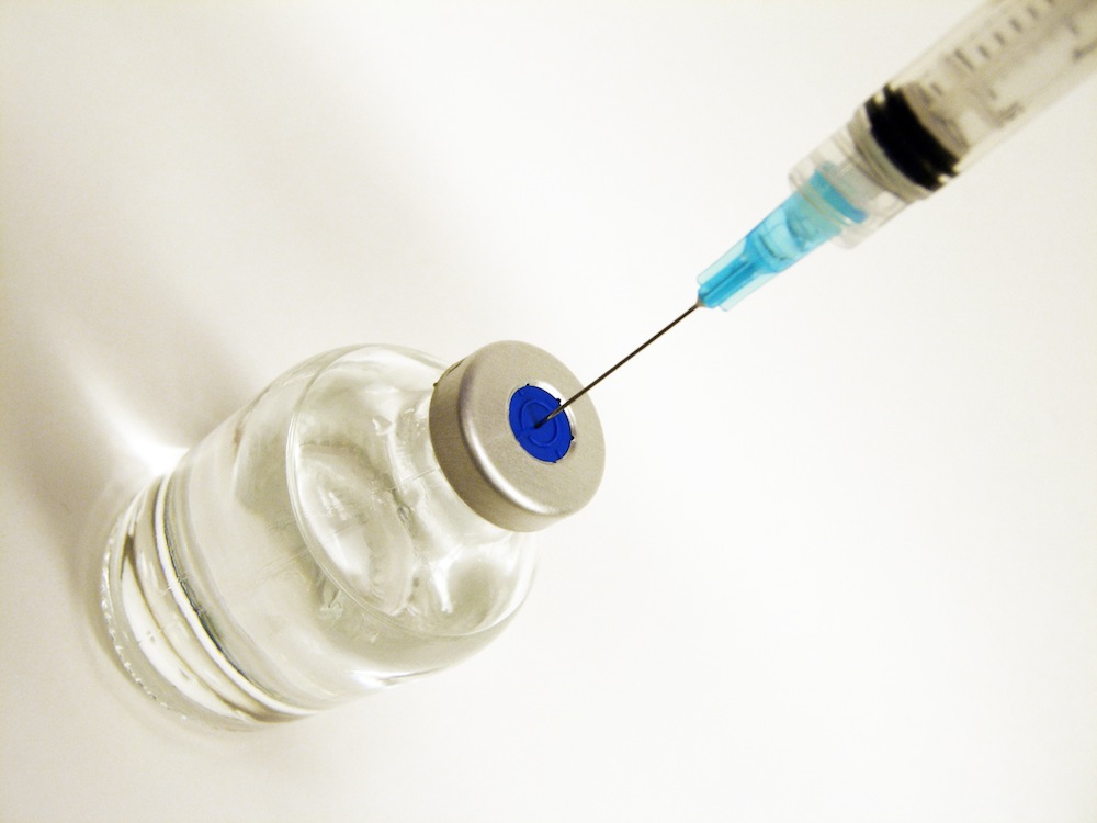
Glibenclamide is a sulfonylurea used in the management of diabetes. It antagonises ATP-dependent potassium (KATP) channels in pancreatic β-cells, stimulating insulin release. The salient risk associated with glibenclamide is accidentally developing hypoglycaemia, an event of varying seriousness in patients who are not critically ill but a great predictor of mortality in acute sepsis. Furthermore, recent studies comparing glibenclamide with metformin and other sulfonylureas elucidated its significant adverse effects in addition to severe hypoglycaemia, such as increased risk of malignancy and mortality from cardiovascular disease due to interference with ischaemic preconditioning.[51,52] Despite glibenclamide still being a popular choice, a trend in medical opinion has arisen against its use in favour of alternative sulfonylureas.[53,54]

However, for T2DM individuals living in regions endemic with melioidosis, choosing glibenclamide over other antidiabetic drugs has an additional benefit that clinicians must consider. In a 5-year prospective cohort study from 2002 to 2006, Koh et al. followed 1160 adult patients with culture-confirmed melioidosis for 28 days during and after their admission to a major NE Thailand hospital. 71.3% of diabetic individuals taking glibenclamide survived from melioidosis after 28 days, while only 50.7% of diabetics not taking glibenclamide and 47.0% of non-diabetics survived (survival of patients discharged from hospital within 28 days were assumed to have survived). Using a logistics regression model, glibenclamide treatment reduced case fatality with an adjusted odds ratio (AOR) of 0.34 when compared to diabetics not taking glibenclamide and an AOR of 0.47 compared to non-diabetics. Incidences of hypotension (AOR 0.48) and respiratory failure (AOR 0.50) were also reduced in these patients.[55] Therefore, this protective effect must be factored into the drug’s risk-benefit comparison with metformin and with other sulfonylureas. If it informs clinicians’ drug choices for glycaemic control in management of T2DM, glibenclamide can contribute to tertiary prevention of melioidosis.

Although preventing hyperglycaemia in sepsis improves patient outcomes, glibenclamide does not rely on its glucose-lowering effect.[56] Using a mouse model, Koh et al. attributed the mechanism of glibenclamide’s protective effect to two main anti-inflammatory pathways. Firstly, glibenclamide reduces IL-1β secretion, attenuating IL-8 production and neutrophilic and monocytic influx into the lungs (without completely ablating the neutrophilic response).[55,57] In vitro evidence suggests that glibenclamide does this by partially blocking NLRP3 inflammasome formation and/or by directly inhibiting the secretion of mature IL-1β (Figure 1).[57-59] Glibenclamide may also attenuate transcription and translation of caspase-1, NLRP3, and IL-1β genes.[57] Secondly, by reducing systemic vasodilation and maintaining normal peripheral vascular resistance, glibenclamide restores systemic mean arterial pressure in the event of septic shock. KATP channels in vascular smooth muscle preferentially open during sepsis.[60] When they do, they hyperpolarise the cell, inhibiting Ca2+ influx (via voltage-dependent Ca2+ channels) and causing muscular relaxation. Glibenclamide opposes this effect by blocking the KATP channels. However, the only evidence for this is found in animal models.[61,62]

Conclusion

Melioidosis remains a significant public health concern due to its high incidence, mortality and case fatality rates particularly in the Top End of Northern Territory and NE Thailand. T2DM is the largest risk factor for melioidosis. The antidiabetic drug, glibenclamide, reduces morbidity and mortality from melioidosis-induced septic shock and so plays a potential role in tertiary prevention of melioidosis. In the debate over glibenclamide use, consideration of the drug’s protective effect for diabetics living in melioidosis-endemic regions, particularly those that are resource-limited, is critical.

Acknowledgements

None

Conflict of Interests

None declared

References

1. Loveleena, Chaudry R, Dhawan B. Melioidosis; the remarkable imitator: recent perspectives. J Assoc Physicians India. 2004;52:417-20.

2. Suputtamongkol Y, Hall AJ, Dance DA, Chaowagul W, Rajchanuvong A, Smith MD, et al. The epidemiology of melioidosis in Ubon Ratchatani, northeast Thailand. Int J Epidemiol. 1994;23(5):1082-90.

3. Kanoksil M, Jatapai A, Sharon PJ, Limmathurotsakul D. Epidemiology, microbiology and mortality associated with community-acquired bactaremia in Northeast Thailand: a multicenter surveillance study. PLoS One. 2013 Oct;8(1):e54714.

4. Suputtamongkol Y, Chaowagul W, Chetchotisakd P, Lertpatansuwun N, Intaranongpai S, Ruchutrakool T, et al. Risk factors for melioidosis and bacteraemic melioidosis. Clin Infect Dis. 1999;29(2):408-13.

5. Blaney DD, Gee JE, Theresa SL. Chapter 3: infectious diseases related to travel: melioidosis. Centers for Disease Control and Prevention 2011.

6. Currie BJ, Dance DA, Cheng AC. The global distribution of Burkholderia pseudomallei and melioidosis: an update. Trans R Soc Trop Med Hyg. 2008;102 Suppl 1:S1-4.

7. Raja NS, Ahmed MZ, Singh NN. Melioidosis: an emerging infectious disease. J Postgrad Med. 2005;51(2):140-5.

8. Limmathurotsakul D, Wongratanacheewin S, Teerawattanasook N, Wongsuvan G, Chaisuksant S, Chetchotisakd P, et al. Increasing incidence of human melioidosis in Northeast Thailand. Am J Trop Med Hyg. 2010;82(6):1113-7.

9. Currie BJ, Ward L, Cheng AC. The epidemiology and clinical spectrum of melioidosis: 540 cases from the 20 year Darwin prospective study. PLoS Negl Trop Dis. 2010;4(11):e900.

10. Parameswaran U, Baird RW, Ward LM, Currie BJ. Melioidosis at Royal Darwin Hospital in the big 2009-2010 wet season: comparison with the preceding 20 years. Med J Aust. 2012;196(5):345-8.

11. Currie BJ, Jacups SP, Cheng AC, Fisher DA, Anstey NM, Huffam SE, et al. Melioidosis epidemiology and risk factors from a prospective whole-population study in northern Australia. Trop Med Int Health. 2004;9(11):1167-74.

12. Currie BJ, Jacups SP. Intensity of rainfall and severity of melioidosis, Australia. Emerg Infect Dis. 2003;9:1538-42.

13. Howe C, Sampath A, Spotnitz M. The Pseudomallei group: A review. J Infect Dis. 1971;124(6):598-606.

14. Allwood EM, Devenish RJ, Prescott M, Adler B, Boyce JD. Strategies for intracellular survival of burkholderia pseudomallei. Front Microbiol. 2011;2:170.

15. Easton A, Haque A, Chu K, Lukaszewski R, Bancroft GJ. A critical role for neutrophils in resistance to experimental infection with Burkholderia pseudomallei. J Infect Dis. 2007;195(1):99-107.

16. Wiersinga WJ, Catharina WW, Mark DC. Toll-like receptor 2 impairs host defense in Gram-Negative sepsis caused by Burkholderia pseudomallei (melioidosis). PLoS Med. 2007;4(7):e248.

17. Ceballos-Olvera I, Sahoo M, Miller MA, Del Barrio L, Re F. Inflammasome-dependent pyroptosis and IL-18 protect against Burkholderia pseudomallei lung infection while IL-1β is deleterious. PLoS Pathog. 2011;7(12):e1002452.

18. Koh GC, Schreiber MF, Bautista R, Maude RR, Dunachie S, Limmathurotsakul D, et al. Host responses to melioidosis and tuberculosis are both dominated by interferon-mediated signaling. PLoS ONE. 2013;8(1):e54961. doi:10.1371/journal.pone.0054961.

19. Ware LB, Matthay MA. The acute respiratory distress syndrome. N Engl J Med. 2000;342(18):1334-49.

20. Flynn JL, Chan J. Immune evasion by Mycobacterium tuberculosis: living with the enemy. Curr Opin Immunol. 2003;15(4):450-5.

21. Boehm U, Klamp T, Groot M, Howard JC. Cellular responses to interferon-gamma. Annu Rev Immunol. 1997;15:749-95.

22. Wiersinga WJ, van der Poll T, White NJ, Day NP, Peacock SJ. Melioidosis: insights into the pathogenicity of Burkholderia pseudomallei. Nat Rev Microbiol. 2006;4(4):272-82.

23. Chin CY, Monach DM, Nathan S. Genome wide transcriptome profiling of a murine acute melioidosis model reveals new insights into how Burkholderia pseudomallei overcomes host innate immunity. BMC Genomics. 2011;11:672.

24. Takeuchi O, Akira S. Pattern recognition receptors and inflammation. Cell. 2010;149(6):805-20.

25. Wiersinga WJ, Currie BJ, Peacock SJ. Melioidosis. N Engl J Med. 2012;367(11):1035-44.

26. Bone RC, Balk RA, Cerra FB, Dellinger RP, Fein AM, Knaus WA, et al. Definitions of sepsis and organ failure and guidelines for the use of innovative therapies in sepsis. The ACCP/SCCM Consensus Conference Committee. American College of Chest Physicians/Society of Critical Care Medicine. Chest. 1992;101(6):1644-55.

27. Meumann EM, Cheng AC, Ward L, Currie BJ. Clinical features and epidemiology of melioidosis pneumonia: results from a 21-year study and review of the literature. Clin Infect Dis. 2012;54:362-9.

28. Muller LMAJ, Gorter KJ, Hak E, Goudzwaard WL, Schellevis FG, Hoepelman AIM, et al. Increased risk of common infections in patients with type 1 and type 2 diabetes mellitus. Clin Infect Dis. 2005;41:281-8.

29. Martin GS. Sepsis, severe sepsis and septic shock: changes in incidence, pathogens and outcomes. Expert Rev Anti Infect Ther. 2012;10(8):701-6.

30. Martin GS, Mannino DM, Eaton S, Moss M. The epidemiology of sepsis in the United States from 1979 through 2000. N Engl J Med. 2003;348(16):1546-54.

31. Hodgson KA, Morris JL, Feterl ML, Govan BL, Ketheesan N. Altered macrophage function is associated with severe Burkholderia pseudomallei infection in a murine model of type 2 diabetes. Microbes Infect. 2011;13(14-15):1177-84.

32. Morris J, Williams N, Rush C, Govan B, Sangla K, Norton R, et al. Burkholderia pseudomallei triggers altered inflammatory profiles in a whole-blood model of type 2 diabetes-melioidosis comorbidity. Infect Immun. 2012;80(6):2089-99.

33. Williams NL, Morris JL, Rush C, Govan BL, Ketheesan N. Impact of streptozotocin-induced diabetes on functional responses of dendritic cells and macrophages towards Burkholderia pseudomallei. FEMS Immunol Med Microbiol. 2011 Mar;61(2):218-27.

34. Chin CY, Monack DM, Nathan S. Delayed activation of host innate immune pathways in streptozotocin-induced diabetic hosts leads to more severe disease during infection with Burkholderia pseudomallei. Immunology. 2012;135(4):312-32.

35. Hodgson KA, Govan BL, Walduck AK, Ketheesan N, Morris JL. Impaired early cytokine responses at the site of infection in a murine model of type 2 diabetes and melioidosis comorbidity. Infect Immun. 2013;81(2):470-7.

36. Wade CE. Hyperglycemia may alter cytokine production and phagocytosis by means other than hyperosmotic stress. Crit Care. 2008;12(5):182.

37. Bopp C, Bierhaus A, Hofer S, Bouchon A, Nawroth PP, Martin E, et al. Bench-to-bedside review: The inflammation-perpetuating pattern-recognition receptor RAGE as a therapeutic target in sepsis. Crit Care. 2008;12(1):201.

38. Van den Berghe G, Wilmer A, Hermans G, Meersseman W, Wouters PJ, Milants I, et al. Intensive insulin therapy in the medical ICU. N Engl J Med. 2006;354(5):449-61.

39. Liu IM, Chang CK, Juang SW, Kou DH, Tong YC, Cheng KC, et al. Role of hyperglycaemia in the pathogenesis of hypotension observed in type-1 diabetic rats. Int J Exp Pathol. 2008;89(4):292-300.

40. Lemkes BA, Hermanides J, Devries JH, Holleman F, Meijers JC, Hoekstra JB. Hyperglycemia: a prothrombotic factor? J Thromb Haemost. 2010;8(8):1663-9.

41. Patel N, Conejero L, De Reynal M, Easton A, Bancroft GJ, Titball RW. Development of vaccines against Burkholderia pseudomallei. Front Microbiol. 2011;2:198.

42. Centers for Disease Control and Prevention (CDC). Melioidosis: prevention. Atlanta: CDC; 2012.

43. Australian Centre for Disease Control. Melioidosis. Darwin: ACDC; 2012.

44. Currie B, Anstey N. Treatment and prognosis of melioidosis. UpToDate; 2012.

45. White NJ, Dance DA, Chaowagul W, Wattanagoon Y, Wuthiekanun V, Pitakwatchara N. Halving of mortality of severe melioidosis by ceftazidime. Lancet. 1989;2(8665):697-701.

46. Panomket P, Chetchotisakd P, Sermswan RW, Pannengpetch P, Wongratanacheewin S. Use of a low-dose steroid as an adjunct in the treatment, in mice, of severe sepsis caused by Burkholderia pseudomallei. Ann Trop Med Parasitol. 2009;103(7):635-46.

47. Mohammad RA. Use of granulocute colony-stimulating factor in patients with severe sepsis or septic shock. Am J Health Syst Pharm. 2010;67(15):1238-1245.

48. Cheng AC, Stephens DP, Anstey NM, Currie BJ. Adjunctive granulocyte colony-stimulating factor for treatment of septic shock due to melioidosis. Clin Infect Dis. 2004;38(1):32-7.

49. Cheng AC, Limmathurotsakui D, Chierakul W, Getchalarat N, Wuthiekanun V, Stephens DP, et al. A randomized controlled trial of granulocyte colony-stimulating factor for the treatment of severe sepsis due to melioidosis in Thailand. Clin Infect Dis. 2007;45(3):308-14.

50. Stephens DP, Thomas JH, Higgins A, Bailey M, Anstey NM, Currie BJ, et al. Randomized, double-blind, placebo-controlled trial of granulocyte colony-stimulating factor in patients with septic shock. Crit Care Med. 2008;36(2):448-54.

51. Monami M, Balzi D, Lamanna C, Barchielli A, Masotti G, Buiatti E, et al. Are sulphonylureas all the same? A cohort study on cardiovascular and cancer-related mortality. Diabetes Metab Res Rev. 2007;23(6):479-84.

52. Schramm TK, Gislason GH, Vaag A, Rasmussen JN, Folke F, Hansen ML, et al. Mortality and cardiovascular risk associated with different insulin secretagogues compared with metformin in type 2 diabetes, with or without a previous myocardial infarction: a nationwide study. Eur Heart J. 2011;32(5):1900-8.

53. WHO Expert Committee. The selection and use of essential medicines. World Health Organ Tech Rep Ser. 2008;(950):backcover, vii-174.

54. Riddle MC. Editorial: sulfonylureas differ in effects on ischaemic preconditioning – is it time to retire glyburide? J Clin Endocrinol Metab. 2003;88(2):528-30.

55. Koh GC, Maude RR, Schreiber MF, Limmathurotsakul D, Wiersinga WJ, Lee SJ, et al. Glyburide is anti-inflammatory and associated with reduced mortality in melioidosis. Clin Infect Dis. 2011;52(6):717-25.

56. Koh GC, Weehuizen TA, Breitbach K, Krause K, de Jonh HK, Kager LM, et al. Glyburide reduces bacterial dissemination in a mouse model of melioidosis. PLoS Negl Trop Dis. 2013;7(10):e2500.

57. Kewcharoenwong C, Rinchai D, Utispan K, Suwannasaen D, Bancroft GJ, Ato M, et al. Glibenclamide reduces pro-inflammatory cytokine production by neutrophils of diabetes patients in response to bacterial infection.

58. Lamkanfi M, Mueller JL, Vitari AC, Misaphi S, Fedorova A, Deshayes K, et al. Glyburide inhibits the Cryopyrin/Nalp3 inflammasome. J Cell Biol. 2009;187(1):61-70.

59. Hamon Y, Luciani MF, Becq F, Verrier B, Rubartelli A, Chimini G. Interleukin-1beta secretion is impaired by inhibitors of the Atp binding cassette transporter, ABC1. Blood. 1997;90(8):2911-5.

60. Sorrentino R, d’Emmanuele di Villa Bianca R, Lippolis L, Sorrentino L, Autore G, Pinto A. Involvement of ATP-sensitive potassium channels in a model of a delayed vascular hyporeactivity induced by liposaccharide in rats. Br J Pharmacol. 1999;127(6):1447-53.

61. Vanelli G, Hussain SN, Aguggini G. Glibenclamide, a blocker of ATP-sensitive potassium channels, reverses endotoxin-induced hypotension in pig. Exp Physiol. 1985;80(1):167-70.

62. Landry DW, Oliver JA. The ATP-sensitive K+ channel mediates hypotension in endotoxaemia and hypoxic lactic acidosis in dog. J Clin Invest. 1992;89(6):2071-4.

Categories
Review Articles

The α-5 subunit-containing GABA-A receptor: a target for the treatment of cognitive defects

Amnesic effects of benzodiazepines are in part the result of the activity of α5-subunit containing GABAA receptors (GABRA5). Negative modulators at this receptor could improve cognition. In order to explore this beneficial effect, this article reviews the evidence on the effects of GABRA5 negative modulators and searches potential uses for such drugs. A literature search found a number of GABRA5 negative modulators. These drugs generally improve hippocampal-dependant learning via an increase in longterm potentiation (LTP) in the hippocampus. Passive avoidance learning was also improved. In addition, the compounds examined demonstrated minimal side effects partly due to lack of binding to different alpha subunit-containing GABAA types. Due to its beneficial properties, there is potential for such a drug in treating Alzheimer’s, alcohol-related amnesia and Down syndrome. Despite the myriad animal studies that utilised GABRA5 negative modulators, only three human studies were found. Due to its cognitive enhancing properties and minimal side effects, further human trials should be conducted in order to ascertain the potential of such drugs in treating cognitive deficits.


Introduction
The α5 subunit-containing GABAA receptor: a target for the treatment of cognitive defectsγ-aminobutyric acid (GABA) is the major inhibitory neurotransmitter in the brain and is responsible for regulating neuronal excitability. There are at least three different receptors that it targets — GABAA, GABAB and GABAC. [1] The GABAA receptor is the main target for the popular class of drugs, the benzodiazepines. This receptor is an ionotropic membrane receptor, which facilitates the movement of chloride into cells. In neurons, this increases the threshold needed to excite them.[2] Benzodiazepines are positive modulators at this receptor and exert their effects by binding to the interface between the γ2 and α subunits on the GABAA receptor. [3] Since they are modulators and not agonists, they do not work in the absence of GABA. [4] They only bind to the receptors containing the α5, α3, α2, and α1 subunits. [5] Each of these subunits mediates different effects. In knockout mice, the α1 subunit has shown hypnotic/sedative effects, whilst the α3, α2 and α5 subunits have anti-anxiolytic effects and these have been exploited for therapeutic use.[6]

Despite their uses in treating various conditions, traditional benzodiazepines have numerous side effects. The main side effects associated with therapeutic use are amnesia, confusion, impaired coordination and dizziness. There may be tolerance due to rapid escalation in the dose needed to provide the required effect. There are also long-term issues with dependence. In acute overdose the most life-threatening effect is respiratory depression, especially when combined with alcohol. [7]

Interestingly, it is thought that positive modulators at the α5 subunit-containing GABAA receptors (GABRA5) produce the anterograde amnesia associated with benzodiazepine use. [7] Most of these receptors are found in the hippocampus (a brain region associated with memory) and provide tonic inhibition in this region. [8] The exploitation of this receptor has led to the increasing use of the infamous ‘date rape’ drug flunitrazepam, which is a positive modulator at the GABRA5 in addition to its other functions. Because of its amnestic effects, victims are unable to recall events following intoxication and this provides a major challenge for prosecutors. [7] Despite these negative properties, by using a GABRA5 negative modulator the opposite effect might The α5 subunit-containing GABAA receptor: a target for the treatment of cognitive defects be achieved and cognition improved. The use of such a drug could potentially improve the quality of life for those living with cognitive defects and could also counteract drug-induced amnesia (for example, alcoholic ‘blackout’). However, this must be balanced with the inverse activity of non-selective negative modulators which could produce
convulsant or anxiogenic effects. [4]

Based on the premise that a GABRA5 negative modulator could improve cognition, the aim of this literature review was to review the evidence on (1) the effects of GABRA5 negative modulators on cognition; and (2) investigate the potential of GABRA5 negative modulators in managing conditions involving cognitive defects.

Effects of GABRA5 selective negative modulators

A number of GABRA5 negative modulator compounds were examined. Many of the studies used animals as subjects. Studies in animals provide a solid starting platform for understanding the various physiological changes that a drug induces. [9] Of particular importance is the avoidance of potential side effects as a result of non-selective actions at other GABAA receptors. Side effects could include an increase in anxiety, aggressiveness, motor impairment, inability to sleep and proconvulsant effects. [7] Ultimately, the knowledge gained from animal experimentation can be used to conduct safe and effective clinical trials.

Dawson et al. [10] examined a compound named α51A (3-(5-Methylisoxazol-3-yl)-6-[(1-methyl-1,2,3-triazol-4-yl)methyloxy]-1,2,4-triazolo[3,4-a]phthalazine), which has selective negative modulator effects at GABRA5. The authors found that α51A reversed the inhibiting effects of GABRA5 in the hippocampus in rats and mice. This resulted in an increase in performance in a memory test named the “delayed matching-to-position version of the Morris water maze”, which is a hippocampus-dependant cognitive test. [11] In addition, ‘long-term potentiation’ (LTP), which is thought to underlie the synaptic changes that take place during memory formation, was found to be enhanced in the hippocampus. [11,12] Benzodiazepine (agonist) effects and non-selective GABAA negative modulator effects were also examined. The authors found no anxiogenic, convulsant, withdrawal or motor-impairing effects from the drug. [10]

Only certain components of memory have shown to be improved by GABRA5 negative modulators. Collinson et al. [13] extrapolated on the results obtained by Dawson et al. [10] and looked at the effect of a modified version of α51A, α51A-II. They separated memory into three components — encoding, consolidation (conversion into long-term memory) and recall. Results were obtained by measuring performance in the delayed matching-to-position (DMTP) version of the Morris water maze in rats. The authors found that the compound improved encoding and recall but not consolidation in this hippocampaldependant memory test. [13]

The effect on cognition by another selective GABRA5 negative modulator was examined by Ballard et al. [14] This was done by the use of an imidazo-triazolo-benzodiazepine compound named RO4938581. The effect of this compound on cognition was examined in rats. [15] This compound demonstrated similar effects to those found by Dawson et al. [10] in that they found no convulsant or anxiogenic effects. In addition, similar to Dawson et al. [10], there was an increase in hippocampal LTP. The authors also found that working memory was enhanced since RO493881 reversed scopolamine-induced working memory impairment. [14] This was shown by an increase in performance in the DMTP task, which is used to assess spatial working memory. [14,15] It also reversed diazepam-induced spatial impairment. This was demonstrated by an increase in performance in the Morris water maze task. [14]

‘Moderate’ GABRA5 negative modulators improve passive avoidance learning but generally have no effect on active learning. [16] This was shown by an experiment conducted by Savic et al. [16] in which they examined effects of PWZ-029 (a ‘moderate’ GABRA5 negative modulator) on passive and active learning avoidance in rats. The result was obtained through various shuttle-box based behavioural experiments. This experiment proved that even at ‘moderate’ efficacy a GABRA5 negative modulator can induce memory formation. The compound also had no effect on muscle tension and anxiety (nonselective side effects). [16] Although promising, this study was limited by the fact that the compound only had ‘moderate’ negative modulator activity at GABRA5 so using a more efficient compound may display different effects on avoidance learning. Despite this limitation, this shows that the use of a ‘moderate’ GABRA5 negative modulator would be beneficial in the treatment of disease due to its limited side effects and its memory-enhancing properties. [16]

Application in management

The compounds examined in this review show that selective GABRA5 negative modulators have nootropic effects without any serious side effects, which are seen in non-selective negative modulators at the alpha subunit of the GABAA receptor. [17] Thus, there is strong potential for the use of GABRA5 negative modulators in healthcare settings. One major limitation is that most of the data obtained for this review was from animals. Further human trials need to be conducted to ascertain the potential of this drug. Drawing on the literature, possible future uses for a GABRA5 negative modulator are detailed below.

GABRA5 negative modulators could be used to treat Alzheimer’s disease since GABRA5 is preserved in Alzheimer’s disease patients.
[18] Alzheimer’s disease is commonly characterised by the gradual worsening of ability to remember new information. [19] Administration of a GABRA5 negative modulator could help with the ‘encoding’ and ‘recall’ of this information. [8] It could also be used to treat mild cognitive impairment (MCI), which is a risk factor for later developing the disease. [20] Administration of such a drug to patients may provide relief to older caregivers, who often show signs of sleep detriment. [21]

A review by Attack [22] in 2010 found two human trials on the GABRA5 negative modulator α5Ia and one trial on MRK-016. Since then no human trials were found and this could be an area of future research. The first study found that a potential application of GABRA5 negative modulators is the treatment of alcohol-induced amnesia. Nutt et al. [23] found that pre-treatment reduces alcohol’s amnestic effects in humans. This was measured by word list learning which is linked to hippocampal processing. Alcohol-induced amnesia has been shown to predict future alcohol-related injury. [24] Therefore, the use of a GABRA5 negative modulator may help in reducing this risk. In addition, it may also reduce alcohol-related stress since it has been found that amnestic episodes related to alcohol have resulted in moderate psychological stress. [25]

Unfortunately, GABRA5 does not improve age-related cognitive defects. In fact it has been found that α5IA significantly impairs cognition in the elderly despite having positive effects on the young. Attack [22] found that young subjects (mean age 22 years) performed much better than older subjects (mean age 72 years) on the paired associates learning test, which is sensitive to age-related cognitive decline. Therefore, this trial showed no potential in reversing age-related cognitive decline. This demonstrates that careful consideration based on age should be taken in to account when using this drug.

MRK-016 (3-tert-butyl-7-(5-methylisoxazol-3-yl)-2-(1-methyl-1H-1,2,4-triazol-5-ylmethoxy)-pyrazolo[1,5-d]-[1,2,4]triazine) is another negative modulator and showed greater LTP in rat hippocampal slices than α51A. It also enhanced performance in the DMTP and Morris water maze tasks, which are used to test spatial memory. In humans, it was well tolerated in young adults with a maximum tolerated dose of 5 mg with 75% occupancy. In elderly subjects, however, it was poorly tolerated even at 10% of the maximum dosage in young adult males. Therefore, this particular drug has been precluded for development. [26]

Recent trials of GABRA5 agonists, in particular L-655,708 and MRK-016, have focused on restoring post-anaesthetic cognitive deficits. Lecker et al. [27] found that L-655,708 and MRK-016 reduced the potentiation of GABRA5 post-inhalation of isoflurane and sevoflurane. A further study by Zureck et al. [28] found that short-term memory assessed by the novel object recognition task was fully reversed by L-655,708 after isoflurane anaesthesia. This demonstrates the potential use of L-655,708 in reducing post-anaesthetic amnesia. However, further studies which include those performed on humans are needed to validate the potential of MRK-016 and other GABRA5 negative modulators in reducing post-anaesthetic amnesia.

The use of GABRA5 negative modulators could help with treating cognitive deficits related to Down syndrome. A recent review by Martínez-Cué et al. [29] investigated this specific application. It identified two studies that examined the effects of a GABRA5 inverse modulator on a Down syndrome mouse model (Ts65Dn). Braudeau et al. [30] found that acute treatment with the GABRA negative modulator α5IA improved learning deficits in the Morris water maze task. The second study also showed that chronic administration of a similar drug, RO4938581 has also been shown to have memory-promoting effects in the Morris water maze task on Ts65Dn mice. [31] In a practical sense, administration of such a drug could improve performance in learning a wide range of functional skills in those living with Down syndrome. For example, in children this may include learning how to use the toilet and administering self-care. [32]

Conclusion

The literature supporting the use of a GABRA5 negative modulator in the treatment of cognitive deficits is promising. GABRA5 negative modulators exert their actions by enhancing hippocampal dependant memory formation. There are minimal side effects as no withdrawal symptoms, convulsant, anxiogenic or motor-impairing effects were found. There is great potential for the use of GABRA5 negative modulators as they have been shown to reduce alcohol-related amnesia and may have potential in the treatment of Alzheimer’s disease. They could also treat cognitive deficits in Down syndrome patients, increasing the speed at which they learn functional skills. Due to these favourable findings, there is an increased need for human clinical trials in order to validate the potential for this important receptor target.

Acknowledgements

A/Prof Zoltan Sarnyai, for reviewing this article when I submitted it as an assignment.

Conflict of interest

None declared.

Correspondence

A Bhandari: abhishta.bhandari@my.jcu.edu.au

References

[1] Lonstein JS, Maguire J, Meinlschmidt G, Neumann ID. Emotion and mood adaptations in the peripartum female: complementary contributions of gamma-aminobutyric acid and oxytocin. J Neuroendocrinol. Forthcoming 2014. DOI: 10.1111/jne.12188.

[2] D’Hulst C, Atack JR, Kooy RF. The complexity of the GABAA receptor shapes unique pharmacological profiles. Drug Discov Today. 2009;14(17-18):866-75.

[3] Sigel E, Buhr A. The benzodiazepine binding site of GABAA receptors. Trends Pharmacol Sci. 1997;18(11):425-9.

[4] Rudolph U, Knoflach F. Beyond classical benzodiazepines: novel therapeutic potential of GABAA receptor subtypes. Nat Rev Drug Discov. 2011;10(9):685-97.

[5] Atack JR. GABAA receptor subtype-selective modulators. II. α5-selective inverse agonists for cognition enhancement. Curr Top Med Chem. 2011;11(9):1203-14.

[6] Mohler H. GABAA receptor diversity and pharmacology. Cell Tissue Res. 2006;326(2):505-16.

[7] Rang HP, Dale M. Rang and Dale’s Pharmacology: Churchill Livingstone; 2007.

[8] Collinson N, Atack JR, Laughton P, Dawson GR, Stephens DN. An inverse agonist selective for α5 subunit-containing GABAA receptors improves encoding and recall but not consolidation in the Morris water maze. Psychopharmacology (Berl). 2006;188(4):619-28.

[9] Ferreira LM, Hochman B, Barbosa MVJ. Modelos experimentais em pesquisa. Acta Cirurgica Brasileira. 2005;20:28-34.

[10] Dawson GR, Maubach KA, Collinson N, Cobain M, Everitt BJ, MacLeod AM, et al. An inverse agonist selective for α5 subunit-containing GABAA receptors enhances cognition. J Pharmacol Exp Ther. 2006;316(3):1335-45.

[11] Nakazawa K, Sun LD, Quirk MC, Rondi-Reig L, Wilson MA, Tonegawa S. Hippocampal CA3 NMDA receptors are crucial for memory acquisition of one-time experience. Neuron. 2003;38(2):305-15.

[12] Cooke SF, Bliss TV. Plasticity in the human central nervous system. Brain. 2006;129(Pt 7):1659-73.

[13] Collinson N, Kuenzi FM, Jarolimek W, Maubach KA, Cothliff R, Sur C, et al. Enhanced learning and memory and altered GABAergic synaptic transmission in mice lacking the alpha 5 subunit of the GABAA receptor. J Neuro Sci. 2002;22(13):5572-80.

[14] Ballard TM, Knoflach F, Prinssen E, Borroni E, Vivian JA, Basile J, et al. RO4938581, a novel cognitive enhancer acting at GABAA α5 subunit-containing receptors. Psychopharmacology (Berl). 2009;202(1-3):207-23.

[15] Goto K, Kurashima R, Watanabe S. Delayed matching-to-position performance in C57BL/6N mice. Behav Process. 2010;84(2):591-7.

[16] Savic MM, Clayton T, Furtmuller R, Gavrilovic I, Samardzic J, Savic S, et al. PWZ-029, a compound with moderate inverse agonist functional selectivity at GABAA receptors containing α5 subunits, improves passive, but not active, avoidance learning in rats. Brain Res. 2008;1208:150-9.

[17] Navarro JF, Buron E, Martin-Lopez M. Anxiogenic-like activity of L-655,708, a selective ligand for the benzodiazepine site of GABAA receptors which contain the α5 subunit, in the elevated plus-maze test. Prog Neuropsychopharmacol Biol Psychiatry. 2002;26(7-8):1389-92.

[18] Howell O, Atack JR, Dewar D, McKernan RM, Sur C. Density and pharmacology of α5 subunit-containing GABAA receptors are preserved in hippocampus of Alzheimer’s disease patients. Neuroscience. 2000;98(4):669-75.

[19] Daulatzai MA. Early stages of pathogenesis in memory impairment during normal senescence and Alzheimer’s disease. J Alzheimers Dis: JAD. 2010;20(2):355-67.

[20] Grundman M, Petersen RC, Ferris SH, Thomas RG, Aisen PS, Bennett DA, et al. Mild cognitive impairment can be distinguished from Alzheimer disease and normal aging for clinical trials. Arch Neurol. 2004;61(1):59-66.

[21] Rowe MA, McCrae CS, Campbell JM, Benito AP, Cheng J. Sleep pattern differences between older adult dementia caregivers and older adult noncaregivers using objective and subjective measures. J Clin Sleep Med. 2008;4(4):362-9.

[22] Atack JR. Preclinical and clinical pharmacology of the GABAA receptor α5 subtypeselective inverse agonist α5IA. Pharmacol Ther. 2010;125(1):11-26.

[23] Nutt DJ, Besson M, Wilson SJ, Dawson GR, Lingford-Hughes AR. Blockade of alcohol’s amnestic activity in humans by an α5 subtype benzodiazepine receptor inverse agonist. Neuropharmacology. 2007;53(7):810-20.

[24] Mundt MP, Zakletskaia LI, Brown DD, Fleming MF. Alcohol-induced memory blackouts as an indicator of injury risk among college drinkers. Inj Prev. 2012;18(1):44-9.

[25] Buelow G, Koeppel J. Psychological consequences of alcohol induced blackout among college students. J Alcohol Drug Educ. 1995.

[26] Atack JR, Maubach KA, Wafford KA, O’Connor D, Rodrigues AD, Evans DC, et al. In vitro and in vivo properties of 3-tert-butyl-7-(5-methylisoxazol-3-yl)-2-(1-methyl-1H-1,2,4-triazol-5-ylmethoxy)- pyrazolo[1,5-d]-[1,2,4]triazine (MRK-016), a GABAA receptor α5 subtype-selective inverse agonist. J Pharmacol Exp Ther. 2009;331(2):470-84.

[27] Lecker I, Yin Y, Wang DS, Orser BA. Potentiation of GABAA receptor activity by volatile anaesthetics is reduced by α5-GABAA receptor-preferring inverse agonists. Bri J Anaesth. 2013;110 Suppl 1:i73-81.

[28] Zurek AA, Bridgwater EM, Orser BA. Inhibition of α5 γ-Aminobutyric Acid Type A Receptors Restores Recognition Memory After General Anesthesia. Anesth Analg. 2012;114(4):845-55 DOI: 10.1213/ANE.0b013e31824720da.

[29] Martínez-Cué C, Delatour B, Potier M-C. Treating enhanced GABAergic inhibition in Down syndrome: Use of GABA α5-selective inverse agonists. Neurosci Biobehav Rev. Forthcoming 2014. DOI: 10.1016/j.neubiorev.2013.12.008.

[30] Braudeau J, Delatour B, Duchon A, Pereira PL, Dauphinot L, de Chaumont F, et al. Specific targeting of the GABAA receptor α5 subtype by a selective inverse agonist restores cognitive deficits in Down syndrome mice. J Psychopharmacol. 2011;25(8):1030-42.

[31] Martinez-Cue C, Martinez P, Rueda N, Vidal R, Garcia S, Vidal V, et al. Reducing GABAA α5 receptor-mediated inhibition rescues functional and neuromorphological deficits in a mouse model of down syndrome. J Neurosci. 2013;33(9):3953-66.

[32] Dolva A-S, Coster W, Lilja M. Functional performance in children with Down syndrome. Am J of Occup Ther. 2004;58(6):621-9.

Categories
Review Articles Articles

Factors that influence Australian medical graduates to become General Practitioners

Aim: To determine the factors that influence Australian medical graduates to become general practitioners. Method: A literature review was conducted. Medline, PubMed and Cochrane Library were searched using the terms; “Australia”, “medical”, “graduates”, “interns”, “students”, “choice”, “specialty”, “general”, “practice”, “factors” and “influencing”. Results: The factors were grouped into intrinsic (age, gender, personality and skill set,) and extrinsic influences (lifestyle, income, stress, location and role models), with extrinsic influences regarded as the most influential. Most importantly, 72% of the Australian medical graduates viewed work culture as important, while 56% prioritised flexibility of working arrangements and hours of work. Conclusion: There are a variety of both intrinsic and extrinsic factors influencing medical graduates to choose General Practice over others. This can be seen as an opportunity for Australian workforce planners and policy makers to target the extrinsic factors with the aim of balancing the medical workforce to combat the shortage of rural general practitioners.

Introduction

In the field of medicine, a specialty is simply a specific study of medical science. [1] Dermatology, Obstetrics and Gynaecology, Cardiology, Neurosurgery and General Practice are just a few of the vast array of medical specialties that medical graduates must decide between before embarking on a long, strenuous but nevertheless, highly rewarding journey. Students endure four to six years of medical school, only to begin a new journey as junior doctors. Internship is followed by residency, and the pathway after this depends upon the choice of specialty. [2] Medical students and junior doctors are faced with the tough challenge of selecting a specialty. This review seeks to precisely identify the factors influencing medical graduates to undertake General Practice.

This narrative review aims to explore the intricate complexities that invade the mind of medical graduates faced with the dilemma of choosing a specialty; in particular, what influences them to choose General Practice. The aims of this literature review are to highlight the spectrum of factors that play a role in medical students and interns choosing to undertake General Practice, and present medical colleges, recruitment agencies, workforce planners and national organisations with a platform upon which they can correct the imbalances in the medical workforce.

Methodology

This literature review covered recent literature that has focused on the factors influencing choice of medical specialisation (in particular General Practice) in Australia. Medical and social science databases were searched for publications from 1990-2013. Medline, PubMed and Cochrane Library were searched using these terms; “Australia”, “medical”, “graduates”, “interns”, “students”, “choice”, “specialty”, “general”, “practice”, “factors” and “influencing”. 7670 papers were identified through the database searches. These were then reviewed to only include studies conducted in Australia and concerning Australian medical graduates, which narrowed it down to 25 papers. 9 of these papers were excluded because they were not completely relevant to the topic. In addition, the bibliographies of articles were searched for further relevant publications. Studies referred to in this review vary widely and include both qualitative and quantitative studies.

Results

The primary influential factors involved in the selection of a particular specialisation can be separated into intrinsic and extrinsic factors.

Intrinsic Factors

Intrinsic factors include age, personality and gender. Individuals have little or no control over such factors. [8]

Age is an intrinsic factor that plays a role in the selection of a particular specialty. The majority of medical students in Australian universities are under the age of 24. [9] Despite this, there has been an increase in the number of ‘mature age’ students over the past two decades. One Australian study, that compared the career choices of medical graduates, found that older students were more likely to specialise in a primary care field such as General Practice. [3]

An individuals’ personal set of skills and the satisfaction they receive from using these skills are intrinsic factors which play a large role in influencing medical graduates to undertake General Practice. In 2005, Harris et al examined the factors influencing the choice of specialty of 4259 Australian medical graduates. [3] The study showed that 79% of graduates considered their own skills and aptitude to be of importance when selecting General Practice. Personal satisfaction was also linked to choosing General Practice as a specialty. Laurence and Elliot supported this in their 2007 study which concluded that personal satisfaction arises from procedural skills, activities involved and patient contact. [4] All of the 54 Pre-Registration Junior Medical Officers (PMJOs) interviewed in South Australia, agreed with this however, this was only a small sample size.

Gender has been shown by studies to be a vital influencing factor for Australian medical graduates when choosing General Practice as a speciality. Whilst the responsibilities of raising children have evolved over the past few decades, Prideaux et al found that Australian female medical graduates are more likely to become specialists in General Practice due to child bearing responsibilities and family commitments. [10] It has been noted the Australian literature that female doctors tend to work shorter hours and have a preference for working shorter hours due to family commitments. [11] Despite this, there has been a rise in male doctors choosing to work fewer hours due to family reasons. [10] This reflects the fact that both partners now commonly work.

Extrinsic Factors

Extrinsic factors include stress, work hours, family commitments, lifestyle and mentors. They are variables that may be controlled. [8]

Lifestyle plays the greatest role in influencing medical graduates to choose particular specialties over others. Laurence and Elliot found that 100% of the 54 South Australian PMJOs interviewed regarded lifestyle as a vital factor in choosing General Practice as a specialty. [4] This included hours worked, stress, career potential and potential for travel (55%). Most participants described their ideal job as having shorter working hours and less time on call. Most PMJOs also wanted a certain amount of control over hours and hence chose anaesthesia and GP practice. Wanting a life outside of medicine (85%) for example, spending more time with family and friends was also important. [4] Similarly, Harris et al also rated extrinsic factors as the most influential factors of choosing a medical specialty in Australia. [3] Seventy two percent of the Australian medical graduates viewed work culture as important, while 56% prioritised flexibility of working arrangements and hours of work. In contrast, Thomas concluded that only 28% of Australian medical graduates saw work life balance and lifestyle as important to selecting General Practice as their specialty. [7] However, this study had a small sample size and focused on only one speciality (General Practice).

Location is of importance when choosing a medical specialty. Stagg et al found that key influences on choosing a rural pathway specialty were mentors and undergraduate rural exposure. [6] In contrast, Ward et al, in a longitudinal study that followed 229 UWA medical graduates, showed that a rural background is the most important predicator of rural general practice. [5] Clearly, there factors that influence an individual to undergo a rural generalist pathway are multifactorial and more research is needed in this area.

The studies used in this literature review all stressed the importance of role models in influencing Australian medical graduates to choose particular specialties. Laurence and Elliot studied when, what and how SA pre-registration junior medical officers made their career choice. [4] Fifty four percent of the 54 graduates perceived the role of mentors, supervisors and consultants to be of importance in selecting General Practice as a specialty. Their interaction with ‘mentors’ was through observing and asking questions. Role models were seen to demonstrate specific characteristics admired by the students. [12]

Discussion

The results confirm that choosing General Practice as a specialty is a complex decision strongly influenced by personal qualities (intrinsic factors), individual experiences and opportunities (extrinsic factors), and domestic circumstances. Whilst parenting dynamics have changed over the last century, there is still a trend for females to choose General Practice over other specialties due to flexibility and option of part time employment, which may be helpful when choosing to start a family. [13]

The most important extrinsic factors include lifestyle, work experience since graduation, flexible hours, influence of mentors and hours of work. [3] In Australia, it was concluded that factors relating to lifestyle and job satisfaction were the most important influencing factor. [4] This is consistent with the belief that recent graduates regard lifestyle factors as more important than income. [14] The younger generation of graduates also prioritise potential for travel. These graduates are influenced by their experience of General Practice and confirm that work experience is helpful for developing knowledge within medical. [3]

The results also suggested that trainees in different specialties prioritised certain influencing factors over others. Surgical trainees viewed mentors and role models as more important than trainees in other specialties. Additionally, General Practice trainees were more likely to prioritise flexibility, whilst this factor was of less importance to trainees in Adult Medicine. [15] Compounding this notion is the fact that surgical and emergency trainees found it extremely important to do procedural work compared to trainees in other programs. [3]

Clearly, there is not one single factor that influences an individual to undertake a particular career path, rather a vast array of factors. A medical graduate’s choice of career is dependent upon a wide range of intrinsic and extrinsic factors. [3] Whilst there is not much chance of altering intrinsic factors, the nature of extrinsic factors allows for an interventionist approach.  [16]

The main goal of medical workforce agencies and groups is to ensure a balance of doctors across a vast array of specialties to provide equal, effective and holistic medical care to the community. For 80% of doctors, the decision about choice of specialty has to be made by the end of the third postgraduate year (PGY3). [3] As such, training programs, teaching facilities and recruitment agencies involved in medical workforce planning should aim to educate medical students and graduates up until PGY3 and allow them to make an informed decision. Given the importance of extrinsic factors, there should be a review of the work culture typical of specialties that are under-represented. [13] Training providers can therefore implement strategies that attempt to increase entry to less well-represented specialties. [4]

This literature review has a number of limitations. Firstly, this review was limited to articles concerning the Australian medical workforce. This review excluded international medical graduates, which may have given greater insight into the factors influencing the specialty choice of graduates. A comparison study could be done in the future, comparing the mindset of Australian medical graduates to overseas graduates. Secondly, whilst certain conclusions can be made concerning the factors which influence choice of General Practice as a specialisation, this review did not focus on the factors that influence doctors to change specialties or even the percentage of medical students that graduate without having made a decision about their future career. Research on the factors influencing General Practice specialty choice could be improved by including a larger number of schools and students, studying trends over several years, and using validated measures and outcomes.

Conclusion

The main factors which were identified as influencing medical graduates to choose General Practice included both intrinsic and extrinsic factors. Additionally, a career choice that is made when an individual is young may not represent how they will feel as they progress through life and their priorities will often change. While there remains a continuous need for valuable research in the area of factors affecting medical specialisation, it appears that we need to use this information to prevent imbalances and skews in medical workforce planning. There is a great opportunity for governments, health authorities and the medical profession to influence extrinsic determinants of choice of specialty. [13]

Acknowledgements

I was given the opportunity to conduct this research through the GPSN First Wave Scholarship and Tropical Medical Training (TMT). I am thankful to Dr. Aileen Traves who provided me with support, encouragement and feedback throughout the course of this review.

Conflict of interest

None declared.

Correspondence

K Singh: karan.singh@my.jcu.edu.au

References

[1] Ellsbury, K. E., & Stritter, F. T.. A study of medical students’ specialty-choice pathways: trying on possible selves. Acad Med. 1997; 72, 534-541.

[2] Zurn, P., Dal Poz, M. R., Stilwell, B., & Adams, O. Imbalance in the health workforce. Hum Resour Health. 2004; 2(1), 13.

[3] Harris, M. G., Gavel, P. H., & Young, J. R. Factors influencing the choice of specialty of Australian medical graduates. Med Educ. 2005; 183(6), 295.

[4] Laurence, C. and Elliott, T. When, what and how South Australian pre-registration junior medical officers’ career choices are made. Medical Educ. 2007; 41: 467–475. doi: 10.1111/j.1365-2929.2007.02728.x

[5] Ward, A. M., Kamien, M. and Lopez, D. G. Medical career choice and practice location: early factors predicting course completion, career choice and practice location. Medical Educ. 2004; 38: 239–248. doi: 10.1046/j.1365-2923.2004.01762.x

[6] Stagg, P., Greenhill, J., & Worley, P. A new model to understand the career choice and practice location decisions of medical graduates. Rural Remote Health. 2009; 9(4):1245.

[7] Thomas, T. Factors affecting career choice in psychiatry: a survey of RANZCP trainees. Australas Psychiatry. 2008; 16(3), 179-182.

[8] Belfer, B. Stress and the medical practitioner. Stress Medicine. 1989; 5(2), 109-113.

[9] Joyce C, McNeil J & Stoelwinder J, ‘More doctors, but not enough: Australian medical workforce supply 2001–2012’, Med J Australia 2006; 185(3):182

[10] Prideaux D, Saunders N, Schofield K, Wing, L, Gordon J, Hays R, Worley P, Martin A, Paget N, ‘Country report: Australia’, Med Educ. 2001;35:495-504.

[11] Firth-Cozens J. and Lema VC. ‘Specialty choice, stress  and  personality:  Their relationships over time’, Hosp Med. 1999; 60(10):751-55

[12] Dunbabin, J., & Levitt, L. (2003). Rural origin and rural medical exposure: their impact on the rural and remote medical workforce in Australia. Rural Remote Health, 3(1), 212.

[13] Australian Medical Workforce Advisory Committee. Career decision making by doctors in their postgraduate years — a literature review. Sydney: AMWAC, 2002. (AMWAC Report 2002.1.)

[14] Joyce, C. M., & McNeil, J. J. Fewer medical graduates are choosing general practice: a comparison of four cohorts, 1980-1995. Med J Australia. 2006;185(2):102.

[15] Mowbray, R. Research in choice of medical  speciality: A review of the literature 1977-87. Aust NZ J Med. 1989;19(4):389-399.

[16] Joyce, C. M., Stoelwinder, J. U., McNeil, J. J., & Piterman, L. Riding the wave: current and emerging trends in graduates from Australian university medical schools. Med J Australia. 2007;186(6): 309.

Categories
Review Articles Articles

Examining the pathological nature of Hepatitis C and current drug therapies used in an Australian general practice context

Aim: This review aims to examine the pathological nature of Hepatitis C and review current drug therapies relevant to Australian health practitioners. Methods: Terms hepatitis C, Australia, pathogenesis and current treatment were searched using MEDLINE and CHINAL databases to identify research articles and systematic reviews. Constraints were used when researching drug developments to include only full-length papers, on humans published between 2009 and 2013.  Literature was analysed to identify shared themes. Sixty-eight articles were analysed and fifty-two chosen based on relevance to objective, reputable data sources and current information. Two websites and five books were included upon cross referencing data to journal articles. Four Australian guideline publications were included due to relevance to topic and general practitioners. Results: The aetiology, clinical significance and molecular pathogenesis of hepatitis C virus were examined to provide Australian practitioners with a basis of knowledge for presentation of both acute and chronic stages of hepatitis C infection. This understanding was further linked to current drug treatments available in Australia and potential future therapeutic options. Conclusion: The consequences of Hepatitis C infections will burden the Australian healthcare system in the next few decades as the chronic nature of HCV infection leads to complications of liver failure, cirrhosis and hepatocellular carcinoma in many patients. Practitioners must equip themselves with knowledge of HCV pathogenesis which forms the basis of current and future treatments in order to provide best quality care at all levels of prevention and management.

Introduction

The recognition of viral hepatitis can be dated as far back as the fifth century BC to Babylonian records. [1] Our understanding of Hepatitis C gained remarkable ground when previously non-A non-B hepatitis infections were attributed to the hepatitis C virus discovered by Choo et al. in 1989. [1,2] Since then efforts have been made to develop drug treatments to combat the virus which progresses to chronic infection in up to 80% of patients, increasing their risk of cirrhosis, liver failure and hepatocellular carcinoma. [3,4,5] Chronic hepatitis C infection is currently the leading cause of liver transplantation in Australia. [6,7,8]

Hepatitis C is a major health concern for Australian practitioners with 260 000 Australians infected in 2010 and an estimated 12 000 new infections occurring annually. [4,7] The dominant mode of transmission of the hepatitis C virus (HCV) is parenteral exposure to infected blood and thus the epidemic of HCV infection in Australia continues to escalate predominantly through people who inject drugs (PWID). [7]

This review aims to summarise the aetiology, transmission and life cycle of HCV as well as examine the most recent literature regarding current and future drug therapies to provide the Australian general practitioner with a contemporary understanding in emerging hepatitis treatments.

Aetiology of Hepatitis C

Transmission in the Australian context

Hepatitis C is a blood-borne viral infection and is most commonly spread in Australia via shared injecting equipment (up to 80% acquiring the infection via this route). [7] Other means of transmission include unsterile tattooing, needle-stick injuries and vertical transmission from mother-to-infants from trauma during pregnancy and/or birth. [9,10] About 5% of all cases in Australia arise from HCV contaminated blood transfusions and blood products prior to screening introduced in February 1990. [7]

Virological Structure

Hepatitis C is caused by a small, positive-stranded RNA virus of the Flaviviridae family. The RNA strand is enveloped by a protein capsid which is further surrounded by a lipid bilayer envelope studded with E1 and E2 heterodimer proteins. The genome contains a 5’ noncoding region required for viral translation, followed by an open reading frame terminated by a 3’ noncoding region necessary for replication. The open reading frame translates into a 3000 amino acid polyprotein which is cleaved into structural (core, E1, E2) and non-structural (p7, NS3, NS4A, NS4B, NS5A, and NS5B) proteins. [11-15]

The NS5B protein is a RNA-dependent RNA-polymerase which lacks proofreading function. This combined with a high replication rate (1012 virions/day) results in rapid mutations driving genetic diversity. [16] Thus within a host, HCV circulates as a population of extremely closely related, but not identical variants called quasispecies. [17] This feature has contributed to difficulty in developing a vaccine as well as implications for pharmacological therapies. HCV is classified into seven major genotypes which differ genetically by at least 30% with over 100 subtypes. [11,13] The prevalence of genotypes differ with geographical distribution. Genotype 1 mostly dominates Australia, the Americas, Japan and Europe with genotypes 2 and 3 also prevalent in these areas. Genotype 7 was only recently discovered in a small proportion of people in Central Africa. Disease association is largely similar across genotypes, however genotype 3 has been correlated with a higher risk of hepatic steatosis and progressive liver disease. [13,18]

Life Cycle

The main stages of the HCV replication cycle are binding and entry, uncoating, translation and replication of RNA, assembly into new particles, maturation and secretion. [11,19] Several host factors have been identified aiding entry of HCV including heparan sulphate and low-density lipoprotein receptor. Other host factors CD81, scavenger receptor B1 and tight junction proteins claudin-1 and occludin allow for clathrin-dependent endocytosis which delivers the virus to early endosomes, which become acidified causing fusion of the viral envelope, uncoating and release of the viral RNA into the cytoplasm. [12,19] HCV replication induces a membranous web concentrating lipid-rich structures that aid the replication process. Hepatocyte -specific microRNA-122 has been shown to bind to target sites on the 5’ untranslated region of the HCV genome which forms a complex that protects the HCV genome from nucleolytic degeneration and innate host immune responses. [20] Lipid droplets interact with the core and NS5A viral proteins allowing viral assembly. The newly synthesised viral proteins can then exit the cell in a manner similar to the hosts’ very low-density lipoprotein (VLDL) export pathway by utilising cofactor ApoB and microsomal triglyceride transfer protein to form low-density viral particles termed lipo-viral particles. [11,12,18] ApoE is involved in HCV particle morphogenesis and infectivity. HCV particles exist in the serum as a mixture of complete low-density infectious lipo-viral particles and an excess of apoB-associated empty non-infectious particles complexed with anti-HCV envelope antibodies. [18]

The mechanism responsible for onset and progression of chronic hepatitis are not fully understood but it is currently believed that HCV establishes persistent infection by impairing host innate and adaptive immunity. [1,21] The infected hepatocytes recognise Pathogen Associated Molecular Patterns (PAMPs) through receptors known as Pattern Recognition Receptors (PRRs) which include Toll like receptors (TLRs) and RIG-1 like receptors (RLRs). Upon sensing HCV via TLR3 and RIG-1, intracellular signalling cascades result in the induction of type I and type III interferon and pro-inflammatory cytokines which establish an antiviral state in infected and neighbouring cells. [21,22] Resident antigen presenting cells, such as dendritic cells residing in the liver migrate from infected tissue to lymph nodes where they prime T and B cell activation to induce adaptive immunity. [11]

Recent studies have established multiple routes whereby HCV impairs immune functioning so as to coexist and replicate in the host. [23] NS3/4A protease cleaves intracellular pathway protein TRIF and CARDIF to impair TLR3 and RIG-1 receptors. [11] Furthermore, the HCV enveloped particle is not detected by TLR-2 and TLR-4 which also contribute to antiviral states. HCV has been shown to up regulate MHC Class I molecules on infected hepatocytes which suppresses Natural Killer cell activity. [23] Finally generation of quasispecies carry mutations to evade B and T-cell recognition as well induce hypermutation in B cell receptors to lower affinity allowing the virus to escape immune surveillance. [11]

Clinical Implications

The World Health Organisation estimates 170 million people are infected with Hepatitis C globally. [1,24] Hepatitis C is thus  the leading cause of chronic liver disease worldwide and is a growing burden on healthcare systems,  including within Australia. [3,25] Infection is characterised by a wide range of clinical manifestations and propensity to develop into chronicity. Up to 80% of infected patients will develop a chronic infection. [1,3] Persistent infection and chronic hepatitis are the hallmarks of HCV infection with severity varying widely from asymptomatic chronic infection with normal liver function tests to severe cases leading to cirrhosis and hepatocellular carcinoma. [2,14]

Acute Hepatitis

Acute HCV infection is often asymptomatic due to a mild immune response and reversible cellular injury seen at the microscopic level. [9] Where symptoms occur they tend to be minimal involving jaundice and flu-like malaise. [5] Strong immune responses during the acute infection are associated with clearance of the virus however in the majority of cases milder initial infections lead to chronic viral persistence. [5,26]

Chronic Hepatitis

Cirrhosis develops in as many as 20% of chronic HCV patients and is associated with hepatocellular failure. Patients may present with portal hypertension manifested as splenomegaly, variceal bleeding or ascites. [9,26] Primary hepatocellular carcinoma is thought to result from the continual division of infected hepatocytes attempting to regenerate in the presence of injury. [5] Once cirrhosis has established, patients have a 5% annual risk of developing hepatocellular carcinoma. [27] Extra-hepatic diseases such as mixed cryoglobulinemia and glomerulonephritis are also believed to be caused by HCV induced antibody complex depositions in small vessels causing vasculitis, however the pathogenesis of this is not fully understood providing an area for investigation in the future. [28,29]

Current drug therapies

Current standard of care treatment of HCV genotype 1 is triple therapy with pegylated interferon-α (cytokine), ribavirin (antiviral) and a direct acting antiviral (NS3/4A protease inhibitor) – either telaprevir or boceprevir. [2,8,30] The combination of pegylated interferon (PEG-IFN) and ribavirin remain the recommended treatment for HCV infection with genotypes 2, 3, 4, 5 and 6. [19] The aim of treatment is a sustained virological response (SVR), defined as the absence of detectable HCV RNA for 6 months after treatment cessation. [31] SVR is associated with crucial end points, particularly survival and protection from the complications of chronic hepatitis C such as cirrhosis and hepatocellular carcinoma. [19,30]

For reasons that remain elusive, interferon-based therapies result in a SVR of 80% in genotype 2 and 3 infections but only 45% in genotype 1 and 4 infections. [4,13] With the approval of boceprevir and telaprevir in 2011 by the US Food and Drug Administration, triple therapy has enabled the SVR to increase in patients with genotype 1 from 45% in 2010 to ~66% in 2011 and is expected to be >75% by 2014. [4,26]

The SVR is also influenced by a myriad of host factors such as ethnicity, gender, age and insulin resistance. [13,32] Furthermore, new biomarkers such as serum IP10 levels and genetic tests to determine polymorphisms in the gene encoding IFNL3 (formerly known as IL28B or IFN-λ3) and recently discovered IFNL4 show strong value with respect to interferon-based therapy as predictors of treatment outcome. [1,13,30]

In Australia, hepatitis C treatment is available for all eligible patients over 18 years of age who have chronic HCV infection with compensated liver disease and are using effective forms of contraception. Treatment is subsidised by the government under the Highly Specialised Drugs (HSD) program, section 100 (S100) of the National Health Act 1953 (Cwlth). [8]

Pegylated Interferon-α

Interferons are naturally produced by immunological cells in response to tumour or infectious organism. They are glycoproteins with antiviral, anti-proliferative and immunomodulatory functions. [31]

Upon administration of IFN- α, the type I interferon binds to IFNAR-1 and IFNAR-2 receptors on cell surfaces initiating a complex intracellular signalling pathway resulting in activation of genes coding for proteins which inhibit intracellular viral replication. Proteins include RNA-dependant protein kinase (PKR) which inhibits RNA translation and oligoadenylate synthetase (OAS) which mediates RNA degradation. IFN- α also stimulates TH1 cell production while reducing suppressor TH2 cells as part of its immunomodulation. [31,33]

Pathogenesis of HCV occurs as a result of the virus’ ability to prevent host cells from responding to natural levels of interferon. As previously discussed, HCV blocks TLR3 and RIG-1 receptors reducing type I IFN production. [5,11] Thus overwhelming host cells with high levels of injected IFN allow normal cellular mechanisms to control the virus. [5,13] Replacement of standard interferon with pegylated interferon (interferon-α conjugated to polyethylene glycol) improves pharmacokinetics and efficacy and has allowed its administration as a once weekly subcutaneous injection. [30]

Ribavirin

Unlike pegylated interferon-α, whose function was unravelled due to developments in cell culture models, the mechanism of action of ribavirin against HCV is unknown. [33] Ribavirin was originally synthesised as a guanosine analogue that could inhibit viral polymerases by chain termination. The process by which this is thought to occur is when the polymerase incorporates the nucleotide but cannot add more after inserting the analogue, hence preventing viral replication and transcription. [5]

However there is much debate about the mechanism of ribavirin activity in chronic hepatitis C.  Despite showing in vitro activity against some RNA and DNA molecules, studies conducted with ribavirin as a monotherapy against HCV reflect no effect on HCV RNA levels or improvement of hepatic histology following 12 months of therapy. [33] Yet analysis of the current literature shows multiple studies where combination therapy of IFN- α and ribavirin is significantly more effective then IFN- α alone. [34-36] Furthermore, the anti-HCV activity of ribavirin occurs at much lower doses then expected for the chain termination theory to occur. [5] This suggests other mechanisms of action are at work.

Greenblatt presents two possibilities. [5] One involves ribavirin as depleting the cell’s reservoir of normal guanosine to interfere with viral RNA synthesis. Secondly she proposes a mutagenic theory in which ribavirin incorporation into viral genomes renders them funtionless. [5] Other theories propose that ribavirin induces IFN-stimulated genes or may have immunomodulatory functions which like IFN- α, push patient cytokine profiles towards TH1 types which are more effective against viral infections then type 2 Helper T cells. [13,31]

Telaprevir and Boceprevir

Telaprevir and boceprevir are first generation peptidomimetic, reversible inhibitors of NS3/4A protease. [30] The HCV NS3/4A serine protease is essential for viral replication by cleaving polyproteins into mature non-structural proteins. [13] Thus by inhibiting this protease, telaprevir and boceprevir are the first direct-acting antivirals (DAAs) approved for use against HCV genotype 1.

Despite both drugs having similar mechanisms of action and thus sharing most clinically relevant strengths and weaknesses, there are discrepancies between telaprevir-based regimens and boceprevir-based regimens. [1,30] These differences are in the timing and duration of combined therapy. Typically, telaprevir is given in triple therapy with PEG-IFN and ribavirin for the first 12 weeks of therapy, PEG-IFN and ribavirin are then continued for the remainder of treatment (either 24 or 48 weeks) without the protease inhibitor. Duration of treatment is dependent on virological response (response-guided therapy). Boceprevir, however is started 4 weeks after commencement with PEG-IFN and ribavirin and is continued for the remaining treatment duration of 28 or 48 weeks depending on response. [19] Telaprevir-based regimen is stopped in patients with a HCV RNA level greater than 1000 IU/ml at week 4 or 12 and all three drugs should be discontinued. For the boceprevir-based regimen, patients with HCV RNA levels greater than 100 IU/ml at week 12 should discontinue treatment. For both treatments, if HCV RNA is detectable at 24 weeks of therapy, all three drugs should be stopped. [19]

Side effects profile – pegylated interferon-α and ribavirin

These can be quite distressing and contribute to low tolerance and compliance in patients. The major effects of interferon include depression, constant flu-like symptoms, thrombocytopenia, leucopenia, thyroid dysfunction, retinopathy and alopecia. [25,37] Ribavirin is highly teratogenic and can lead to haemolytic anaemia and autoimmune disorders. [37]

Psychiatric status as well as full blood count, kidney and liver function tests should be monitored continuously throughout therapy. Furthermore, precautions should be taken with patients with depressive histories, thyroid dysfunctions, diabetes, autoimmune disorders and renal impairment. [38] Finally pregnancy in female patients or the partners of male patients must be avoided during treatment, and owing to the long half life of ribavirin, also 6 months after cessation of treatment. [39]

Side effects profile – telaprevir and boceprevir

Although triple therapy is more efficacious in HCV genotype 1 infections, there are additional side effects compared to traditional dual therapy and thus management of hepatitis C patients has become more complex. Common side effects of telaprevir include rash and anorectal discomfort while dysgeusia (altered taste sensation) and neutropaenia are associated with boceprevir. The most challenging side effect of both drugs is marked anaemia (haemoglobin level < 10 g per decilitre) occurring in 36-50% of patients. [19,30] Erythrocyte-stimulating agents have some success in managing this complication however are not approved for routine use in chronic hepatitis C patients due to serious side effects and cost. Some studies have shown that reduction in the dose of ribavirin can effectively manage anaemia in this setting and this is the current recommended first line approach. [19]

Due to the highly variable nature of HCV with the error-prone RNA polymerase, drug resistance is also an issue with these protease inhibitors and can develop as early as day 4 upon use in monotherapy. Consequently, these drugs are not to be used in isolation. Because of the similar mechanism of action, resistance to one protease inhibitor can result in other drugs within the same class to be ineffective.  Once the drug is stopped, the frequency of resistance-associated variants within the quasispecies slowly decreases until they disappear, most likely because they do not replicate as effectively as the wild-type virus. [19,30] General practitioners can play a crucial role in patient education to ensure adherence to the prescribed regimen in order to limit the development of resistance-associated variants.

The third major consideration with these new drugs is the issue of drug-drug interactions. Both telaprevir and boceprevir are inhibitors of the cytochrome P450 3A (CYP3A). CYP3A enzymes are involved in the metabolism of numerous drugs such as statins, antidepressants, antiarrhythmics, anticonvulsants, analgesics and sedatives. [19] As such, these are all contraindicated in patients undergoing treatment with telaprevir and boceprevir. This has important implications for general practitioners who are frontline prescribers of such agents. Efforts are being made to make such complex information widely available to the medical community through platforms such as the ‘Hepatitis Drug Interactions’ website from the University of Liverpool, UK. [30,40]

The future of hepatitis C

With the exponential increase in knowledge of life cycle and replication of HCV due to breakthroughs in cell culture systems in 2005, there is fierce competition to develop medicines that will replace PEG-IFN, ribavirin and first generation protease inhibitors. [30] About two-thirds of agents in Phase II and III trials are directed against the NS3/4A and NS5B viral proteins called second generation protease inhibitors and polymerase inhibitors respectively. [19,30] The current challenges in drug development are decreasing side effects and drug interactions, exploring combinations for genotypes 2-6, exploring individualised drugs to specific genetic polymorphisms, and eradicating the need for interferon and ribavirin in treatment.

A number of drugs are currently being developed for genotypes 2-6. A preliminary phase 2a study in New Zealand involved combining sofosbuvir, an oral nucleotide inhibitor of HCV polymerase, and ribavirin in various interferon and interferon-sparing regimens for 12 weeks. [41] Patients with HCV genotype 1, 2 and 3 were investigated. In this early trial sofosbuvir showed a promising result with 100% rate of SVR among patients with genotype 2 or 3 infection. [19,26,41] However phase 3 studies of sofosbuvir fall short of the results produced by the phase 2a study. [42,43,44] One noninferiority trial looked at sofosbuvir plus ribavirin compared to standard peginterferon alfa-2a plus ribavirin in 499 patients with HCV genotype 2 or 3 infection. The results revealed the same SVR rate of 67% in both the sofosbuvir-ribavirin and peginterferon-ribavirin group at 12 weeks after cessation of therapy. [43] Two further phase 3 trials (POSITRON and FUSION studies) investigated sofosbuvir-ribavirin therapy in patients for whom peginterferon treatment was not an option (due to pre-existing psychiatric or autoimmune disorders) and in those who did not have a response to previous interferon treatment. The POSITRON trial was a randomized, blinded, placebo-controlled study that compared 12 weeks of sofosbuvir-ribavirin treatment with matching placebo in patients who had previously discontinued interferon therapy due to adverse events or concurrent medical condition. In this group, 78% had a SVR at 12 weeks after treatment compared to 0% in the placebo group. [42] The FUSION study looked at patients who had failed a sustained response to interferon-based therapy and compared 12 and 16 week regimens of sofosbuvir-ribavirin therapy. Results showed that four additional weeks of therapy made a difference with an increase in the SVR from 86% to 94% in patients with genotype 2 infection and from 30% to 63% in genotype 3 infections. [42,44] All three phase 3 studies with sofosbuvir-ribavirin showed better SVR rates in genotype 2 infections compared to genotype 3 infections. [42,43,44]

Nonstructural 5A (NS5A) protein is also a target of recent drug development. [45] The functions of the NS5A protein are not yet fully understood with in vitro studies suggesting is has a role in viral replication and assembly and release of infectious particles. [45] Daclatasvir is a potent NS5A inhibitor which has shown early promising results for use in interferon-free combinations with rapid decline of extracellular HCV titres upon administration. [45] In a phase 2a trial, patients who had not had a response to previous therapy received daclatasvir and a protease inhibitor (asunaprevir) for 24 weeks. [46] Four out of eleven patients had a SVR at 12 and 24 weeks after treatment ended, suggesting a cure may be possible with an all-oral interferon-ribavirin free treatment. [45,46]

Another group of host targeting antiviral agents are arising. Miravirsen is a drug undergoing development which targets miR-122. Liver specific miR-122, as discussed previously is a microRNA which all strains of HCV use to survive and replicate in liver cells. [11,12] A recent phase 2a study by Janssen et al, dose-dependent reductions in HCV RNA levels were found without viral resistance. [20] The study was limited by a small sample of 36 patients and only moderate levels HCV RNA reduction which rebounded once miravirsen was stopped in patients who had not begun interferon and ribavirin. [20,47] Normally miR-122 is involved in controlling cholesterol levels independent of its effect on HCV. In the study, there was a sustained decrease of serum cholesterol levels by ~25% which lasted 14 weeks after the final injection. [47] Given statins are contraindicated in the current triple therapy treatment of HCV genotype 1 infections, there is potential in the future to develop liver-targeting nucleic acid drugs which can be used intermittently for both HCV treatment and other co-morbid conditions.

Furthermore, the future of HCV treatment is trending towards highly individualised regimens which consider not only the viral genotypes but also the patient’s genetic polymorphisms. For instance, easy-to-treat patients have been identified as treatment-naïve, IL28B CC. [30] Patients with a favourable interlukin-28B genotype (CC variant as opposed to CT or TT) have shown sustained virologic responses up to 80%. [19] It is theorised that these patients could receive PEG-IFN and ribavirin first, minimising the adverse effects of triple therapy. [19] Although testing for the IL28B genotype is not currently the approved standard of care, in the future Australian general practitioners may be managing care of these ‘easy-to-treat’ patients while more complex cases, such as patients with IL28B TT with decompensated cirrhosis are managed at tertiary centres using a cocktail of tailor made drug regimens. [30]

Implications for Australian health practitioners

Accessing and treating hepatitis C infection in PWID – the role of the general practitioner

Advances made in the development of better tolerated interferon free HCV treatment will remain negligible as long as access to therapy cannot be expanded to the most affected and underserved risk groups. [48,49,50] People who inject drugs act as a virus reservoir, as the burden of HCV-related liver disease in this group is increasing but treatment uptake remains low. [49,50] There are a number of barriers to accessing care at the level of the patient, practitioner and system. [48,50,51] New guidelines have been published with recommendations for the management of HCV infection among PWID which aim to overcome these barriers by providing evidence-based treatment recommendations.[50] Analysis of the literature revealed a common theme supported by high quality evidence which was the use of multidisciplinary care teams in enhancing treatment uptake in PWID. [49,50] General practitioners can play a crucial role in co-ordinating multidisciplinary care between specialists, drug and alcohol support services, psychiatric services, social work and other social supports such as peer-based groups. [49] In an Australian community-based study, hepatitis C positive patients who had seen a general practitioner about HCV in the last 6 months were four times more likely to be assessed for therapy by a specialist. [50] Furthermore, a prospective cohort study using telehealth technology in supporting and training GPs was compared to HCV treatment provided at a tertiary centre. Similar rates of treatment success were achieved in both groups. [49,51] From these studies, it was seen that general practitioners not only co-ordinated care but provided a more patient-centred approach necessary in dealing with the complex psychiatric and substance abuse co-morbidities which required individualised models of care. Enhanced personal contact provides an ideal environment for pre-therapeutic assessment of housing, education, cultural and social issues, supports, finances, nutrition, drug and alcohol use and psychiatric evaluation. [50] Merging different disciplines into one general practice model may be a simple and effective model in the future for a sub-population of PWID with HCV but will require commitment by motivated and supported GPs who have undergone further training in addiction and HCV medicine. [49]

The role of general practitioners in assessment and management of HCV infection

As stated previously, hepatitis C may be present in patients unknowingly for decades before symptoms of liver failure prompt a seeking of treatment. At this stage of irreversible cellular damage, treatment options are limited, often associated with distressing side effects and yield less efficient results in certain genotypes. The onus is on health practitioners, armed with an understanding of the pathogenesis of HCV, to identify high risk patients and test for anti-HCV antibodies and HCV RNA levels as a secondary preventative strategy to provide early detection and referral. There are a number of useful international guidelines which can assist general practitioners in managing newly diagnosed hepatitis C patients in regards to indications for treatment as well as first-line treatment recommendations. [3,24] Furthermore, primary prevention strategies whereby educating the public about transmission, symptoms and progression of the disease can be effectively implemented in a consultation setting.

In regards to treating chronic hepatitis C patients, this review aims to equip the general practitioner with an up-to-date understanding of the molecular and immunological aspects of HCV pathogenesis to aid in diagnostic tools as well as provide a platform of knowledge for future pharmaceuticals. We are entering an exciting new era of hepatitis C treatment where interferon-free therapies are likely to dominate the therapeutic landscape within the next 5 years [19] and so an understanding of their mechanism of action in hepatitis C is crucial for continuing treatment and management.

Conclusion

From the ancient observations to the discovery of hepatitis C virus 24 years ago, up until recent advances in cell model systems, our understanding of the pathological nature of hepatitis C has grown exponentially. With this growth have come parallel developments in treatment, both in understanding mechanisms behind current drug therapies but also providing a platform for future pharmaceuticals to target aspects of HCV pathogenicity. These developments come at a timely point, as the burden of an escalating epidemic of hepatitis C in Australia will have major impacts on our healthcare system within the next few decades as the chronic nature of the disease will come into play.

Conflict of interest

None declared.

Correspondence

J Aslanidis: jaimie.aslanidis@gmail.com

References

[1] Boyer N, Marcellin P. Pathogenesis, diagnosis and management of hepatitis C.  J Hepatol. 2000;32(1 Suppl):98-112.

[2] Liang TJ. Current progress in development of hepatitis C virus vaccines. Nat Med. 2013;19(7):896-78.

[3] EASL Clinical Practice Guidelines: Management of hepatitis C virus infection. J Hepatol. 2011;55:245-64.

[4] Boonwaat L, Haber PS, Levy MH, Lloyd AR. Establishment of a successful assessment and treatment service for Australian prison inmates with chronic hepatitis C. Med J Aust. 2010;192(9):496-500.

[5] Greenblatt R. Hepatitis C: Progress and Challenges. Clinical Microbiology Newsletter. 2006;28(15):113-118.

[6] Hepatitis Australia. Hepatitis C burden set to explode in decades to come if government doesn’t act [Internet]. 2010 [updated 2010 April 1; cited 2011 April 4]. Available at: http://www.hepatitisaustralia.com/media-centre/media-releases/media-releases-items/hepatitis-c-burden-set-to-explode-in-decades-to-come-if-government-doesnt-act

[7] Dore GJ, Law M, MacDonald M, Kaldor JM. Epidemiology of hepatitis C virus infection in Australia. J Clin Virol. 2003;26(2):170-184.

[8] Gidding HF, Law MG, Amin J, Macdonald GA, Sasaduesz JJ, Jones TL et al. Predictors of deferral treatment for hepatitis C infection in Australian clinics. Med J Aust. 2011;194(8):398-402.

[9] Bisceglie A. Hepatitis C. Lancet. 1998;351(9099):351-5.

[10] Sompayrac L. How Pathogenic Viruses Work. London: Jones and Barlett Publishers; 2002. 56-59.

[11] Georgel P, Schuster C, Zeisel MB, Stoll-Keller F, Berg T, Bahram S et al. Virus-host interactions in hepatitis C virus infection: implications for molecular pathogenesis and antiviral strategies. Trends Mol Med. 2010;16(6):277-86.

[12] Fischer R, Baumert T, Blum HE. Hepatitis C virus infection and apoptosis. World J Gastroenterol. 2007;13(36):4865-72.

[13] Scheel TK, Rice CM. Understanding the hepatitis C virus life cycle paves the way for highly effective therapies. Nat Med. 2013;19(7):837-49.

[14] Pawlotsky JM. Pathophysiology of hepatitis C virus infection and related liver disease. Trends Microbiol. 2004;12(2):96-102.

[15] Kumar V, Abbas AK, Aster JC, Fausto N. Robbins and Cotran Pathological Basis of Disease. 8th ed. Philadelphia: Saunders Elsevier; 2010. p. 847-53.

[16] Wyles DL. Moving beyond interferon alfa: Investigational drugs for hepatitis C virus infection. Top HIV Med. 2010;18(4):132-6.

[17] Fishman SL, Branch AD. The quasispecies nature and biological implications of the hepatitis C virus. Infect Genet Evol. 2009;9(6):1158-67.

[18] Bassendine MF, Sheridan DA, Bridge SH, Felmlee DJ, Neely RDG. Lipids and HCV. Semin Immunopathol. 2013;35:87-100.

[19] Liang TJ, Ghany MG. Current and future therapies for hepatitis C virus infection. N Engl J Med. 2013;368(20):1907-17.

[20] Janssen HL, Reesink HW, Lawitz EJ, Zeuzem S, Rodriguez-Torres M, Keyur P et al. Treatment of HCV infection by targeting microRNA. New Engl J Med. 2013;368(18):1685-95.

[21] Saito T, Gale MJ. RIG-1 mediated hepatic innate immune signalling that controls HCV infection. Uirusu. 2008;58(2):105-15.

[22] Barth H. Insights into the role of interferon lambda in hepatitis C virus infection. J Hepatol. 2011;54:844-847.

[23] Rehermann B. Hepatitis C virus versus innate and adaptive immune responses: a tale of coevolution and coexistence. J Clin Invest. 2009;119(7):1745-54.

[24] Ghany MG, Strader DB, Thomas DL, Seeff LB. American Association for the Study of Liver Diseases: Diagnosis, management, and treatment of hepatitis C: an update. Hepatology. 2009;49:1335-74.

[25] Suzuki T. A Hepatitis C virus-host interaction involved in viral replication: toward the identification of antiviral targets. Jpn J Infect Dis. 2010;63(5):307-11.

[26] Thomas DL. Global control of hepatitis C: where challenge meets opportunity. Nat Med. 2013;19(7):850-8.

[27] Bode JG, Brenndorfer ED, Haussinger D. Subversion of innate host antiviral strategies by the hepatitis C virus. Arch Biochem Biophys. 2007;462(2):254-65.

[28] Eisen-Vandervelde AL, Yao ZQ, Hahn YS. The molecular basis of HCV-mediated immune dysregulation. Clin Immunol. 2004;111(1):16-21.

[29] Craxi A, Laffi G, Zignego AL. Hepatitis C virus (HCV) infection: a systemic disease. Mol Aspects Med. 2008;29(1-2):85-95.

[30] Manns MP, von Hahn T. Novel therapies for hepatitis C – one pill fits all? Nat Rev Drug Discov. Forthcoming 2013 June.

[31] Vezali E, Aghemo A, Colombo M. Interferon in the treatment of chronic hepatitis C: a drug caught between past and future. Expert Opin. Biol. Ther. 2011;11(3):301-13.

[32] Cua IHY, Hui JM, Kench JG, George J. Genotype-specific interactions of insulin resistance, steatosis, and fibrosis in chronic hepatitis C. Hepatology. 2008;48:723–731.

[33] Clark V, Nelson DR. The role of ribavirin in direct acting antiviral drug regimens for chronic hepatitis C. Liver Int. 2012 Feb;32(Suppl 1):103-7.

[34] Berg T, Hoffmann RM, Teuber G, Leifeld L, Lafrenz M, Baumgarten R et al. Efficacy of a short-term ribavirin plus interferon alpha combination therapy followed by interferon alpha alone in previously untreated patients with chronic hepatitis C: a randomised multicenter trial. Liver. 2000;20(6):427-36.

[35] Tsubota A, Fujise K, Namiki Y, Tada N. Peginterferon and ribavirin treatment for hepatitis C virus infection. World J Gastroenterol. 2011;17(4):419-32.

[36] Foster GR. Pegylated interferons for the treatment of chronic hepatitis C: pharmacological and clinical differences between peginterferon-alpha-2a and peginterferon-alpha-2b. Drugs. 2010;70(2):147-65.

[37] Murtagh J. Murtagh’s general practice companion handbook. 4th ed. Sydney. McGraw-Hill Australia. 2008. p. 262-3.

[38] MIMS Australia. St Leonards NSW: UBM Medica Pty. Ltd: 2009 Dec. Chap. Immunomodifiers.

[39] Chapter 5, Anti-infectives. In: Australian Medicines Handbook [online]. 2011. [cited 2011 April 11]. Available from: http://amh.hcn.net.au.elibrary.jcu.edu.au/view.php?page=chapter5/monographribavirin-interferon.html#ribavirin-interferon

[40] University of Liverpool Drug Interaction Charts [Internet] 2013 [updated 2013 April 17; cited 2013 July 14]. Available from: http://www.hep-druginteractions.org

[41] Gane EJ, Stedman CA, Hyland RH, Ding X, Svarovskaia E, Symonds WT et al. Nucelotide polymerase inhibitor sofosbuvir plus ribavirin for hepatitis C. New Engl J Med. 2013;368(1):34-44.

[42] Jacobson IM, Gordon SC, Kowdley KV, Yoshida EM, Rodriguez-Torres M, Sulkowski MS, et al. Sofosbuvir for hepatitis C genotype 2 or 3 in patients without treatment options. N Engl J Med. 2013;368:1867–1877.

[43] Lawitz E, Mangia A, Wyles D, Rodriguez-Torres M, Hassanein T, Gordon SC, et al. Sofosbuvir for previously untreated chronic hepatitis C infection. N Engl J Med. 2013;368:1878–1887.

[44] Drenth JPH. Editorial: HCV treatment–no more room for interferonologists? N Engl J Med. 2013;368:1931–1932.

[45] Guedj J, Dahari H, Rong L, Sansone ND, Nettles RE, Cotler SJ et al. Modeling shows that the NS5A inhibitor daclatasvir has two modes of action and yields a shorter estimate of the hepatitis C virus half-life. Proc Natl Acad Sci. 2013;110(10):3991-6.

[46] Lok AS, Gardiner DF, Lawitz E, Martorell C, Everson GT, Ghalib R et al. Preliminary study of two antiviral agents for hepatitis C genotype 1. N Engl J Med. 2012;366:216-224.

[47] Lieberman J, Sarnow P. Micromanaging hepatitis C virus. New Engl J Med. 2013;368(18):1741-3.

[48] Bruggmann P. Accessing hepatitis C patients who are difficult to reach: it is time to overcome barriers. J Viral Hepat. 2012;19(12):829-35.

[49] Bruggmann P, Litwin AH. Models of care for the management of hepatitis C virus among people who inject drugs: one size does not fit all. Clin Infect Dis. 2013;57(Suppl 2):S56-61.

[50] Robaeys G, Grebely J, Mauss S, Bruggmann P, Moussalli J, de Gottardi A et al. Recommendations for the management of hepatitis C virus infection among people who inject drugs. Clin Infect Dis. 2013;57(Suppl 2):S129-137.

[51] Grebely J, Dore GJ. An expanding role for primary care providers in the treatment of hepatitis C virus in the community. Hepatology. 2011;54(6):2258-2260.

[52] Batey R. Managing hepatitis C in the community. Aust Prescr. 2006;29(2):36-9.

Categories
Original Research Articles Articles

Melioidosis in the Torres Strait Islands: an 11 year audit 2001-2012

Melioidosis is an infection of concern to global health. It is caused by the intracellular gram-negative bacterium Burkholderia pseudomallei, which is found in the soil and fresh waters of endemic regions. This study identified the average annual incidence of melioidosis in the Torres Strait region between 2001-2012, and compared this to one other similar study, which identified the average annual incidence between 1995-2000. Patient demographics, clinical presentation, outcomes and risk factors were compared to other available studies. In this retrospective study of melioidosis in the Torres Strait, 31 cases were identified over an 11-year period, representing an annual incidence of 37 cases per 100,0000 population. Of these cases, 84% recovered, 16% required intensive care unit (ICU) admission, 3% had a relapse and two patient deaths occurred. The mortality rate was 6.4%. Pneumonia accounted for fifteen presentations (48%) and splenic abscesses for ten presentations (32%), with nine patients presenting with septic arthritis of a joint (29%). Other presentations included hepatic (19%), prostatic (19%), renal (10%), skin (6%), pancreatic (3%), scrotal (3%) and spinal abscesses (3%). Four presented with bacteraemia alone (13%) and one case presented with urethritis (3%). Risk factors included diabetes mellitus (68%), excessive alcohol intake (35%), renal disease (12%), autoimmune disease (6%), malignancy (4%) and the use of immunosuppressive medication (2%).

Introduction

Melioidosis is an infection caused by the intracellular gram-negative bacterium Burkholderia pseudomallei, which is found in the soil and fresh waters of endemic regions. [1] Endemic regions include Southeast Asia and Northern Australia, with peaks of infection occurring during the wet seasons. [2] Melioidosis is of global public health significance, and may be thought of as an emerging infection across tropical regions. [3]

The Torres Strait is a tropical region comprised of 274 islands between the Cape York Peninsula of mainland Australia and Papua New Guinea (PNG). According to the 2006 Australian Bureau of Statistics (ABS) census data, the region has a total population of 7,624, with 82.5% identifying as Indigenous. [4] Half of this population is clustered within the central island group located closest to Thursday Island (TI), which is the commercial and governmental centre of the region. Hospital services are also centralised at TI, however, the closest tertiary referral centre for the Torres Strait region is the Cairns Base Hospital, located 800km south of TI.

Melioidosis is endemic in the Torres Strait, with the most recent average annual incidence reported to be 42.7 cases per 100,000. [5] This is significantly greater than other centres such as Darwin, where the annual incidence was noted to be 19.6 cases per 100,000 between 1986 and 2008. [6] However, during periods of extreme climate, such as during years of significant heavy rainfall, this incidence dramatically increases. This was observed in Darwin between 2009 and 2010, during which the annual incidence increased to 50.2 cases per 100,000, as a result of a heavy wet season. [7] The variability in annual incidence highlights the significant relationship between the transmission of melioidosis and certain environmental factors, such as rainfall level. [1,2]

The transmission of melioidosis most commonly occurs through percutaneous inoculation, and less commonly through inhalation, aspiration and ingestion. [2] A range of host and environmental factors must also exist for an individual to be infected. This includes reduced host immunity and the significant environmental exposure to the pathogen which occurs in endemic regions. [1] This was demonstrated in the study by Kanaphun et al. conducted in northeast Thailand, in which serological studies of 80% of the population exhibited positivity for antibodies against B. pseudomallei by four years of age. [18] There is clear significant environmental exposure in populations of northeast Thailand, yet only 20% of these children developed a symptomatic infection. [1,18] In addition, of the adults infected with symptomatic melioidosis, over 80% displayed reduced host immunity, with most affected by diabetes mellitus or renal failure. [8] In comparison, studies conducted in Australia demonstrated that most individuals were affected by excessive alcohol consumption and diabetes mellitus. [9]

The clinical syndrome associated with the infection of B. pseudomallei is diverse and can affect a variety of organs. Both domestic and international literature overwhelmingly demonstrated the lung as the most commonly affected organ, with pneumonia being the most common clinical presentation of melioidosis. [7,9] Other clinical presentations include symptoms of septicaemia such as fever, malaise, pain in the joints or abdomen, which may be the result of abscess formation in the liver, prostate, kidney, skin or pancreas.

The incubation period varies, as B. pseudomallei can remain dormant for a prolonged period of time. This makes it difficult to establish the exact period of infection. In most cases, a diagnosis of melioidosis is made through positive cultures demonstrating the growth of B. pseudomallei. Serological evidence can also be used to demonstrate past infections, or the presence of rising titres can provide a diagnosis in the absence of positive cultures. [2]

Recurrence of melioidosis can occur in 15% of individuals within ten years of the primary infection, with 50% of these occurring within the first twelve months. [10] Overall, 25% of individuals with recurrence will die. [10] Risk factors for recurrence include severity of initial infection, treatment regime and compliance, and short treatment duration. [10]

Within Australia, it was noted that the mortality rate was similar across the Torres Strait, North Queensland and Darwin. The most recent study conducted in the Torres Strait demonstrated a 22% mortality rate, [5] whilst a larger study in Darwin exhibited a similar mortality rate of 19%. [6,9]

The literature demonstrates the importance of melioidosis to the Torres Strait region. Its seasonal, wet, tropical location and its burden of chronic disease make it a prime location for B. pseudomallei. Furthermore, the most recent examination of this condition in the area is ten years old, highlighting the need for more recent data. This study aims to retrospectively examine all melioidosis cases between the year 2001 and 2012, in order to understand the current burden, risk factors and disease pattern of melioidosis in the Torres Strait.

Methods

This study aimed to conduct a retrospective audit of all patient data between 2001 and 2012, with diagnosis of melioidosis confirmed by isolation of B. pseudomallei. Patients who had been coded as having a diagnosis of a melioidosis infection within this period were identified. All patients who had a positive culture or serology for melioidosis were identified through Queensland Health Pathology. Electronic records were accessed for confirmation of diagnosis and to collect patient medical history, social history and medication lists. Electronic data was accessed through the Queensland Health Electronic Discharge Summary (EDS) program and via clinical notes in Best Practice. AusCare was accessed to confirm positive blood or swab cultures.

Positive serological diagnoses without supporting positive cultures were excluded. Patients with negative pus or blood cultures were excluded. Patient records were de-identified and analysed for demographical data, risk factors, clinical presentation and outcomes. All cases identified were acquired within the Torres Strait region.

Patient transfer to a tertiary hospital for further management and treatment did not result in exclusion of the patient. Once stabilisation was achieved in tertiary centres, patients returned to the TI Hospital to complete treatment, and were not listed as an additional case in the study.

Annual incidence rates were calculated using the 2006 ABS population census data of the Torres Strait region. [4] Recognised risk factors for melioidosis were utilised to aid in analysis of patient records. Data were compared to previous studies from both the Torres Strait and other similar regions within Australia, to determine similarities and differences across these areas.

Patient occupational data was not included in this study, as they were not reflective of environments which would cause significant increased exposure to B. pseudomallei.

Results

Melioidosis was confirmed in 31 cases by isolation of B. pseudomallei from any clinical sample. Of the 31 cases, 28 cases were confirmed by blood culture and two cases were confirmed by swab culture of pus, one from a septic ankle and the other from an epidural abscess. These two cases did not culture B. pseudomallei in serological samples. This represented an average annual incidence of 37 cases per 100,000 of melioidosis within the Torres Strait region.

The majority of individuals affected were male (65%), of Torres Strait Islander decent (Figure 1), with a median age of distribution between 40-49 years (Figure 2). Most patients presented from outer islands (71%), in particular Badu Island. Ninety percent of presentations occurred during the wet season months of the Torres Strait, between January and May.

Many patients had more than one risk factor, and diabetes mellitus was by far the most common, present in 21 cases (68%). Excess ethanol intake (35%) and renal disease (12%) were also identified as significant risk factors in this study. Autoimmune disease (6%), malignancy (4%) and the use of immunosuppressive medication (2%) were considered minor risk factors (Figure 3). Significant risk factors were defined as those that represented a higher percentage in the population as extrapolated from data. Obesity, heart disease and COPD were not identified as significant risk factors in this study.

Of all cases, pneumonia was the most common presentation (48%), closely followed by splenic abscesses (32%) and septic arthritis of a joint (29%). Other presentations included hepatic (19%), prostatic (19%), renal (10%), skin (6%), pancreatic (3%), scrotal (3%) and spinal abscesses (3%). Four presented with bacteraemia alone (13%), and one case presented with urethritis (3%) (Figure 4).The majority of patients (84%) recovered with a total of five ICU admissions (16%), two patients had long- term disability and there were two deaths, giving a mortality rate of 6.4%. Two of the 31 cases occurred in children, one in a five-week old infant with the outcome of death, and one in a five year old child who was not of Aboriginal or Torres Strait Islander origin, who presented with pneumonia and septicaemia. One case of relapse was identified in a 30-year old male from Badu Island who was non-adherent with treatment following discharge from ICU at the Cairns Base Hospital.

Discussion

To date, this study is the largest retrospective study of melioidosis within the Torres Strait. It highlights that melioidosis continues to be a disease of significance in the region, despite a larger public profile in recent years. [3] The average annual incidence reported in this study is 37 cases per 100,000 between 2001 and 2012, less than that reported in a previous study examining 1995 to 2000, in which a total of 24 cases were reported, giving an annual incidence of 42.7 cases per 100,000. [5] The incidence of melioidosis in the Torres Strait remains one of the highest in Australia, excluding the seasonal peak in Darwin between 2009 and 2010. [6] The finding of this study is high compared to international figures, such as those reported in Thailand, where the annual incidence of melioidosis is 5.5 per 100,000. [10]

The high incidence reported here could be attributed to a number of environmental and population factors unique to the Torres Strait region. Climate is a major factor, as the Torres Strait region has very high rainfall during the seasonal wet months from December to May. [5] This reinforces the seasonal distribution of melioidosis, as 90% percent of cases identified occurred within these months. This strong association between the incidence of melioidosis and the rainfall patterns highlights an opportunity for public health intervention to be focussed on the annual wet period in the Torres Strait. In addition, melioidosis incidence was highest on TI (26%), closely followed by Badu Island (23%), which creates additional target locations for public health intervention.

In alignment with the literature, our study demonstrated an over-representation of the Indigenous population presenting with melioidosis. Out of the 31 cases, 28 identified as Indigenous (93.5%), and only three as non-Indigenous. This could be attributed to the generally poorer health status of Indigenous Australians, and the higher rates of chronic illness such as diabetes mellitus, resulting in this population being more susceptible to melioidosis infection. [11] The higher incidence of melioidosis may also be attributed to the cultural differences in the Indigenous population of the Torres Strait. This population has been anecdotally noted to rarely wear shoes, and to spend much of their time in outdoor activities such as fishing. This results in a significant higher environmental exposure to B. pseudomallei.

The incidence of diabetes mellitus within the Torres Strait is one of the highest in Australia, with one third of the population affected. [11] Diabetes mellitus is considered to be one of the most significant risk factors for melioidosis infection, both in Australia and abroad. [10] This was confirmed in this study, with 68% of the case patients identified as having type 2 diabetes mellitus. In comparison to studies in Thailand and Malaysia, 60.3% and 70.4% of their case participants, respectively, were identified as having diabetes mellitus. [12,13] Alternatively, in Darwin, a 20-year prospective melioidosis study from 1989 to 2009, which included 540 cases, identified 39% with underlying type 2 diabetes mellitus. [3] Similarly, a 10-year prospective study in northern Australia identified 37% with diabetes mellitus. [4] Other important risk factors for melioidosis identified were excessive alcohol use (35%) and renal disease (12%). These have been previously identified however alcohol use was considered to be a more significant risk factor for melioidosis in Australia, and renal disease a more significant risk factor in South East Asia. [7,10]

Melioidosis presentation in this study commonly included pneumonia, septic arthritis, and hepatic, splenic and prostatic abscesses. Of these, pneumonia was the most common form of presentation (48%), which reinforces aerosol inoculation as an important transmission route. This finding was similar to that found in numerous studies [3,4]; however, genitourinary infections (3%) were not as common in our study. Genitourinary infections represented 15% of presentations in studies conducted in northern Australia [4] and 14% of presentations in studies conducted in Darwin. [3] Further differences existed in cases presenting with bacteraemia. In our study, 13% of cases presented with bacteraemia alone, compared with 46% of cases presenting with bacteraemia in a north Australian 10-year study. [3] The mortality rate in our study was 6.4%, which was also lower than the north Australian study which reported a mortality rate of 19%. [3] International mortality rates are significantly higher than in Australia, and were reported to be 63% in a Malaysian study and 49% in a Thailand study. [10]

For future follow up and extension of this study, another similar audit would be beneficial to complete for 2012-2022. A future audit would create continuity of data, and would provide further analysis of melioid disease patterns. Melioid disease patterns may be observed to decrease in incidence as a result of increased public awareness, improved access to health care and improved infrastructure, as most of the outer islands currently consist of unpaved roads. Alternatively, the suggested effects of climate change on weather patterns and increased rainfall could lead to an increase in melioidosis incidence, due to the strong environmental links. [1,15-17]

Another type of study that would be beneficial for the Torres Strait region would be to investigate the levels of B. pseudomallei exposure patterns. This would involve environmental sampling, to determine the concentration of B. pseudomallei present in the soils, waters and grasses across different islands. [14] This data could then be cross-referenced with our study data, which identified specific islands as having a higher number of clinical cases. Environmental studies could provide and explanation for the higher incidence of melioidosis present on particular Torres Strait islands. For example, if a decreased concentration of B. pseudomallei was found in the soils, waters and grasses of Badu Island, the health status of the population may be considered as a more weighted risk factor for melioidosis relative to environmental exposure.

Finally, a cost analysis study of the financial burden of melioidosis could be completed. Our study identified that the majority of patients required long stay admissions at TI Hospital and tertiary centres, and that a significant proportion of cases (16%) required ICU stay. This financial burden of melioidosis on the public health system needs to be addressed, as it may provide further incentive to fund greater public health programs aimed at the primary prevention of melioidosis.

Acknowledgements

We acknowledge and appreciate the support and input from all the staff at the TI Hospital. We would like to particularly thank medical records and the pathology department at TI Hospital.

Conflict of interest

None declared.

Correspondence

K Rac: kathrinrac@gmail.com

[1] Wiersinga WJ, van der Poll T, White NJ, Day NP, Peacock SJ. Melioidosis: insights into the pathogenicity of Burkholderia pseudomallei.  Nature Review: Microbiology 2006;4:272-282.

[2] Wiersinga WJ, Currie BJ, Peacock SJ. Melioidosis. The New England Journal of Medicine 2012; 367: 1035-1044.

[3] Dance DAB. Meliodosis as an emerging global problem.  Acta Tropica 2000;74:115-119.

[4] Australian Bureau of Statistics. National regional profile: Torres Strait Islands [Internet]. 2007 [cited 2012 April 24]. Available from:  http://www.censusdata.abs.gov.au

[5] Faa AG, Holt PJ. Melioidosis in the Torres Strait Islands of far North Queensland. Communicable Diseases Intelligence 2002;26:279-283.

[6] Parameswaran U, Baird RW, Ward LM, Currie BJ. Melioidosis at Royal Darwin Hospital in the big 2009-2010 wet season: comparison with the preceding 20 years.  Medical Journal Australia 2012; 196(5):345-8.

[7] Currie BJ, Fisher DA, Howard DM, Burrow JN, Selvanayagam S, Snelling PL, Anstey NM, Mayo MJ. The epidemiology of melioidosis in Australia and Papua New Guinea” Acta Tropica 2000;74:121-127.

[8] Malczewski AB, Oman KM, Norton RE, Ketheesan N. Clinical presentation of Melioidosis in Queensland, Australia. Royal Society of Tropical Medicine and Hygiene 2005; 99(11):856-60.

[9] Currie BJ, Fisher DA, Howard DM, Burrow JN, Lo D, Selva-Nayagam S, Anstey NM, Huffam SE, Snelling PL, Marks PJ, Stephens DP, Lum GD, Jacups SP, Krause VL. Endemic Melioidosis in tropical northern Australia: a 10 year prospective study and review of the literature.  Clinical Infectious Diseases 2000; 31:981-6.

[10] Limmathurotsakul D, Chaowagul W, Chierakul W, Stepniewska K, Maharjan B, Wuthiekanun V, Day NP, Peacock SJ. Risk Factors for Recurrent Melioidosis in Northeast Thailand. Clinical Infectious Disease 2006; 43:979-986.

[11] McDermott RA, McCulloch BG, Campbell SK, Young DM. Diabetes in the Torres Strait of Australia: Better clinical systems but significant increase in weight and other risk conditions among adults, 1999-2005. Medical Journal of Australia 2007; 186:505-508.

[12] Leelarasamee A. Meliodosis in Southeast Asia. Acta Tropica 2000; 74:129-132.

[13] Hansan DZ, Suraiya S. Clinical characteristics and outcomes of bacteraemic melioidosis in a teaching hospital in a north-eastern state of Malaysia: a five year review. Journal of Infections in Developing Countries 2010; 4(4):430-435.

[14] Currie BJ, Ward L, Cheng AC. The epidemiology and clinical spectrum of melioidosis: 540 cases from the 20 year Darwin prospective study. Neglected Tropical Diseases 2010; 30:4-11.

[15] Suppiah R, Collier MA, Kent D. Climate change projections for the Torres Strait region [Internet] 2011. [Cited 2012 April 14]. Available from: http://www.mssanz.org.au/modsim2011/F5/suppiah.pdf

[16] Costello A, Abbas M, Allen A, Ball S, Bellamy R, Friel S, Groce N, Johnson A, Kett M, Lee M, Levy C, Maslin M, McCoy D, McGuire B, Montgomery H, Napier D, Pagel C, Patel J, de Oliveira JA, Redclift N, Rees H, Rogger D, Scott J, Stephenson J, Twigg J, Wolff J, Patterson C. Managing the health effects of climate change. The Lancet 2009; 373:1693-1733.

[17] Kaestli M, Schmid M, Mayo M, Rothballer M, Harrington G, Richardson L, Hill A, Hill J, Tuanyok A, Keim P, Hartmann A, Currie BJ. Out of the ground: aerial and exotic habitats of melioidosis bacterium Burkholderia pseudomallei in grasses in Australia. Environmental Microbiology 2012; 14(8):2058-70

[18] Kanaphun P, Thirawattanasuk N, Suputtamongkol Y, Naigowit P, Dance ABD, Smith MD, White NJ. Serology and Carriage of Pseudomonas pseudomallei: A prospective Study in 1000 Hospitalized Children in Northeast Thailand.  Journal of Infectious Diseases 1993; 167(1): 230-233.
Social phobia in children – risk and resilience factors

Categories
Letters Articles

Adult pertussis vaccinations as a preventative method for infant morbidity and mortality

Pertussis, or whooping cough, is a potentially fatal respiratory illness caused by the Bordetella pertussis bacteria. It commonly occurs in infants who have not completed their primary vaccination schedule. [1]

Since 2001, Australia’s coverage rate with the three primary doses of the diphtheria, tetanus and acellular pertussis-containing vaccine (DTPa) at twelve months has been greater than 90%. [2] Despite this high coverage rate, there has been a sharp increase in the incidence of pertussis. In 2008, the Victorian Government received notification of a 56% increase in reported cases (1,644 cases in 2008 compared to 1054 cases in 2007). That same year, New South Wales also reported over 7,500 cases, more than tripling their 2007 total. [3] Given these startling statistics, we must ask ourselves why we are seeing such a significant rise in the incidence of pertussis.

One well researched explanation for this increase is that the pertussis vaccine is not conferring lifelong immunity. A North American study investigating the effectiveness of the pertussis vaccine found that there was a significant increase in laboratory-confirmed cases of clinical pertussis in children aged eight to 13 years. This correlated to the interval after the end of the preschool vaccinations. [4] Other studies have suggested that immunity can wane anywhere between three to 12 years post vaccination,

creating ambiguity as to when we become susceptible again. [5,6] This limitation is due to the current non-existence of a clear serologic marker correlating with protection from pertussis. Approximately two years after vaccination, pertussis toxin antibodies have reached minimal levels; however, protection from the disease remains. This suggests immunity is multifactorial. [5]

Despite this, there is widespread agreement that adults with waning immunity and who are in close contact with non-immune infants are a major source of transmission. [6,7] In 2001, a study was published which analysed the source of infection in 140 infants under the age of twelve months who had been hospitalised for pertussis. In the 72 cases where the source of infection could be identified, parents were the source in 53% of cases and siblings accounted for another 22%. [8] The Australian paediatric surveillance unit study of 110 hospitalised infants with pertussis demonstrated adults to be the source in 68% of cases, 60% of which were the parents of the infant in question. [9] Other potential sources that have been identified include grandparents and paediatric health workers. [6]

Since the establishment in 2001 of the international collaboration, the Global Pertussis Initiative (GPI), strategies to decrease the incidence of pertussis have been extensively discussed, with particular emphasis on reducing adult transmission to unprotected infants. [6] In general it has been noted that the control of pertussis requires an increase in immunity in all age groups, especially in adults. [10] Although the GPI agrees that universal adult vaccination would be an effective strategy to protect nonimmune infants, this would be too difficult to implement. [2,8] Furthermore, we must be aware that the success of herd immunity is dependent upon the level of population coverage and also the degree of contact between the infected and the non-immune infants. [11]

Due to the difficulties with implementing universal adult vaccinations, more targeted vaccination strategies have been proposed. [10] The concept of a ‘cocoon’ strategy, in which adults in close contact with unprotected infants are given booster vaccinations, [11] has been trialed throughout Australia in various forms. [12] This strategy is simpler to implement, as new parents and family members are easier to access via their contact with health services and their motivation to protect their children. [6]Moreover, because of this motivation, it may be reasonable to assume new parents would be willing to pay for this vaccine out of their own pockets, reducing the economic burden of the increased use of vaccines on our health system.

One model has suggested routine adult vaccination every ten years from the age of 20 years, combined with the ‘cocoon’ strategy of vaccination, would best reduce the rate of infant pertussis infections. However, to date there are no clinical data confirming this strategy to be effective. [11] Furthermore, this particular model is unlikely to receive public funding due to the large expense required.

Another strategy, recently recommended by the Advisory Committee on Immunisation Practices (ACIP), is that of implementing maternal vaccinations. The ACIP reviewed data in 2011 that showed preliminary evidence that there were no adverse effects after the administration of the pertussis vaccine to pregnant women. This strategy would significantly reduce the risk of infection to infants before they were even born. [13]

As one can see, the question of how to increase immunity in our community is complex, given that current strategies are expensive and difficult to implement. As infant deaths from pertussis are easily avoidable, developing effective preventive strategies should be of high priority.

Conflict of interest

None declared.

Correspondence

T Trigger: talia.trigger@my.jcu.edu.au

References

[1] World Health Organisation. Pertussis vaccines: WHO position paper. WHO. 2010; 40: 385-400.

[2] Chuk LR, Lambert SB, May ML, Beard F, Sloots T, Selvey C et al. Pertussis in infants: how to protect the vulnerable. Commun Dis Intell. 2008; 32(4): 449-455.

[3] Fielding J, Simpson K, Heinrich-Morrison K, Lynch P, Hill M, Moloney M et al. Investigation of a sharp increase in notified cases of pertussis in Victoria during 2008. Victorian Infectious Diseases Bulletin. 2009; 12(2): 38-42.

[4] Witt MA, Katz PH, Witt DJ. Unexpectedly limited durability of immunity following acellular pertussis vaccination in pre-adolescents in a north American outbreak. Clinical Infectious Diseases. 2012; 54(12): 1730-1735.

[5] Wendelboe AM, Van Rie A, Salmaso S, Englund J. Duration of immunity against pertussis after natural infection or vaccination. The Paediatric Infectious Disease Journal. 2005; 24(5).

[6] Forsyth KD, Campins-Marti M, Caro J, Cherry J, Greenberg D, Guiso N et al. New pertussis vaccination strategies beyond infancy: recommendations by the global pertussis initiative. Clinical Infectious Diseases. 2004; 39:1802-1809.

[7] Spratling R, Carmon M. Pertussis: An overview of the disease, immunization, and trends for nurses. Pediatric Nursing. 2010; 36(5): 239-243.

[8] Jardine A, Conaty SJ, Lowbridge C, Staff M, Vally H. Who gives pertussis to infants? Source of infection for laboratory confirmed cases less than 12 months of age during an epidemic, Sydney, 2009. Commun Diss Intell. 2010; 34(2):116-121.

[9] Wood N, Quinn HE, McIntyre P, Elliott E. Pertussis in infants: preventing deaths and hospitalisations in the very young. Jounal of Paediatrics and Child Health. 2008; 44(4):161-165.

[10] Hewlett EL, Edwards KM. Pertussis – not just for kids. The New England Journal Of Medicine. 2005; 352(12):1215-1223.

[11] McIntyre P, Wood N. Pertussis in early infancy: disease burden and preventive strategies. Current Opinion In Infectious Diseases. 2009; 22: 215-223.

[12] Australian Government Department of Health and Ageing. Pertussis. Australian Immunisation Handbook 9th Edition [Internet]. 2008[cited 2013 Feb19]; 227-239. Available from: http://www.immunise.health.gov.au/internet/immunise/publishing.nsf/Content/23041983E698DFB7CA2574E2000F9A05/$File/3.14%20Pertussis.pdf

[13]. Advisory Committee on Immunization Practices(ACIP). Updated recommendations for use of tetanus toxoid, reduced diphtheria toxoid and acellular pertussis vaccine (Tdap) in pregnant women and persons who have or anticipate having close contact with an infant aged <12 months. Centers for Disease Control and Prevention Morbidity and Mortality Weekly Report. 2011; 60(41):1424-1426

Categories
Feature Articles Articles

Fiction and psychiatry: The tale of a forgotten teacher

“Wherever the art of medicine is loved, there is also a love of humanity.” -Hippocrates

Reading this declaration today, conjures an unsettling, or almost unpleasant feeling that this once foundational concept may today be lost in time.  A ‘love for humanity’, whilst still lingering in the minds of some clinicians has been largely side-lined by science, research, evidence based practice,  being mindful of patient’s rights, family criticisms and practicing medicine with caution against being sued or criticised ourselves. We may benefit to keep in mind the words of American, philosopher Will Durant, “Every science begins as philosophy and ends as art.”

Ironically, it seems that science may provide answers to the apparent diminution of humanity in medicine. A recent editorial advocated that empathy in medicine may have a neurobiological basis and therefore can be up-regulated through specific neurobiological correlated education methods, in order to enhance professionalism and compassion.[1,2] Suggestions such as these reflect society’s growing dependence, or perhaps near enslavement to science which may soon become a surrogate for what were once regarded as inextricable, innate, features of our humanity – empathy and wisdom.

Beveridge reminds us that, “Doctors need a deeper understanding of their patients that takes account of emotional and existential aspects.”[3] Literature offers us a multitude of human experiences that may serve to deepen our appreciation of the breadth of human consciousness. As T.S. Eliot stated, “We read many books, because we cannot know enough people.”[3]

Psychiatry is one field of medicine which demands a strong level of empathy and a sophisticated level of interpersonal communication. Psychiatry studies the human mind with its complexities of emotions, behaviours, motives, experiences and reactions. Crawford describes the existence of a ‘synergism’ between literature and psychiatry as they both focus on the human mind from two separate paradigms: first, a scientific, biomedical framework of medicine; second, an artistic, creative medium of fiction. He questions, despite this congruence, why does literature still ‘remain the poor relation of the medical textbook?’[4] The concept of two paradigms, or a dualisms of brain and mind is explored further by Australian philosopher David Chalmers who describes how these two entities are different and how understanding each one requires a unique method.[5]

It is at this junction, between clinical psychiatry and fictional literature that our journey begins. This essay will explore some of the reasons for why we, as students and health professionals should and should not engage in fictional reading. We will then delve into some literary examples that provide insight into mental illness.

Benefits of fiction

Reading fiction may allow us to better connect with individuals such as our patients emotionally by first connecting with fictional characters. Evans proposes that when we read for enjoyment, “Our defences are down – and we hide nothing from the great characters of fiction.”[6] He contrasts this to a doctor-patient interaction where doctors, “do [their] best to hide everything beneath the white coat, or the avuncular bedside manner.” Over many years, all that is left is a professional, clinical interaction at the cost of a personal connection with the patient. He reminds us that, “It is at this point where art and medicine collide, that doctors can re-attach themselves to the human race and re-feel those emotions which motivate or terrify our patients.”[6]

Psychiatry tends to place a greater emphasis on thought form when making diagnoses, whilst the patients are more concerned with thought content, even though a doctor may at times miss the subtleties in form too. Literature can make us more aware of the importance of the content to the individual and connect more closely with the patient’s experience. Sims illustrates this:

“The patient is only concerned with the content, ‘that I am pursued by ten thousand hockey sticks.’ The doctor is concerned with both form and content, […] in this case a false belief of being pursued. As far as the form is concerned the hockey sticks are irrelevant. The patient finds the doctor’s interest in form unintelligible and a distraction from what he regards as important. […] The nature of the content is irrelevant to the diagnosis.”[7]

The analysis of thought form leads us to a clinical diagnosis which justifies clinical interest in form over content; thought process over a patient’s narrative. Crawford hypothesises that with the increasing biomedical dominance in psychiatry, there is bound to be further marginalisation of content. He argues that in fiction, the content, which encompasses all human experiences, emotional responses and behaviours, is more valuable and effective in conveying an understanding about the narrative than the form.[4]

Shortcomings of fiction

Not everyone values literature in the context of medical progress – Wassersug proclaimed that, “Real medical progress has not been made by humanitarians but by doctors equipped with microscopes, scalpels, dyes [and] catheters, […] similarly psychiatry should be seen as a branch of the natural sciences.”[3] He argues that literature has nothing to offer to psychiatry, a field which should be led by advances in neurosciences, not narrative.

Reading has been described as a ‘selfish’ activity that can expand individual intellect but cannot instil a spirit of altruism or increased sensitivity towards others.[3] The physician Raymond Tallis illustrates how reading may in fact make us less empathetic; he quotes Tolstoy’s tale of an aristocratic woman weeping over a theatrical tragedy, while outside a real tragedy eventuates as her faithful coachman freezes to death. This anecdote highlights the ability of the arts to delude the woman, to believe herself to be sensitive, when in fact she is actually being inconsiderate.[3]  Some arguments against reading fiction may be valid. However, they are not sufficient to completely discredit the opportunity fiction provides us to expand our sense of enlightenment, self-development and inspiration in a way that a factual textbook simply cannot.

Fiction and mental illness

Literature is an instrument to present facets of mental illness that may not be captured through textbooks, lectures or case presentations. Oyebode suggests that fictional narrative achieves its aims by magnifying or exaggerating specific aspects of characters such as their mannerisms, behaviour or speech to make them stand out to the reader. Oyebode analyses Patrick McGrath’s Asylum to illustrate how delusional jealousy may be the result of multiple trivial everyday occurrences:

“Driven by the morbid processes to suppose that his wife was betraying him with another man, he had reasoned first, that they must have ways of signalling their arrangements, and second, that their activities must leave traces. He had then manufactured evidence of such signals and traces from incidents as banal as her opening a window as a motorbike was going past in the street below, and from phenomena as insignificant as a crease in a pillow or a stain on a skirt.”[8]

Oyebode presents a detailed, focussed magnification on the protagonist’s paranoid, obsessive thoughts about everyday occurrences. This allows the reader unrestricted access into the thoughts that occupy a person’s mind suffering with delusional jealousy.

Oyebode provides a glimpse into nihilistic thoughts through McGrath’s Spider:

“I was contaminated by it, it shrivelled me, it killed something inside me, made me a ghost, a dead thing, in short it turned me bad […] I wonder…what they will find when they cut me open (if I’m not dead)? An anatomical monstrosity surely.”[8]

The emotive and dark imagery in this writing serves to illuminate the depth of nihilism; the torment the protagonist faces at the mercy of his own mind. Once again, the account provides important insight into a paradigm of thought that may otherwise be foreign to an external observer.

Jenny Diski describes the experience of depression beautifully in Nothing Natural:

“Here it was again. Unmistakeably it. […] A physical pain in her diaphragm, a weight as if she had been filled with lead, the absurd difficulty of doing anything – automatic actions having to be thought out to be achieved: how do you get across the room, make the legs move, keep breathing, think carefully about it all. […] The unreasonable difficulty of everything made more unreasonable, more difficult knowing that nothing physical was wrong. […] Depression was an excess of reality: intolerable and unliveable.”[4]

This detailed deconstruction of depression exposes the destructive power of depression to render a person physically powerless whilst they are mentally completely aware of what is happening to them. Literature forms a bridge between the internal world of our patients and our global comprehension of their condition. This bridge elevates us from helpless bystanders to active and effective treating practitioners.

Sometimes the words of fictional characters may attack the reader directly, encouraging the reader to engage in self-reflection. The protagonist, from Kristin Duisberg’s The Good Patient, expresses that mental health practitioners:

“have chosen their profession to deny a terrifying truth other doctors accept – there are ills for which there is no cure.”[4]

At first glance this idea seems completely bizarre, as no psychiatrist believes they can cure all psychiatric conditions. However, it is hard to completely dismiss it without the thought lingering on in our subconscious. This attack stimulates some introspection to determine whether there may be any truth behind it at all. The words of this fictional character have the power to leap off the pages, and into our subconscious to question our role and limitations when treating patients with mental illness.

Literature tries to ‘de-pathologise’ mental illness as evidenced by Sally Vickers in The Other Side of You:

“We are most of us badly cracked and afraid that if we do not guard them with our lives the cracks will show, and will show us up, which is why we are all more or less in a state of vigilance against one another.”

By addressing this ‘cracked’ nature inherent in all of us, the author indirectly places all of us on a spectrum of mental illness. She implies, the only difference between mentally healthy and ill is where we stand on this spectrum. Concepts such as this break down the differences between ‘normal’ and ‘mentally ill’ and help liberate us from our own inbuilt stigmas against mental illness.

An extensive list of texts and their relation to mental illness can be found at www.madnessandliterature.org

Commentary

Literature, good TV and theatre for that matter, may not completely depict psychiatric psychosis or other psychiatric conditions in their entirety and complexity. They do however give us a glimpse into the differences between internalising (major depression, generalised anxiety and panic disorders, phobias) and externalising (alcohol and drug dependence, antisocial personality and conduct disorders) disorders.

We find ourselves in an age where the growth of information, triumphs of science and expansion of technology appears to be propelling us into a biomedical dominated practice of medicine. The question that we must ask ourselves is whether this scientific dominance is encroaching on our capacity for empathy, understanding and appreciation? Fictional literature may provide us with an opportunity to re-connect to our humanity in a way that no other medium can. Literature may not make us better diagnosticians, or change the value system of our profession, but it will make us question ourselves, our thoughts, and our perception of others. This new level of reflection and understanding can result in a more wholesome interaction with patients which will strengthen the therapeutic alliance between patient and doctor.

The study of humanities should not take priority over crucial clinical elements, but it can be used as an adjunct to clinical education. There is evidence to suggest benefit of reading already, and a number of medical schools have implemented medical humanity subjects and faculties because of their appreciation of its inherent value.[9-11] However, reading is something that is not restricted to the classroom; it cannot be tamed by our teachers and it has a timeless ability to touch us if we let it. Alexandra Trenfor writes, “The best teachers are those who show you where to look but don’t tell you what to see,” and fiction is like this teacher – it provides us with a narrative, but leaves its meaning and essence for us to discover ourselves.

Conflict of interest

None declared.

Correspondence

K Makhija: karan.makhija88@gmail.com

Reference

[1] Kaptein AA. et al., ‘Why, why did you have me treated?’: The psychotic experience in a literary narrative. Med Humanit. 2012; 37: 123-26.

[2] Riess H. Empathy in medicine-a neurobiological perspective. JAMA. 2010;304: 1604-5.

[3] Beveridge A. Should psychiatrists read fiction? Bri Jour of Psychiatry. 2003; 182: 385-87.

[4] Crawford P,Baker C, Literature and madness: fiction for students and professionals. J Med Humanit. 2009; 30: 237-51.

[5] Chalmers DJ. The puzzle of conscious experience. Scientific American. 1995;volume?: 62-68.

[6] Evans M, Greaves D, Exploring the medical humanities. BMJ. 1999: 319: 1216.

[7] Sims A. Symptoms in the Mind. 2003; Philadelphia: Saunders/Elsevier Science Ltd.

[8] Oyebode F. Fictional narrative and psychiatry. Advances in Psychiatric Treatment. 2004;10: 140-45.

[9] Shafer A, Borkovi T,  Barr J. Literature and medical interventions: An experiential course for undergraduates. Fam Med. 2005; 37(7): 469-71.

[10] State of the Field Committee, Arts in healthcare. Washington DC: Society for the Arts in Healthcare, 2009.

[11] Bonebakker V. Literature & medicine: Humanities at the heart of health care: A hospital-based reading and discussion program developed by the Maine Humanities Council. Academic Medicine. 2003; 78(10): 963-67.