Categories
Review Articles

MicroRNA-34a: a novel treatment approach for hepatocellular carcinoma

Aim:  To ascertain the function of miRNA-34a in hepatocellular carcinoma (HCC) and to assess its use as a therapeutic agent through the analysis of pre-clinical and clinical trials.


Discussion:  Multiple studies found that miRNA-34a was down-regulated in the majority of human HCC samples and subsequently had a tumour suppressor role via the inhibition of a number of target genes essential for carcinogenesis. MRX34, a miRNA-34a mimic, is currently in an ongoing phase I clinical trial. Interim data has indicated that this therapy has a manageable safety profile, with a partial response observed in one patient. The combination of miRNA-34a with other agents has also proven to exert enhanced anti-tumour effects. Conversely, many studies have reported that miRNA-34a was up-regulated in HCC samples, particularly in those with activation of the beta-catenin pathway.


Conclusion: Pre-clinical studies have shown promising results in the use of a miRNA-34a mimic in HCC as a single agent or as a combination therapy, however, the results from the phase I trial are yet to be fully established. The mechanisms of miRNA-34a in HCC remain to be elucidated, with further research required into its proposed oncogenic role, especially relating to the clinical implications of this interaction.


Introduction

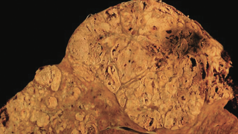
Hepatocellular carcinoma (HCC) is the second most common cause of cancer-related deaths worldwide and is the most common form of liver cancer, accounting for between 85% and 90% of primary liver cancers [1]. The major risk factors for hepatocellular carcinoma include hepatitis B (HBV) or C virus (HCV) infection, smoking, alcohol, and aflatoxin [2]. Currently the prognosis for HCC is poor with Australian statistics indicating that the five year relative survival rate for primary liver cancers is only 16% [3]. The non-specific tyrosine kinase inhibitor sorafenib currently represents the only effective treatment against HCC [4]. This poor prognostic outlook and the limited availability of targeted molecular agents for HCC has led to the development of new therapies such as microRNAs (miRNAs).

miRNAs are short (19-24 nucleotides), non-coding RNA molecules that are post-transcriptional regulators of gene expression. Initially, miRNA is transcribed as primary miRNA (pri-miRNA), which is processed into precursor miRNA (pre-miRNA). This is then transported from the nucleus into the cytoplasm where it is processed into its mature form by the enzyme Dicer. The mature miRNA forms part of the RNA-induced silencing complex (RISC), which is responsible for regulating the output of protein-coding genes. These miRNAs interact with the 3’ untranslated regions (UTRs) of the protein-coding genes to result in a decrease in protein output via mRNA degradation or translational repression [5]. Alternatively, miRNAs can result in post-transcriptional stimulation of gene expression via a multitude of direct and indirect mechanisms [6]. It is estimated that a single miRNA can target hundreds to over one thousand different mRNAs, ultimately resulting in miRNAs being responsible for the regulation of around 20-30% of all protein-encoding genes [7]. These miRNAs have been reported to have key roles in cancer initiation, progression, and metastasis [8]. Oncogenic miRNAs are miRNAs that are up-regulated in cancer cells and promote carcinogenesis via the inhibition of tumour suppressor genes. Conversely, the miRNAs that are decreased in cancer cells are known as tumour suppressor miRNAs, as they normally inhibit proto-oncogenes to prevent cancer from developing [9].  The mammalian miRNA-34 family consists of miRNA-34a, which is encoded via its own individual transcript, and miRNA-34b and miRNA-34c, which possess a common primary transcript [10]. Due to the promising and extensive research conducted on miRNA-34a, this review article focused specifically on this particular isoform. Dysregulation of miRNA-34a has been implicated in a wide variety of cancers, including prostate, colon [11], and HCC [12]. The purpose of this review is to analyse the specific role of miRNA-34a in HCC, including addressing contradictory findings and investigating the recent clinical trials.

Table 1. Percentage of human HCC samples with decreased miRNA-34a expression compared to surrounding non-cancerous liver tissue.

The tumour suppressor role of miRNA-34a in hepatocellular carcinoma

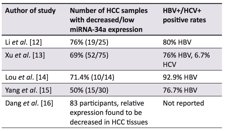
A range of studies established that in the majority of human HCC samples, miRNA-34a expression was decreased in comparison to the surrounding non-cancerous liver tissue (Table 1) [12-16]. A murine model of hepatocarcinogenesis induced by a methyl-deficient diet also resulted in the down-regulation of miRNA-34a [17]. Low expression of miRNA-34a in HCC samples has been correlated with a shorter overall [13-15] and disease-free survival [14], as well as higher recurrence rates [13] when compared with samples that displayed up-regulation of miRNA-34a. The decreased expression of miRNA-34a is thought to be caused by genetic alterations such as deletions, point mutations, or chromosomal translocations of its genomic region 1p36 [18], which is common in HCC [19, 20]. Alternatively, this decreased expression has been linked to inactivating mutations of the p53 gene [18], as the induction of miRNA-34a is correlated with p53 status [21]. Furthermore, epigenetic silencing of miRNA-34a has been implicated with these decreased expression levels in multiple forms of cancer, via abnormal CpG methylation in its promoter region [22].

The administration of a miRNA-34a mimic (MRX34) has been shown to cause inhibition of a number of genes within multiple oncogenic pathways such as Wnt/ beta-catenin, c-MET, VEGF, hedgehog, and MAPK (all of which have been implicated in hepatocarcinogenesis), as well as stimulating multiple genes of the p53 pathway [23]. Daige et al. explored a diverse range of HCC related pathways, demonstrating how miRNA-34a exerts its anti-cancer effects by modulation of a number of genes responsible for processes such as metastasis, cellular proliferation, cell cycle regulation, apoptosis, and cellular senescence [23].


miRNA-34a and cellular proliferation, cell cycle regulation, and apoptosis

A number of cell culture studies investigated the effects of ectopic expression of miRNA-34a in the HepG2 cell line, with contradicting results [12,16,18]. Ectopic expression of miRNA-34a caused significant inhibition of cellular proliferation at 72 hours [18] and 96 hours [16] post- transfection. In addition, miRNA-34a was demonstrated to regulate the cell cycle via inducing G1 arrest [18]. Furthermore, it has been found that miRNA-34a can induce apoptosis, as determined by increased caspase 3/7 activity [16]. In contrast, other reports claimed that there was no effect on cellular proliferation, G1 arrest, or apoptosis, [12,18], highlighting the conflicting information within the current literature. The discrepancies between these three studies could partially be explained by the varying methods used to express miRNA-34a, and the different measurement times post-transfection (48, 72 or 96 hours) [16]. Additional research has found that miRNA-34a induces apoptosis in HCC cells via binding to the 3’ UTR of the Bcl-2 mRNA, causing inhibition of its translation [15]. Over-expression of miRNA-34a has also been correlated with a decreased expression of Bcl-2 in a number of other HCC studies [14,24].


miRNA-34a and metastasis

Down-regulation of miRNA-34a expression has been associated with metastatic HCC [12,16,25]. Multiple studies have established that the ectopic[1] expression of miRNA-34a in the HepG2 cell line inhibits tumour cell migration and invasion via silencing of the c-Met gene, which subsequently decreases the c-Met-induced phosphorylation of extracellular signal-regulated kinases 1 and 2 (ERK1/2) [12,16]. Cheng et al. also proposed that miRNA-34a prevents tumour migration, invasion, and metastasis by decreasing cathepsin D [18], a protease that contributes to the degradation of the basement membrane [26]. Furthermore in the HCC cell line Hca-F (high lymphatic metastatic potential) it was determined through in vitro and in vivo studies in mice that the ectopic expression of miRNA-34a caused a reduction in the metastatic potential [27].


miRNA-34a and cellular senescence

Earlier studies showed that miRNA-34a induced cellular senescence via cell cycle arrest in pathways that were telomere-independent [13, 28]. Recently, miRNA-34a over-expression has been shown to induce senescence in HCC cells in a telomere-dependent manner, regulated by p53.  This cellular senescence occurs by the inhibition of FoxM1 and c-Myc, which causes the inactivation of telomerase activity, resulting in telomere shortening (Figure 1) [13].

Figure 1. Telomere dependent pathway of miRNA-34a induced cellular senescence (adapted from Xu et al. [13]). High levels of miRNA-34a, which is regulated by p53, results in the inhibition of FoxM1 and c-Myc, causing the inactivation of telomerase, leading to telomere shortening and subsequently cellular senescence.

miRNA-34a delivery methods

There is a vast array of delivery systems, both viral and non-viral, that are used to increase miRNA expression. Both mechanisms are associated with advantages and disadvantages [29]. Rubone and MRX34 are two prominent non-viral delivery methods that have been investigated in HCC studies [23,24].  Rubone is a small molecular modulator of miRNA-34a that was shown to induce miRNA-34a expression selectively in HCC cells (although only those with wild-type or mutant p53, not with p53 deletion), causing inhibition of tumour growth both in vitro and in vivo in the HepG2 xenograft mouse model.  Xiao et al. also found that this miRNA-34a modulator displayed similar or even greater anti-HCC activity than sorafenib, the current treatment for advanced HCC [24]. MRX34 is a double-stranded miRNA-34a mimic that is delivered by liposomes [30]. The systemic delivery of this molecule resulted in tumour regression during in vivo studies in two different xenograft mouse models (Hep3B and HuH7) of liver cancer [23]. An oncolytic adenoviral vector that co-expressed miRNA-34a and IL-24 has also been studied in a HCC model. This was found to cause increased anti-tumour activity both in vitro and in vivo, predominantly via the downregulation of SIRT1 and Bcl-2 [14].


Clinical trials

Currently there is an ongoing phase I trial of MRX34 [31], which commenced in April 2013 and was originally indicated for patients with primary liver cancer or cancers with metastasis to the liver [32]. It was then gradually expanded to include patients with other advanced solid tumours (with or without liver metastasis) and haematological malignancies (lymphoma and multiple myeloma) [31]. There were 75 patients with advanced solid tumours enrolled in this study and 30 of these had HCC [33]. This trial’s data revealed that partial responses to the treatment, as per the RECIST guidelines (Table 2) [34], were observed in one patient with HCC, one with melanoma, [33] and one with advanced renal cell carcinoma [35]. Furthermore a number of patients in this trial were found to have attained a stable disease state [35]. This interim phase I data also determined that MRX34 has a manageable safety profile [33]. A recent press release from MIRNA therapeutics has indicated that phase II clinical trials will commence by the end of 2016 and will consist of two studies, one on renal cell carcinoma and the other on melanoma [35]. In terms of HCC-specific trials with MRX34, the future direction is currently unclear and with limited data available at the present time it is difficult to draw any definitive conclusions.

Table 2. Revised RECIST guidelines [34]

Combination therapy

Yang et al. demonstrated that ectopic expression of miRNA-34a resulted in the sensitisation of HCC cells to sorafenib-induced apoptosis and toxicity via inhibiting expression of Bcl-2 [15]. Additionally, the administration of miRNA-34a was found to sensitise HCC cells to chemotherapy (cisplatin) in vitro through the AXL pathway [36]. The combination of a miRNA-34a mimic and C-met inhibitor also resulted in a greater inhibition of cell growth and induction of apoptosis in vitro than either of these two therapies alone [16]. However, to establish more definitive results, further research is required in this field, particularly in regards to clinical trials.


The oncogenic role of miRNA-34a in hepatocellular carcinoma

Conversely, a number of other studies have shown increased expression of miRNA-34a in both murine and human HCC tissues [37-41], suggesting it may have an oncogenic role in addition to its tumour suppressor role. A recent article has investigated these claims and found that miRNA-34a displays oncogenic properties in liver tumours with beta-catenin activation [40]. Increased beta-catenin activation is most commonly caused by mutations in the CTNNB1 gene (the gene encoding  beta-catenin), and this is estimated to occur in 20-40% of HCCs [42]. Gougelet and colleagues demonstrated using ApcKO mice (ApcKO causes activation of the Wnt/beta-catenin pathway [43]) that administration of a miRNA-34a inhibitor (LNA-34a) caused increased expression of hepatocyte nuclear factor 4a. This leads to increased apoptosis predominantly via caspase 2 activation and decreased cell proliferation through inhibition of cyclin D1 (Figure 2).  This theory was then postulated to complement the data of the studies listed in Table 1, by accounting for those samples that showed up-regulation of miRNA-34a expression. The disparity in results between studies is thought to be due to the varying causes of HCC [40]. For example, the majority of HCC samples from the studies in the Table 1 were HBV+, and this was found to be associated with a lower frequency of CTNNB1 mutations [44]. Conversely HCC resulting from HCV infection has been shown to have a higher rate of CTNNB1 mutations [45].  However, these findings contradict the theory that miRNA-34a has a tumour suppressor function, and with relatively limited research on this oncogenic pathway, further investigation is required. Studies investigating the concept of miRNA-34a having a tumour suppressor or oncogenic function depending on the cause of the tumour would also be important, as well as an investigation of the clinical implications of this relationship.

Figure 2. Role of miRNA-34a inhibitor in counteracting the oncogenic action of miRNA-34a in HCC with beta-Catenin activation (Adapted from Gougelet et al. [40]). The miRNA-34a inhibitor LNA-34a causes a reduction in β-catenin induced miRNA-34a expression which leads to an increased expression of HNF-4α. Subsequently cyclin D1 levels are reduced, leading to decreased cell proliferation and increased caspase 2 levels, leading to increased apoptosis. The combined effects of these two actions then leads to the decreased progression of the hepatocellular carcinoma.

Limitations

A number of limitations were identified within this review, particularly relating to the conflicting information and the limited availability of clinical trial results. Contradictory information was noted on a number of occasions, especially with the use of a miRNA-34a inhibitor for HCC, making it difficult to evaluate a clear clinical benefit to this potential therapy. The data surrounding the clinical trial was also restricted as the trial is ongoing.  Subsequently, all the data had to be sourced from press releases and abstracts from presentations at conferences, which were all funded by MIRNA therapeutics; thus a potential conflict of interest was noted.


Conclusion

Results have indicated that miRNA-34a has a tumour suppressor function in HCC and is responsible for the down-regulation of a number of genes involved in carcinogenesis. There is, however, contradicting information described in studies investigating these parameters, highlighting the complexity of this topic. Rubone and MRX34 are two prominent miRNA-34a delivery systems that were shown to exert anti-tumour activity in pre-clinical models. Additionally, MRX34 has been commenced in a clinical phase I trial that is still currently ongoing, with a partial response already observed in one patient. However, the future of HCC-specific MRX34 trials remains unclear as limited information is currently available. Based on the promising results of miRNA-34a as a combination therapy, this is an area that requires further investigation through clinical trials. Conversely, other sources have found that miRNA-34a plays an oncogenic role in HCC, particularly in those with beta-catenin activation. Subsequently, it was demonstrated that miRNA-34a inhibitors should be used in these instances. Further research is necessary in order to ascertain the clinical implications of using a miRNA-34a mimic or inhibitor depending on the beta-catenin mutation status of the patient.


Conflicts of interest

None declared.


References

  1. Park J-W, Chen M, Colombo M, Roberts LR, Schwartz M, Chen P-J, et al. Global patterns of hepatocellular carcinoma management from diagnosis to death: the BRIDGE Study. Liver Int. 2015;35(9):2155-66.
  2. Chuang S-C, Vecchia CL, Boffetta P. Liver cancer: Descriptive epidemiology and risk factors other than HBV and HCV infection. Cancer Lett. 2009;286(1):9-14.
  3. Australian Institute of Health and Welfare. Cancer in Australia: an overview 2014 [Internet]. Canberra: Australian Institute of Health and Welfare; 2014 [updated 2015 Apr 16; cited 2016 Apr 2]. 220p. Available from: http://www.aihw.gov.au/WorkArea/DownloadAsset.aspx?id=60129550202
  4. Yim HJ, Suh SJ, Um SH. Current management of hepatocellular carcinoma: an Eastern perspective. World J Gastroenterol. 2015;21(13):3826-42.
  5. Ling H, Fabbri M, Calin GA. MicroRNAs and other non-coding RNAs as targets for anticancer drug development. Nat Rev Drug Discov. 2013;12(11):847-65.
  6. Vasudevan S. Posttranscriptional upregulation by microRNAs. Wiley Interdiscip Rev RNA. 2012;3(3):311-30.
  7. Felekkis K, Touvana E, Stefanou C, Deltas C. MicroRNAs: a newly described class of encoded molecules that play a role in health and disease. Hippokratia. 2010;14(4):236-40.
  8. Lin S, Gregory RI. MicroRNA biogenesis pathways in cancer. Nat Rev Cancer. 2015;15(6):321-33.
  9. Misso G, Di Martino MT, De Rosa G, Farooqi AA, Lombardi A, Campani V et al. Mir-34: a new weapon against cancer? Mol Ther Nucleic Acids. 2014;3(9):e195.
  10. Hermeking H. The miR-34 family in cancer and apoptosis. Cell Death Differ. 2010;17(2):193-9.
  11. Saito Y, Nakaoka T, Saito H. MicroRNA-34a as a therapeutic agent against human cancer. J Clin Med. 2015;4(11):1951-9.
  12. Li N, Fu H, Tie Y, Hu Z, Kong W, Wu Y et al. miR-34a inhibits migration and invasion by down-regulation of c-Met expression in human hepatocellular carcinoma cells. Cancer Lett. 2009;275(1):44-53.
  13. Xu X, Chen W, Miao R, Zhou Y, Wang Z, Zhang L et al. miR-34a induces cellular senescence via modulation of telomerase activity in human hepatocellular carcinoma by targeting FoxM1/c-Myc pathway. Oncotarget. 2015;6(6):3988-4004.
  14. Lou W, Chen Q, Ma L, Liu J, Yang Z, Shen J et al. Oncolytic adenovirus co-expressing miRNA-34a and IL-24 induces superior antitumor activity in experimental tumor model. J Mol Med. 2013;91(6):715-25.
  15. Yang F, Li QJ, Gong ZB, Zhou L, You N, Wang S et al. MicroRNA-34a targets Bcl-2 and sensitizes human hepatocellular carcinoma cells to sorafenib treatment. Technol Cancer Res Treat. 2014;13(1):77-86.
  16. Dang Y, Luo D, Rong M, Chen G. Underexpression of miR-34a in hepatocellular carcinoma and its contribution towards enhancement of proliferating inhibitory effects of agents targeting c-MET. PLoS ONE. 2013;8(4):e61054.
  17. Tryndyak VP, Ross SA, Beland FA, Pogribny IP. Down-regulation of the microRNAs miR-34a, miR-127, and miR-200b in rat liver during hepatocarcinogenesis induced by a methyl-deficient diet. Mol Carcinog. 2009;48(6):479-87.
  18. Cheng J, Zhou L, Xie QF, Xie HY, Wei XY, Gao F et al. The impact of miR-34a on protein output in hepatocellular carcinoma HepG2 cells. Proteomics. 2010;10(8):1557-72.
  19. Herath NI, Kew MC, Whitehall VL, Walsh MD, Jass JR, Khanna KK et al. p73 is up-regulated in a subset of hepatocellular carcinomas. Hepatology. 2000;31(3):601-5.
  20. Midorikawa Y, Yamamoto S, Tsuji S, Kamimura N, Ishikawa S, Igarashi H et al. Allelic imbalances and homozygous deletion on 8p23.2 for stepwise progression of hepatocarcinogenesis. Hepatology. 2009;49(2):513-22.
  21. Bommer GT, Gerin I, Feng Y, Kaczorowski AJ, Kuick R, Love RE et al. p53-mediated activation of miRNA34 candidate tumor-suppressor genes. Curr Biol. 2007;17(15):1298-307.
  22. Lodygin D, Tarasov V, Epanchintsev A, Berking C, Knyazeva T, Korner H et al. Inactivation of miR-34a by aberrant CpG methylation in multiple types of cancer. Cell Cycle. 2008;7(16):2591-600.
  23. Daige CL, Wiggins JF, Priddy L, Nelligan-Davis T, Zhao J, Brown D. Systemic delivery of a miR34a mimic as a potential therapeutic for liver cancer. Mol Cancer Ther. 2014;13(10):2352-60.
  24. Xiao Z, Li CH, Chan SL, Xu F, Feng L, Wang Y et al. A small-molecule modulator of the tumor-suppressor miR34a inhibits the growth of hepatocellular carcinoma. Cancer Res. 2014;74(21):6236-47.
  25. Budhu A, Jia HL, Forgues M, Liu CG, Goldstein D, Lam A et al. Identification of metastasis-related microRNAs in hepatocellular carcinoma. Hepatology. 2008;47(3):897-907.
  26. Khalkhali-Ellis Z, Hendrix MJC. Elucidating the function of secreted maspin: inhibiting cathepsin D–mediated matrix degradation. Cancer Res. 2007;67(8):3535-9.
  27. Guo Y, Li S, Qu J, Wang S, Dang Y, Fan J et al. MiR-34a inhibits lymphatic metastasis potential of mouse hepatoma cells. Mol Cell Biochem. 2011;354(1-2):275-82.
  28. Chen F, Hu SJ. Effect of microRNA-34a in cell cycle, differentiation, and apoptosis: a review. J Biochem Mol Toxicol. 2012;26(2):79-86.
  29. Zhang Y, Wang Z, Gemeinhart RA. Progress in microRNA delivery. J Control Release. 2013;172(3):962-74.
  30. Agostini M, Knight RA. miR-34: from bench to bedside. Oncotarget. 2014;5(4):872-81.
  31. Mirna Therapeutics Inc; Cancer Prevention Research Institute of Texas. A multicenter phase I study of MRX34, microRNA miR-RX34 liposomal injection. In: ClinicalTrials.gov [Internet]. Bethesda (MD): National Library of Medicine (US). 2013 [cited 2016 Apr 3]. Available from: https://clinicaltrials.gov/ct2/show/study/NCT01829971?view=results. NLM Identifier: NCT01829971
  32. Bouchie A. First microRNA mimic enters clinic. Nat Biotech. 2013;31(7):577.
  33. Beg MS, Brenner A, Sachdev J, Ejadi S, Borad M, Kang Y-K et al. Safety, tolerability, and clinical activity of MRX34, the first-in-class liposomal miR-34 mimic, in patients with advanced solid tumors [Abstract]. Mol Cancer Ther. 2015;14(12 S2):C43.
  34. Eisenhauer EA, Therasse P, Bogaerts J, Schwartz LH, Sargent D, Ford R et al. New response evaluation criteria in solid tumours: revised RECIST guideline (version 1.1). Eur J Cancer. 2009;45(2):228-47.
  35. MIRNA Therapeutics. Mirna therapeutics provides operational update & reports financial results for fourth quarter and full year ended December 31, 2015 [Internet]. Austin Texas: MIRNA Therapeutics; 2016 Mar 29 [cited 2016 Apr 3]. Available from: http://investor.mirnarx.com/releasedetail.cfm?ReleaseID=962545.
  36. Li X-Y, Wen J-Y, Jia C-C, Wang T-T, Li X, Dong MIN et al. MicroRNA-34a-5p enhances sensitivity to chemotherapy by targeting AXL in hepatocellular carcinoma MHCC-97L cells. Oncol Lett. 2015;10(5):2691-8.
  37. Pineau P, Volinia S, McJunkin K, Marchio A, Battiston C, Terris B et al. miR-221 overexpression contributes to liver tumorigenesis. Proc Natl Acad Sci USA. 2010;107(1):264-9.
  38. Sukata T, Sumida K, Kushida M, Ogata K, Miyata K, Yabushita S et al. Circulating microRNAs, possible indicators of progress of rat hepatocarcinogenesis from early stages. Toxicol Lett. 2011;200(1-2):46-52.
  39. Zhu L, Gao J, Huang K, Luo Y, Zhang B, Xu W. miR-34a screened by miRNA profiling negatively regulates Wnt/β-catenin signaling pathway in Aflatoxin B1 induced hepatotoxicity. Sci Rep. 2015;5:16732.
  40. Gougelet A, Sartor C, Bachelot L, Godard C, Marchiol C, Renault G et al. Antitumour activity of an inhibitor of miR-34a in liver cancer with beta-catenin-mutations. Gut [Internet]. 2015 Mar 19. Available from: http://gut.bmj.com/content/65/6/1024.long [Epub ahead of print].
  41. Pok S, Wen V, Shackel N, Alsop A, Pyakurel P, Fahrer A et al. Cyclin E facilitates dysplastic hepatocytes to bypass G1/S checkpoint in hepatocarcinogenesis. J Gastroenterol Hepatol. 2013;28(9):1545-54.
  42. Tornesello ML, Buonaguro L, Tatangelo F, Botti G, Izzo F, Buonaguro FM. Mutations in TP53, CTNNB1 and PIK3CA genes in hepatocellular carcinoma associated with hepatitis B and hepatitis C virus infections. Genomics. 2013;102(2):74-83.
  43. Colnot S, Decaens T, Niwa-Kawakita M, Godard C, Hamard G, Kahn A et al. Liver-targeted disruption of Apc in mice activates beta-catenin signaling and leads to hepatocellular carcinomas. Proc Natl Acad Sci USA. 2004;101(49):17216-21.
  44. Guichard C, Amaddeo G, Imbeaud S, Ladeiro Y, Pelletier L, Maad IB et al. Integrated analysis of somatic mutations and focal copy-number changes identifies key genes and pathways in hepatocellular carcinoma. Nat Genet. 2012;44(6):694-8.
  45. Li M, Zhao H, Zhang X, Wood LD, Anders RA, Choti MA et al. Inactivating mutations of the chromatin remodeling gene ARID2 in hepatocellular carcinoma. Nat Genet. 2011;43(9):828-9.
Categories
Review Articles

Hepatocellular carcinoma: the potential for an effective genetic screening test

Hepatocellular carcinoma (HCC) is the sixth most common cancer worldwide contributing to approximately 600,000 deaths each year with this number on the rise in the developing world. The aetiology of HCC has been well characterised, including chronic hepatitis B and C infection as well as alcoholic cirrhosis. Current screening programs for patients at high risk of developing HCC, including ultrasound and alpha-fetoprotein analysis, are neither sufficiently sensitive nor specific to detect early HCC.  This reduces the likelihood of detecting HCC when curative treatment is effective. One genetic marker which has been shown to be associated with HCC carcinogenesis is the p16 INK4a/ARF locus. If further research confirms this mutation as a common step in early hepatocarcinogenesis then this maker could be utilised to screen for early HCC lesions in at risk populations. Further research in this area could facilitate the early diagnosis of HCC, improving the efficacy of treatment.

Introduction

Liver cancer is the sixth most common cancer worldwide and the second largest cause of cancer mortality [1]. It has several subtypes including hepatocellular carcinoma (HCC), bile duct cancer, hepatoblastoma, and various other liver sarcomas and carcinomas [2]. Of those subtypes, HCC is the most common, comprising about 78% of all liver cancers [1,2]. HCC is unequally distributed globally with over 80% of cases occurring in either Sub-Saharan Africa or Eastern Asia; predominantly in China [3,4]. When considering Western countries, there is strong evidence from the United States that the incidence of HCC is rising, with nine cancer registries reporting via the National Cancer Institute that there has been a 41% rise in mortality from primary liver cell cancer and a 70% rise overall in incidence between 1980 and 1995 [5].

A similar rise in HCC incidence and death rates in Australia have also been identified; possibly linked to the increased prevalence of Hepatitis B and Hepatitis C infection in Australia [6,7]. Moreover, there is evidence that HCC incidence rates in Australia may be up to two-fold higher than the rates reported by cancer registries such as the Victorian Cancer Registry [8].  A higher rate of HCC has also been reported in Aboriginal and Torres Strait Islander populations; estimated at 2% and 8% in urban and rural areas, respectively, compared with less than 1% for the total Australian population [9].

The aetiology of HCC is well documented in the literature with high rates linked to both hepatitis B and hepatitis C exposure [9]. Other significant causes that may cause patients to present include alcoholic liver disease, non-alcoholic steatohepatitis as well as hereditary conditions such as haemochromatosis, alpha-1 antitrypsin deficiency, and autoimmune disorders (Figure 1) [10,11]. These conditions result in significant parenchymal loss, increased fibrogenesis and inflammatory signalling resulting in cirrhosis; a condition associated with 70-90% of all detectable HCC cases [10,11].

Figure 1. Aetiology of hepatocellular carcinoma

The natural history of HCC growth begins as small asymptomatic nodules which can often take years to develop depending on the aetiological exposure [5]. Small HCCs at detection have relatively long tumour doubling times with tumours of less than five centimetres associated with a survival of 81-100% at one year, and 17-21% at three years without therapy, which suggests that early diagnosis may allow for a greater intervention window [5].

These features make HCC an insidious and difficult disease to clinically detect and investigate. Patients who develop HCC are usually asymptomatic, mostly displaying symptoms related to their chronic liver disease which can become modified with disease progression [6,12]. Examples of this include signs of decompensation such as ascites, encephalopathy, jaundice, and variceal bleeding [12]. Advanced lesions also can present more conspicuously causing obstructive symptoms such as jaundice, diarrhoea, weight loss, and fatigue [13]. These signs are a result of local tumour invasion and growth inside the liver. However, systemic signs can also occur as a result of metastases which can develop in the lung, portal vein, periportal nodes, bone or brain [13]. As a result of these features, HCC is typically diagnosed late in its course with a median survival following diagnosis of 6 to 20 months, and a five-year survival ranging from 12% in those outside major cities and 17% within major cities in Australia. [13,14].


Screening and detection of HCC

Accordingly, screening programs for HCC in at risk groups, those with chronic liver disease or chronic hepatitis infection, is recommended with specialist review forming part of a 6-12 monthly management plan [15]. These programs involve using alpha-fetoprotein (AFP) and ultrasound to screen for the presence of cancer lesions.

AFP is a widely studied screening test marker for HCC with a level above 400 ng/mL regarded as diagnostic [5]. However, two thirds of HCCs less than 4 cm have AFP levels less than 200 ng/mL and up to 20% of HCCs do not produce AFP even when they are very large [5]. Moreover, there is evidence that fluctuating levels of AFP in patients with cirrhosis might reflect flares of HBV or HIV infection or liver disease exacerbation rather than HCC development [16]. Some studies investigating the clinical utility of AFP have suggested it lacks the sensitivity to be useful, with one study suggesting it rarely assisted in a diagnosis [5,17,18]. As a result of this, it has been suggested that AFP testing alone should only be used if ultrasound is unavailable, with the American Association for the Study of Liver Diseases and the European Association for the Study of the Liver recommending it not be used at all [16,18,20]. This therefore limits the use of AFP as a reliable test to screen for HCC in at risk groups. Due to these limitations, other serum markers which include plasma microRNAs such as miR-122 and miR-192, des-gamma-carboxy prothrombin, AFP isoforms and glypican-3 are currently being investigated and evaluated for future use [19,20].

Another modality that is used in HCC screening is ultrasound. Ultrasound can detect large HCCs with high sensitivity and specificity; however, it is less able to reliably identify smaller lesions, which are required if more effective therapy is to be offered [6,19,20]. Ultrasound has been shown to detect 85-95% of lesions 3-5 cm in diameter, but can only achieve a 60-80% sensitivity of 1 cm lesions [5]. A large meta-analysis investigating this in 2009 found similar results demonstrating that surveillance with ultrasound showed a limited sensitivity (64%) for early HCC detection [19]. Combined use of AFP and ultrasound has been shown to increase detection rates, but had a raised combined false positive rate of 7.5% compared to AFP and ultrasound alone at 5.0% and 2.9% respectively [18].  Despite the limitations of these tests, the Royal Australian College of General Practitioners guidelines suggest that patients with chronic liver disease or chronic hepatitis infection should be considered for 6-12 monthly AFP and ultrasound screening [15]. The Asian Pacific Association for the Study of the Liver also recommends surveillance for HCC with both AFP and ultrasound every 6 months [21].

The importance of early HCC detection cannot be understated. The natural history of early tumours is poorly known as most are treated upon diagnosis; however, surgical resection, tranplantation and ablation offer high rates of complete responses and a potential cure in all patients with early HCC [22]. Advanced course HCC has a survival of less than six months without treatment with prognostic factors for survival including anatomical extension of the tumour, performance status and functional hepatic reserve based on the Child-Pugh Score [22-24]. It is from this that researchers are currently investigating superior screening techniques which can identify tumours earlier and with greater sensitivity and specificty to enable earlier intervention and better treatment outcomes.


Cancer biology as the pathway for a HCC screening test

One approach that is currently being investigated in the medical literature focusses on the biology of HCC hepatocarcinogenesis to develop a sensitive early screening test that can guide and detect treatment before the cancer can extend and spread. Mature hepatocytes have an average lifespan of between 200-400 days and rarely proliferate unless stimulated by acute injury [25]. The observation of normally quiescent hepatocytes and cholangiocytes proliferating after partial hepatectomy has highlighted the significant regenerative ability of the liver after acute insult [26]. If this regenerative capacity is compromised, the liver has liver progenitor cells (LPCs) which can expand and regenerate the chronically damaged liver [27,28]. LPCs can propagate and differentiate into two types of liver epithelial cells; hepatocytes and cholangiocytes [27]. These cells have been defined as stem cells because they are clonogenic, with a high growth potential and are able to be induced to differentiate into both types of liver cells and have shown capability in repopulating the liver on transplantation [27-30].

The proliferation and differentiation of LPCs into hepatocytes render them a target population for hepatocarcinogenesis [30]. LPCs have been traced to hepatocytes and are markedly elevated in chronic liver disease [28]. Recent laboratory experimentation has shown a link between induced liver damage in mice and the development of HCC, suggesting a tenable link between LPCs and HCC development [31,32]. LPCs have also been documented in chronic human liver pathologies, such as chronic hepatitis C, which is highly associated with hepatocarcinogenesis [29,33-35]. Analysis of premalignant lesions in HCC have also identified the presence of LPCs, with up to 50% of developed HCCs being shown to express markers of progenitor cells including CK7, CK19, OV6 [35-38] These findings have also been found in further studies on both human and mouse liver cells [38,39]. These results suggest a significant link exists between LPCs and HCC carcinogenesis.

From a molecular analysis of HCC progression, it has been shown that hepatocarcinogenesis is a multistep process that is heterogeneous and not well understood [40]. Progressive genetic alterations have been shown to cause a spectrum of cellular changes starting from cell hyperplasia, proliferation to dysplasia and eventually cancer [28,40]. This model is widely accepted and has been applied to many types of cancer, including HCC [28]. Multiple studies have demonstrated that two tumour suppressor pathways are important in controlling cell proliferation including the retinoblastoma protein pathway and the p53 pathway [28,41]. Most human tumours have genetic mutations, deletions, deregulated methylation or alterations in microRNA signalling in their Retinoblastoma and p53 pathways; making these genes likely candidates in the transformation of non-tumourigenic LPCs to tumourigenic LPCs [42-44].

Future HCC screening with the marker p16 INK4a/ARF

One gene involved in the p53 tumour suppressor pathway which may play a crucial role in hepatocarcinogenesis is the INK4a/ARF locus. Studies have identified variable rates of inactivation of the p16 INK4a/ARF gene in HCC with inactivation ranging in the literature from 35-82% of HCCs depending on the aetiology [45-50]. Studies on HCC mouse models have highlighted the important role of INK4a/ARF in tumourigenesis with concomitant loss of p53 and INK4a/ARF accelerating tumourigenesis and the progression to metastatic lung lesions [51-53]. Furthermore, tumours lacking both p53 and INK4a/ARF demonstrated strong migration and invasion capabilities. This was not demonstrated when p53 itself was inactive; suggesting that INK4a/ARF inactivation may be a critical step in HCC development (Figure 2) [52,53]. Significant evidence suggests that INK4a/ARF are important tumour suppressors encoded at 9p21 [9,46,52-54]. Kaneto et al. suggested that the methylation of the INK4a/ARF locus promoter is an early event in hepatocarcinogenesis, making this gene an ideal candidate for further study in the pursuit for an accurate diagnostic tool for HCC [46].

Figure 2. Stages of hepatocellular carcinoma development

The nature of how genes are inactivated in tumourigenesis is biologically complex. The INK4a/ARF gene can be affected by many different forms of inactivation notably mutations, homozygous deletions or gene methylation [54]. Tannapefel et al. showed that of 71 carcinomas examined, 59% showed aberrant methylation of the INK4a gene [54]. Another recent review suggested that as many as 40-70% of HCCs demonstrate an INK4a methylation resulting in the downregulation of protein expression [55]. The pathogenesis of the ARF gene in HCC was not as clear, with studies finding ARF gene methylations in non-cancerous liver tissue as well as low rates of ARF methylation in human HCC samples [54,55]. Despite this, evidence of promotor methylation has arisen from investigations into the cause of INK4a/ARF inactivation in other tumours such as human cutaneous squamous cell carcinoma and colorectal carcinomas [56,57]. These studies provide evidence that the methylation of the INK4a/ARF gene may be an important step in the hepatocarcinogenesis of HCC and therefore could be an effective clinical marker for the identification of HCC development in at risk patients.


Conclusion

While it is clear from this review that further research needs to be conducted to better understand the role of the INK4a/ARF gene locus in HCC development, the potential for a clincial application remains a potent driver for further research in this area of molecular medicine. While the current screening methods using AFP and ultrasound are considered to be clincially useful in detecting HCC, there exists a space for more accurate modalities to detect early HCC lesions.  If future research shows that the INK4a/ARF gene is a common early mutation in HCC hepatocarcinogenesis, then future tests may be developed which can sample high probability sites in the liver or the blood to investigate for DNA which contains this mutation. This could potentially improve the prognosis of patients with HCC development and allow early directed treatment with the possibility of cure.


Acknowledgements

Thank you to Professor George Yeoh for his assistance in proofreading this article and providing support and advice.

Conflicts of interest

None declared.


References

[1]       Laursen L. A preventable cancer. Nature. 2014;516(7529):S2-3.

[2]       Australian Government. Liver cancer. Australia: Cancer Australia; 2014 [cited 2016 April]. Available from: http://canceraustralia.gov.au/affected-cancer/cancer-types/liver-cancer

[3]       El-Serag HB, Rudolph KL. Hepatocellular carcinoma: epidemiology and molecular carcinogenesis. Gastroenterology. 2007;132(7):2557-76.

[4]       Han ZG. Functional genomic studies: insights into the pathogenesis of liver cancer. Annu Rev Genomics Hum Genet. 2012; 9c;13:171-205.

[5]       Ryder SD. Guidelines for the diagnosis and treatment of hepatocellular carcinoma (HCC) in adults. Gut. 2003;S3:iii1-iii8.

[6]       Law MG, Roberts SK, Dore GJ, Kaldor JM. Primary hepatocellular carcinoma in Australia, 1978–1997: increasing incidence and mortality. Med J Aust. 2000;173:403–5.

[7]       El-Serag HB, Mason AC. Rising incidence of hepatocellular carcinoma in the United States. N. Engl. J. Med. 1999;340:745–50.

[8]       Hong TP, Fink M, Dev A, Roberts S Nicoll A, Lubel J, Kronborg I, et al. Novel population-based study finding higher than reported hepatocellular carcinoma incidence suggests an updated approach is needed. Hepatology. 2016;63(4):1205-1212.

[9]       Cunningham J, Rumbold AR, Zhang X, Condon JR. Incidence, aetiology, and outcomes of cancer in Indigenous peoples in Australia. Lancet Oncology. 2008;9(6):585–95.

[10]     Lee YA, Wallace MC, Friedman SL. Pathobiology of liver fibrosis: a translational success story. Gut. 2015;64(5):830-41.

[11]     Intrahepatic arteriovenous shunting due to hepatocellular carcinoma and cirrhosis, and its change by transcatheter arterial embolization. AU Sugano S, Miyoshi K, Suzuki T, Kawafune T, Kubota M SO Am J Gastroenterol. 1994;89(2):184.

[12]     Schwartz J, Carithers R, Chopra S. Clinical features and diagnosis of primary hepatocellular carcinoma. In: UpToDate, Post TW (Ed), UpToDate, Waltham, MA. (Accessed 2016 June.)

[13]     Saunders, WB. A new prognostic system for hepatocellular carcinoma: a retrospective study of 435 patients: the cancer of the liver Italian program (CLIP) investigators. 1998;28(3):751-5.

[14]     Refshuage A, Kalisch D. Cancer survival and prevalence in Australia period estimates from 1982 to 2010. Australian Government: Australian Institute of Health and Welfare. 2012 [cited 2016 Sep]. Available from: http://www.aihw.gov.au/WorkArea/DownloadAsset.aspx?id=10737422721

[15]     Lusis N. Clinical guidelines: prevention and early detection of liver (hepatocellular) cancer. Royal Australian College of General Practitioners. 2012 [cited 2016 May] Available from: http://www.racgp.org.au/your-practice/guidelines/national-guide/prevention-and-early-detection-of-cancer/prevention-and-early-detection-of-liver-(hepatocellular)-cancer/

[16]     Llovet JM, Ducreux M, Lencioni R, Bisceglie AM, Galle PR, Dufour JF, et al. EASL-EORTC Clinical practice guidelines: management of hepatocellular carcinoma. European Association for the study of liver, European Organisation for Research and Treatment of Cancer. Journal of Hepatology. 2012; 56;908-943.

[17]     Ebara M, Ohto M, Shinagawa T, Sugiura N, Kimura K, Matsutani S, et al. Natural history of minute hepatocellular carcinoma smaller than three centimeters complicating cirrhosis. A study in 22 patients. 1986;90(2):289-98.

[18]     Bruix J, Sherman M. American association for the study of liver diseases: management of hepatocellular carcinoma: an update. Hepatology. 2011;53(3):1020-2.

[19]     Singal A, Volk ML, Waljee A, Salgia R, Higgins P, Rogers M, et al. Meta-analysis: surveillance with ultrasound for early-stage hepatocellular carcinoma in patients with cirrhosis. Alimentary Pharmacology & Therapeutics. 2009; (30):37–47.

[20]     Colombo M, Chopra S. Prevention of hepatocellular carcinoma and recommendations for surveillance in adults with chronic liver disease. In: UpToDate, Post TW (Ed), UpToDate, Waltham, MA. (Accessed 2016 June.)

[21]     Omata M, Lesmana L Tateishi R, Chen PJ, Lin S, Yoshida H, et al. Asian pacific association for the study of liver consensus recommendations on hepatocellular carcinoma. Hepatology International. 2010;4:439-474.

[22]     Okuda K, Ohtsuki T, Obata H, Tomimatsu M, Okazaki N, Hasegawa H, et al. Natural history of hepatocellular carcinoma and prognosis in relation to treatment. Study of 850 patients. Cancer. 1985;56(4):918-28.

[23]     Llovet JM, Bruix J. Systematic review of randomized trials for unresectable hepatocellular carcinoma: chemoembolization improves survival. Hepatology. 2003; 37(2):429-42.

[24]     Llovet JM, Brú C, Bruix J. Prognosis of hepatocellular carcinoma: the BCLC staging classification. Semin Liver Dis. 1999;19(3):329-38.

[25]     Malato Y, Naqvi S, Schurmann N, Ng R, Wang B, Zape J, et al. Fate tracing of mature hepatocytes in mouse liver homeostasis and regeneration. J Clin Invest. 2011;121(12):4850-60.

[26]     Mishra L, Banker T, Murray J, Byers S, Thenappan A, He AR, et al. Liver stem cells and hepatocellular carcinoma. Hepatology. 2009;49(1):318-29.

[27]     Miyajima A, Tanaka M, Itoh T. Stem/Progenitor cells in liver development, homeostasis, regeneration and reprogramming. Cell Stem Cell. 2014;14(5):561-574.

[28]     Chen C, Wang G. Mechanisms of hepatocellular carcinoma and challenges and opportunities for molecular targeted therapy. World J Hepatol. 2015; 7(15):1964-1970.

[29]     Kumar M, Zhao X, Wang XW. Molecular carcinogenesis of hepatocellular carcinoma and intrahepatic cholangiocarcinoma: one step closer to personalized medicine? Cell Biosci. 2011;1(1):5.

[30]     Yamashita T, Wang XW. Cancer stem cells in the development of liver cancer. J Clin Invest. 2013;123(5):1911-8.

[31]     Viebahn CS, Benseler V, Holz LE, Elsegood CL, Vo M, Beroloino P, et al. Invadin macrophages play a major role in liver progenitor cell response to chronic liver injury. J Hepatol. 2010;53(3):500-507.

[32]     Passman A, Strauss R, McSpadden S, Finch-Edmondson M, Woo K, Diepeveen L, et al. A modified choline-deficient, ethionine-supplemented diet reduces morbidity and retains a liver progenitor cell response in mice. Dis Model Mech. 2015;8(12):1635-41.

[33]     Zhang A, London R, Schulz F, Giguère-Simmonds P, Delriviere L, Chandraratana H, et al. Human liver progenitor cell lines are readily established from non-tumorous tissue adjacent to hepatocellular carcinoma. Stem Cells Dev. 2010; 19(8):1277-84.

[34]     Davies R, Knight B, Tian Y, Yeoh G, Olynyk J. Hepatic oval cell response to the choline-deficient, ethionine supplemented model of murine liver injury is attenuated by the administration of a cyclo-oxyenase 2 inhibitor. Carcinogenesis. 2006;27(8):1607-1616.

[35]     Roskams T. Liver stem cells and their implication in hepatocellular and cholangiocarcinoma. Oncogene. 2006;25(27):3818-22.

[36]     Mishra L, Banker T, Murray J, Byers S, Thenappan A, He AR, et al. Liver stem cells and hepatocellular carcinoma. Hepatology. 2009;49(1):318-29.

[37]     Roskams T. Liver stem cells and their implication in hepatocellular and cholangiocarcinoma. Oncogene. 2006; 25(27):3818-22.

[38]     Li C, Wang Y, Dong W, Xiang S, Liang H, Wang H, et al. Hepatic oval cell lines generate hepatocellular carcinoma following transfection with HBx gene and treatment with aflatoxin B1 in vivo. Cancer Letters. 2011;311(1):1-10.

[39]     Mishra L, Banker T, Murray J, Byers S, Thenappan A, He AR, et al. Liver stem cells and hepatocellular carcinoma. Hepatology. 2009;49(1):318-329.

[40]     Cha C, Dematteo RP. Molecular mechanisms in hepatocellular carcinoma. Best Pract Red CL GA. 2004;19(1):25-37.

[41]     Sherr CJ, McCormick F. The RB and p53 pathways in cancer. Cancer Cell. 2002;2(2):103-112.

[42]     Ito T, Nishiada N, Fukuda Y, Nishimura T, Komeda T, Nakao K. Alterations of the p14(ARF) gene and p53 and p53 status in human hepatocellular carcinoma. J Gastroenterol. 2004;39(4):355-361.

[43]     Gramantieria L, Fornaria F, Challengaria E, Sabbioni S, Lanza G, Croce CN, et al. MicroRNA involvement in hepatocellular carcinoma. J Cell Mol Med. 2008; 12(6):2189-2204.

[44]     Tannapfel A, Busse C, Weinans L, Benicke M, Katalinic A, Geissler F, et al. INK4a-ARF alterations and p53 mutations in hepatocellular carcinomas. Oncogene. 2001;20(48):7104-7109.

[45]     Tannapfel A, Wittekind C. Genes involved in hepatocellular carcinoma: deregulation in cell cycling and apoptosis. Virchows Arch. 2002;440(4):345-52.

[46]     Kaneto H, Sasaki S, Yamamoto H, Itoh F, Toyota M, Suzuki H, et al. Detection of hypermethylation of the p16(INK4A) gene promoter in chronic hepatitis and cirrhosis associated with hepatitis B or C virus. Gut. 2001;48(3):372-7.

[47]     Jin M, Piao Z, Kim NG, Park C, Shin EC, Park JH, et al. p16 is a major inactivation target in hepatocellular carcinoma. Cancer. 2000;89(1):60-8.

[48]     Lee S, Lee HJ, Kim JH, Lee HS, Jang JJ, Kang GH. Aberrant CpG island hypermethylation along multistep hepatocarcinogenesis. Am J Pathol. 2003;163(4):1371-8.

[49]     Li B, Liu W, Wang L, Li M, Wang J, Huang L, et al. CpG island methylator phenotype associated with tumor recurrence in tumor-node-metastasis stage I hepatocellular carcinoma. Ann Surg Oncol. 2010;17(7):1917-26.

[50]     Roncalli M, Bianchi P, Bruni B, Laghi L, Destro A, Di Gioia S, et al. Methylation framework of cell cycle gene inhibitors in cirrhosis and associated hepatocellular carcinoma. Hepatology. 2002;36(2):427-32.

[51]     Farazi PA, DePinho RA. Hepatocellular carcinoma pathogenesis: from genes to environment. Nat Rev Cancer. 2006 Sep;6(9):674-87.

[52]     Chen YW, Klimstra DS, Mongeau ME, Tatem JL, Boyartchuk V, Lewis BC. Loss of p53 and Ink4a/ARF cooperate in a cell autonomous fashion to induce metastasis of hepatocellular carcinoma cells. Cancer Res. 2008; 67(16):7589-7596.

[53]     Chen YW, Paliwal S, Draheim K, Grossman SR. p19ARF inhibits the invasion of hepatocellular carcinoma cells by binding to CtBP. Cancer Res. 2009;68(2):476-482.

[54]     Tannapfel A, Busse C, Weinans L, Benicke M, Katalinic A, Geissler F, et al. INK4a-ARF alterations and p53 mutations in hepatocellular carcinomas. Oncogene. 2001;20(48):7104-7109.

[55]     Nishida N, Goel A. Genetic and epigenetic signatures in human hepatocellular carcinoma: a systemic review. Curr Genomics. 2011;12(2):130-137.

[56]     Brown VL, Harwood CA, Crook T, Cronin JG, Kelsell DP, Proby CM. p16INK4a and p14ARF tumor suppressor genes are commonly inactivated in cutaneous squamous cell carcinoma. J Invest Dermatol. 2004;122:1284-1292.

[57]     Esterller M, Tortola S, Toyota M, Capella G, Peinado MA, Baylin SB, et al. Hypermethylation-associated inactivation of p14ARF is independent of p16INK4a methylation and p53 mutational status. Cancer Res. 2000;60:129.