Categories
Review Articles

Does increased geographical distance to a radiation therapy facility act as a barrier to seeking treatment?

Introduction: Radiation Therapy (RT) is a common treatment modality for cancer management. Due to specific licensing and expertise  requirements,  RT  tends  to  be  centralised  in  larger urban  centres  resulting  in  restricted  geographical  access  for many. Several studies conducted have examined the relationship between distance to treatment and utilisation of RT, however there remains a gap in literature with regards to Australian geography, particularly in rural areas where land is vast and treatment facilities are few. This review aimed to address the question: “Does increased geographical distance to a RT facility act as a barrier to seeking treatment?” Methods: The SCOPUS and Cumulative Index of Nursing and Allied Health Literature (CINAHL) databases were searched for articles pertaining to geography, access, and radiotherapy for all cancer diagnoses. Specific inclusion criteria were applied and the quality of the studies were assessed. Results: Twelve studies were eligible for inclusion in the review. Of these, nine studies identified a negative relationship between distance to RT facility and RT treatment, one study determined a positive relationship  between  geographical  distance  and  RT  treatment, and two studies noted public transportation as a barrier to RT treatment. Conclusion: This review suggests that there may be an inverse association between distance to treatment and utilisation of RT. However, studies were limited by retrospective design and prospective studies are required before firm conclusions can be drawn. In order to apply these findings to rural Australian settings, it would be ideal to examine data in local areas to determine if these populations are serviced adequately and where there are areas of underutilisation of RT.
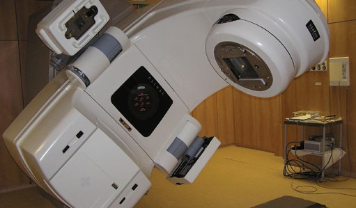
v6_i1_a11a

Introduction

Radiation Therapy (RT) is a common treatment modality for a multitude of cancer diagnoses. RT may be used for radical or palliative intent; to provide disease control or improve quality of life. [1] The radiation dose is fractionated, delivered daily over weeks, and can in some cases take as many as nine weeks to achieve prescribed radiation doses. [2-4] It is a highly technical treatment that uses imaging options such as: Computed Tomography (CT), Magnetic Resonance Imaging (MRI), and Positron Emission Tomography (PET) scanning to accurately delineate the tumour volume. Utilising the skills of radiation oncologists and radiation therapists, a precise dose of radiation is delivered to this targeted volume, destroying cancer cells whilst sparing normal tissue where possible.

Radiotherapy requires multidisciplinary input, for example from nursing, medical oncology, palliative care, dietetics and speech pathology. [5,6] For many patients it is the treatment of choice and yields excellent five year survival rates for localised solid tumours. [7] Due to the specific quality control measures, equipment and licensing requirements, substantial cost of treatment machines and the expertise required, the location of RT facilities tends to be centralised in larger urban centres, subsequently restricting access to those located in more regional and rural areas. [3,8,9]

Despite its therapeutic advantages, there are several factors that patients may consider prior to attending RT facilities, one of which is accessibility. For many patients the distance to a RT facility and the protracted course of treatment means that RT is not a feasible option. Challenges in accessing RT may lead to suboptimal treatment and subsequently poor outcomes for cancer patients. [1,3,7] Several studies have investigated the association between geographical distance to radiotherapy and radiotherapy utilisation, however they are limited by small sample sizes and differ in their conclusions. Accordingly, a systematic review was conducted to assess whether greater geographical distance to a RT facility was a barrier to RT treatment.

Methods

Search-strategy

A search strategy was devised according to the Preferred Reporting Items for Systematic Reviews and Meta-Analyses (PRISMA) Statement. [10] The SCOPUS (incorporating 100% of Medline titles) and Cumulative Index of Nursing and Allied Health Literature (CINAHL) databases were searched using the following search terms: (geograph* distance OR access) AND (radiation therapy OR radiotherapy), from January 1, 2000 to June 26, 2013 applied to abstracts.

Inclusion criteria

Studies were included if factors associated with access to or inequalities in receiving RT or cancer treatment were noted on all diagnoses of cancer. Studies were included if they were in the English language, pertinent to humans and linked to publically available full text articles.

Exclusion criteria

Studies were excluded if the primary objective did not include geographical distance or access barriers to RT facility or cancer treatment; if the study focused on treatments rather than barriers to treatment; or if the data was published prior to 2000.

Data extraction and quality assessment

Studies  were  independently  abstracted  for  quality  assessment  by the primary author with corroboration from co-authors. Quality assessment was based on the study design, sample size, control for confounders, and control of bias. [11,12] The studies were rated as high (H), moderate (M) or low (L) quality based on study design, execution, and reporting. High quality suggested a prospective study design with a large sample size, considerable control of confounders, and little bias, whereas low quality reflected a small sample size, limited control of confounders, and significant bias.

Results

The search of the SCOPUS database yielded 57 results, of which 22 met the eligibility criteria, with 11 that were relevant to geographical distance and variations in access to RT and available in full text. Repeating  the  search  in  CINAHL  provided  39  additional results  of which no articles were deemed relevant to the primary aims of this systematic review. One additional study was identified from grey literature searching and was included in the review, resulting in a total of 12 studies (Figure 1).

v6_i1_a11b

The quality of the included studies is shown in Table 1. As most of the studies were retrospective in study design where data examined was retrieved from cancer registries, they tended to be of moderate quality assessment. Overall, two studies were deemed to be high quality and ten studies were considered to be of moderate quality. None of the included studies were considered to be of poor quality.

v6_i1_a11d

The geographical location of each study is summarised in Table 2. Of the 12 studies, eight were conducted in the United States, one study was conducted in Canada, one study was conducted in the United Kingdom and two studies were conducted in Australia with a mixture of urban and rural settings. Several studies utilised geographic information system (GIS) software to map and measure the distance from the patient’s residence to the RT facility to give an indication of the accessibility of the RT clinics. Distances were calculated using straight line measurements rather than the actual route travelled by the patient via the software.

v6_i1_a11e

Of the 12 studies that met the inclusion criteria, nine identified a negative relationship whereby the greater the distance to the RT facility, the less likely the patient would be to undergo RT. [1,3,4,7-9,13-15] One prospective study with moderate control for confounders determined a positive relationship, whereby the greater the distance to the RT facility, the greater the likelihood of receiving RT. [16] There were two studies that did not address the distance to the RT clinic, but instead noted that lack of public transportation to RT facility was an access barrier, and that the presence of a radiation oncologist reduced mortality rates. [2,17] Although these two studies did not specifically address the primary objective, their results indicate that travel time to RT clinics is a major barrier to patients and that local resources such as radiation specialists can improve prognoses. Synthesis of the study data yielded a list of factors that were considered to influence access to RT (Box 1). The most influential factors contributing to radiotherapy access included: shorter distance to the RT facility, higher socio- economic status (SES), and increased education.

v6_i1_a11c

Discussion

The findings of this review suggest that geographical distance to RT facilities is a barrier to RT treatment. The majority of the studies included found that with increased distance to the RT facility, there was lower utilisation of RT as a treatment. One study conducted in Queensland, Australia reported conflicting findings, suggesting that with increasing distance to RT facilities there was higher utilisation of RT. This study focussed specifically on the prostate cancer population in Queensland, which is often an older population and therefore may have other factors that influence RT accessibility, such as retirement, income, and doctor preferences, whereas other studies often looked at cancer patient populations in younger cohorts. Older populations may not have to factor in time away from employment, and may have family they can reside with that live in regional centres. They may have previous exposure to hospitals and specialists, and therefore may have alternative factors that impact on preferences for location. [16] The findings of this study were potentially also limited by confounding bias as stated by the authors.

It is important to note that there was considerable variability in the geographical setting of the included studies. One study was conducted in a metropolitan city in the USA and the results may not be applicable to Australian settings. Interestingly, at least one study from each nation and the majority of research included in this review found that increased distance to RT facilities can act as a barrier to utilisation of RT, suggesting that this is a global phenomenon.

It would be useful to qualitatively investigate why patients select RT as their treatment option to ascertain insight into the barriers patients subjectively experience. With lower population density and lack of available RT facilities in rural areas such as Northern Queensland, there are great distances that must be traversed in order to receive life- saving treatment. Public transportation alone cannot be considered a barrier in instances where it is not available to patients, as is the case in remote areas. Therefore it is important to investigate area- specific geographical barriers, as rurality may pose other obstacles to overcome. It would also be interesting to explore whether variations exist in the acceptance of RT during the wet season when driving conditions could be challenging. This area of cancer care deserves much attention, especially in areas with vast land and few facilities. Identifying barriers to receiving RT is crucial to addressing the needs of the population.

Limitations of the studies synthesised in this review include the fact that many studies investigated distance to treatment rather than actual road travel times, which can vary significantly in many areas in Australia due to factors such as traffic, road works, the wet season and mountainous regions. There remains controversy in the optimal methodology used to assess accessibility to treatment. The GIS methods that were cited in this review were variable in their measurement of distance, often utilising straight line methods or mile radius buffer zones, which are not representative of the course travelled by the patient and do not give a clear indication of travel time. It is likely that increasing the accuracy of GIS distance measurements, by using round distance or alternatives as opposed to straight line measurements would exaggerate rather than minimise these differences.

This systematic review has a number of limitations. Firstly, two databases were utilised in the literature search and only open-access full text manuscripts were included, therefore restricting the amount of literature reviewed. Secondly, the methodological quality of the majority  of  included  studies  was  moderate.  The  studies  examined were either of retrospective or prospective study design. Most studies identified that the decision to proceed with RT is multifactorial, and many adjusted for a limited number of confounders. An ideal study would follow each patient with a diagnosis of cancer prospectively through a questionnaire or interview to ascertain which factors act as barriers or enablers to the decision for treatment. It would then revisit the patient post treatment to assess for any changes or additional challenges met. This would be a time-intensive process which would involve long follow up of patients, and may potentially be intrusive to  patients  during  an  emotional  and  difficult  period  in  their  life. Finally, the scope of the literature search was expanded to include all geographical locations rather than confining the search to rural areas in Australia alone due to the paucity of literature available. The results are therefore limited in their transferability to Australian settings.

Creating new technologies to deliver better dose profiles to tumour volumes is an integral part of radiation therapy, but however precise these treatments can be, their use is of limited value to populations who are not able to access RT. [18,19] Uniquely, radiotherapy will always need to be delivered in larger centres unlike other areas of oncology where initiatives such as tele-oncology are overcoming geographical access barriers. Therefore, further work in determining the role of innovative strategies to minimise the time patients spend away from home in rural areas and the burden associated with receiving treatment would be useful. [20,21]

Conclusion

Multiple factors are  considered  in the decision  making process to have radiotherapy versus alternative treatments and these remain individual and context specific. Access to the RT facility is one important factor considered in this review. [22] The preference for modalities is important to investigate as studies have indicated a discrepancy between evidence based optimal and actual utilisation rates of RT. [1,23,24] The multitude of factors and social context that influences the patients’ choice for and satisfaction with treatment makes this a complex and significant area of research. [22,25] This review indicates that most likely rurality and increased distance from RT centres are important considerations, thus there is also the requirement for additional research into areas that may improve access for the rural cancer patient population, including travel subsidies, accommodation, and location of treatment facilities. However, there is a need for further studies, ideally prospective, and geography specific, before firm conclusions can be drawn.

Acknowledgements

The author would like to thank Associate Professor Sarah Larkins for her guidance and support. This research was conducted as part of a James Cook University School of Medicine Research Scholarship awarded in 2013.

Conflict of interest

None declared.

Correspondence

D K Sharma: divya.sharma@my.jcu.edu.au

References

[1] Soo J, French J, McGahan CE, Duncan G, Lengoc S. A retrospective study on accessibility of palliative radiation therapy in the management of prostate cancer in British Columbia. Journal of Radiotherapy in Practice. 2011;10(3):159-72.

[2] Aneja S, Yu JB. The impact of county-level radiation oncologist density on prostate cancer mortality in the United States. Prostate Cancer and Prostatic Diseases. 2012;15(4):391-6. [3] Schroen AT, Brenin DR, Kelly MD, Knaus WA, Slingluff Jr CL. Impact of patient distance to radiation therapy on mastectomy use in early-stage breast cancer patients. Journal of Clinical Oncology. 2005;23(28):7074-80.

[4] Voti L, Richardson LC, Reis IM, Fleming LE, MacKinnon J, Coebergh JWW. Treatment of local breast carcinoma in Florida: The role of the distance to radiation therapy facilities. Cancer. 2006;106(1):201-7.

[5] Bomford CK KI. Walter and Miller’s textbook of radiotherapy: radiation physics, therapy and oncology. 6 ed. USA: Churchill Livingstone Elsevier Ltd; 2003.

[6] Khan F. The physics of radiation therapy. 3 ed. USA: Lippincott Williams &Wilkins; 2003. [7] Cetnar JP, Hampton JM, Williamson AA, Downs T, Wang D, Owen JB, et al. Place of residence and primary treatment of prostate cancer: Examining trends in rural and nonrural areas in wisconsin. Urology. 2013;81(3):540-6.

[8] Athas WF, Adams-Cameron M, Hunt WC, Amir-Fazli A, Key CR. Travel distance to radiation therapy and receipt of radiotherapy following breast-conserving surgery. Journal of the National Cancer Institute. 2000;92(3):269-71.

[9] Underhill C, Bartel R, Goldstein D, Snodgrass H, Begbie S, Yates P, et al. Mapping oncology  services  in  regional  and  rural  Australia.  Australian  Journal  of  Rural  Health. 2009;17(6):321-9.

[10]   Moher   D,   Liberati   A,   Tetzlaff   J,   Altman   DG.   Preferred   reporting  items   for systematic reviews and meta-analyses: the PRISMA statement. British Medical Journal. 2009;339:b2535.

[11] Khan, Khalid S, Kunz R, Kleijnen J,   Antes G. Five steps to conducting a systematic review. Journal of the Royal Society of Medicine. 2003;96(March 2003):118-21.

[12] Liamputtong P, editor. Research methods in health education Ch 18: Everything you wanted to know about systematic reviews. Sydney, NSW, Australia; 2010.

[13]  Baldwin  LM,  Patel  S,  Andrilla  CHA,  Rosenblatt  RA,  Doescher MP.  Receipt  of recommended  radiation  therapy  among  rural  and urban  cancer  patients.  Cancer. 2012;118(20):5100-9.

[14] Boscoe FP, Johnson CJ, Henry KA, Goldberg DW, Shahabi K, Elkin EB et al. Geographic proximity to treatment for early stage breast cancer and likelihood of mastectomy. Breast. 2011;20(4):324-8.

[15] Williams MV, Drinkwater KJ. Geographical Variation in Radiotherapy Services Across the UK in 2007 and the Effect of Deprivation. Clinical Oncology. 2009;21(6):431-40.

[16] Baade P, Youlden DR, Gardiner RA, Ferguson M, Aitken JF, Yaxley J et al. Factors associated wth treatment received by men diagnosed with prostate cancer in Queensland, Australia. BJU International. 2012;110:712-9.

[17] Peipins LA, Graham S, Young R, Lewis B, Flanagan B. Racial disparities in travel time to radiotherapy facilities in the Atlanta metropolitan area. Social Science and Medicine. 2013;89:32-8.

[18] Delaney G, Jacob S, Featherstone C, Barton M. The role of radiotherapy in cancer treatment:  Estimating  optimal  utilization  from  a review  of  evidence-based  clinical guidelines. Cancer. 2005;104(6):1129-37.

[19] Mackillop WJ, Groome PA, Zhang-Solomons J, Zhou Y, Feldman-Stewart D, Paszat L et Does a centralized radiotherapy system provide adequate access to care? Journal of Clinical Oncology. 1997;15(3):1261-71.

[20] Sabesan S, Larkins S, Evans R, Varma S, Andrews A, Beuttner P et al. Telemedicine for rural cancer care in North Queensland: Bringing cancer care home. Australian Journal of Rural Health. 2012;20(5):259-64.

[21] Sabesan S, Simcox K, Marr I. Medical oncology clinics through videoconferencing: An acceptable telehealth model for rural patients and health workers. Internal Medicine Journal. 2012;42(7):780-5.

[22] Gauthier DM, Swigart VA. The contextual nature of decision making near the end of life: Hospice patients’ perspectives. American Journal of Hospice and Palliative Medicine. 2003;20(2):121-8.

[23] Delaney G, Barton M, Jacob S. Estimation of an Optimal Radiotherapy Utilization Rate for Breast Carcinoma: A Review of the Evidence. Cancer. 2003;98(9):1977-86.

[24] Delaney G, Jacob S, Barton M. Estimating the optimal external-beam radiotherapy utilization rate for genitourinary malignancies. Cancer. 2005;103(3):462-73.

[25] Resnick MJ, Guzzo TJ, Cowan JE, Knight SJ, Carroll PR, Penson DF. Factors associated with satisfaction with prostate cancer care: Results from Cancer of the Prostate Strategic Urologic Research Endeavor (CaPSURE). BJU International. 2013;111(2):213-20.

Categories
Review Articles

Chronic obstructive pulmonary disease: extrapulmonary manifestations, pulmonary rehabilitation programs and the role of nutritional biomarkers on patient outcomes

Until recently, chronic obstructive pulmonary disease (COPD) received little research attention, as it was perceived as a self- inflicted condition that was difficult to treat. As COPD now affects one in seven Australians over 40 and is a leading cause of disease burden and death, research into this condition has intensified. Traditionally, research focused on the pulmonary effects and yet it is starting to emerge that the condition encompasses a range of   extrapulmonary   manifestations,  such  as  weight  loss  and skeletal muscle dysfunction, which significantly affect the health and functioning capacity of COPD patients. There are many unanswered questions about the disease process and the role of the extrapulmonary manifestations. The aim of the current review is to explore two critical extrapulmonary manifestations of COPD: weight loss and skeletal muscle dysfunction, to investigate how pulmonary rehabilitation aims to improve these pathological processes and, lastly, to investigate the role of nutritional biomarkers and how these may predict outcomes in the pulmonary rehabilitation programs. Ultimately, it is anticipated that research into nutritional biomarkers may lead to the development of a screening tool that can be used to identify COPD patients who may benefit from nutritional supplementation prior to the commencement of a pulmonary rehabilitation program. It is hoped that identifying and managing those patients that require nutritional support will lead to greater improvements in rehabilitation and overall quality of life.

v6_i1_a12a

Introduction

Chronic obstructive pulmonary disease (COPD) is a chronic obstructive lung disease, primarily caused by smoking. It affects one in seven Australians over 40 [1] and is a leading cause of disease burden and death. [2-4] The morbidity and mortality rates associated with COPD are continuing to increase, and it has been predicted that COPD may be the third most common cause of death worldwide by 2020. [5,6] In the past, COPD has been perceived as a self-inflicted condition, which was difficult to treat. Although the perceptions surrounding COPD have changed and research has shed more light on the disease processes, there are still many gaps in the scientific knowledge regarding this condition.

One such gap, which has only recently been explored, is the role of extrapulmonary manifestations associated with COPD. Previously, clinicians and researchers solely focused on the structural and functional changes occurring in the pulmonary system of patients with COPD. However, in recent years, it has become increasingly evident that the disease encompasses a range of other manifestations outside the lungs, including weight loss and skeletal muscle dysfunction. [7] Weight loss, which is a very common manifestation in patients with COPD, causes a reduction in respiratory and skeletal muscle function, which is associated with reduced quality of life and increased mortality rates. [8] Weight loss, skeletal muscle dysfunction and some of the other manifestations associated with COPD can be managed through pulmonary rehabilitation programs, although these are costly, time consuming and individual success is highly variable. [9] Furthermore, there is currently no dedicated funding for these programs in Australia. [10]

In   order  to  improve   the   effectiveness  and  outcomes  achieved through pulmonary rehabilitation programs, researchers have begun investigating the role of nutrition in COPD patients. Ultimately, it is anticipated that the identification of important nutritional biomarkers that  predict  improved  outcomes  in  pulmonary  rehabilitation,  may lead to the development of nutrient supplementation strategies to improve success in rehabilitation programs and optimise the quality of life of patients with COPD. The aim of the current review is to explore two critical extrapulmonary manifestations of COPD: weight loss and skeletal muscle dysfunction, to investigate how pulmonary rehabilitation aims to improve these effects and lastly, to investigate the role of nutritional biomarkers in COPD and how these may predict outcomes in the pulmonary rehabilitation programs.

Extrapulmonary manifestations

Recent research has found that patients with COPD suffer a range of extrapulmonary manifestations that were not previously related to the condition. Two of the major extrapulmonary manifestations seen in COPD patients that have a critical impact on quality of life and prognosis are weight loss and skeletal muscle dysfunction.

Weight loss

Although traditionally research focused on the pulmonary effects of the disease, it was evident as early as the 1960’s that a low body weight and weight loss were associated with an increased mortality rate in COPD patients. [11] However, at the time weight loss was believed to be only associated with the terminal phase of the disease, and hence it was considered inevitable and irreversible. [11] Currently, excessive weight loss, especially loss of fat-free mass, is very common in COPD patients and is associated with poor functional capacity, reduced quality of life and increased mortality. [8] Although the exact cause of excessive weight loss in COPD remains unclear, the proposed mechanisms include low testosterone levels, increased pro-inflammatory cytokines and increased catecholamine synthesis. [12-14]

Skeletal muscle dysfunction

A  common  extrapulmonary  manifestation  that  significantly  affects the quality of life of a COPD patient is skeletal muscle dysfunction. Skeletal muscle dysfunction is characterised by increased muscle fatigability, and a reduction in muscle endurance and strength. [15] In many studies, body mass index (BMI) (which is calculated as weight/ height squared in kg/m2) is used as a basic indicator of weight loss or  possible  muscle  alterations,  although  these  measures  can  be further investigated by evaluating skeletal muscle strength and body composition. [16] COPD patients with skeletal muscle dysfunction have increased mortality rates and are likely to place a significant burden on healthcare resources. [17,18] The precise mechanisms causing skeletal muscle dysfunction in COPD patients are still unclear, although several factors that may contribute include sedentary lifestyle, nutritional abnormalities, tissue hypoxia, systemic inflammation, skeletal muscle apoptosis, oxidative stress, tobacco use and medications. [7] In patients with COPD, skeletal muscle dysfunction is characterised by two different phenomena: (1) net loss of muscle mass; and (2) dysfunction or malfunction of the remaining muscle. [7] One of the key features involved in the loss of muscle mass is increased protein catabolism. The major pathway involved in the degradation of proteins, which relates to muscle wasting, is the ATP-ubiquitin dependent proteolytic system (Figure 1). [19] This system can be activated by several factors such as cytokines, glucocorticoids, acidosis, inactivity or low insulin levels. [20-22] Following the activation of this pathway, proteins are marked for degradation by ubiquitination, and then they are recognised and processed  in  the  proteasome.  [4]  Pro-inflammatory cytokines  may also play a role in muscle deterioration by producing reactive oxygen species, which modify skeletal muscle proteins allowing them to be easily degraded by the proteasome. [4]

v6_i1_a12b

The exact trigger for the development of the extrapulmonary manifestations is unknown, although it is thought that the process is mediated by systemic inflammation. [24] As COPD is a condition primarily caused by smoking, it could be questioned whether smoking is the major cause for the development of systemic inflammation and in turn the extrapulmonary manifestations. As multiple studies have found  that  persistent  inflammation  is  still  present  in  ex-smokers, [25] it is possible that tobacco smoke may initiate the inflammatory process, however it does not explain the sustained inflammatory state evident in COPD patients. [24] Instead, it is possible that the systemic inflammation arises from pathological changes occurring within the lungs of COPD patients. [24] This is supported by other studies that have found that inflammation is still present in COPD patients who have ceased smoking. [26,27] In light of these observations, some researchers have speculated that part of the COPD pathogenesis process involves an autoimmune component. [28]

The discovery that COPD encompasses both pulmonary and systemic manifestations has created new possibilities for rehabilitation and treatment targets. As weight loss and skeletal muscle dysfunction are reversible and treatable, pulmonary rehabilitation programs have been reorientated in order to focus on improving skeletal muscle function and the overall quality of life of COPD patients.

Pulmonary rehabilitation programs to improve patient outcomes

In response to the growing prevalence and burden of COPD, the Australian  Lung  Foundation  and  Thoracic  Society  of  Australia  and New Zealand developed clinical guidelines (COPDX) for the diagnosis and management of COPD. [29] One of the main aims of COPDX is to optimise patient function using pulmonary rehabilitation programs. [29] Pulmonary rehabilitation programs are composed of exercise training, behavioural and psychosocial interventions and nutritional therapy. [30]

Exercise training

Prior to the work by Barach et al. [31] in the 1950’s, who suggested that  exercise  may  be  beneficial,  the  only  recommendations  for the management of respiratory conditions were rest and avoiding breathlessness. [32] Since this suggestion, there have been many experimental findings, randomised controlled trials and observations supporting the benefits of exercise training for patients with COPD. Based on the most recent evidence, patients with COPD undergoing pulmonary rehabilitation should participate in exercise training at least 2-5 days per week, for at least 20-30 minutes per session, over an 8-12 week period. [33, 34] Exercise programs involve endurance and strength training, mainly focusing on the lower limbs. [30] In patients with COPD, exercise training has been shown to significantly improve exercise tolerance and endurance time and it is also able to improve or reverse the physiological, metabolic and structural skeletal muscle abnormalities seen in COPD patients [35], suggesting that pulmonary rehabilitation is an anabolic stimulus. [36] Although, it is unknown how pulmonary rehabilitation improves skeletal muscle dysfunction and the role of specific nutrients during this process.

Nutritional approaches to improving muscle function and body composition in COPD

The role of nutritional therapy in the management of COPD has changed dramatically during the past twenty years. Although it was widely known that a large proportion of COPD patients experienced significant weight loss, it was viewed as irreversible and nutritional support was not considered. [37] This concept has been challenged by recent studies, which have revealed that nutritional depletion affects functional performance and exercise intolerance. [37] After this discovery, many trials have investigated the benefits and effects of nutritional support in patients with COPD, although the initial results from these studies were disappointing. A meta-analysis of the available literature conducted by Ferreira et al. [38] concluded that nutritional support, defined as any caloric supplement administered for more than two weeks, had no significant effect on 6-min walk distance, anthropometric measures, respiratory muscle strength, weight  gain  or  FEV1.  These  results  led  to  the  suggestion  that  in order to improve muscle mass and physiologic function, nutritional support must be combined with an anabolic stimulus such as exercise training.  [39] In  a  large  clinical  trial  combining  nutritional therapy (daily high caloric supplement (420 kcal)) with an 8-week pulmonary rehabilitation program, patients showed an increase in body weight and a significant improvement in fat-free mass and respiratory muscle strength. [40] These results were further supported by a more recent study by Creutzberg et al. [39], who observed that the combination of nutritional therapy with pulmonary rehabilitation was effective in improving physiological measures such as body composition, muscle function,  exercise  capacity,  serum  protein,  as  well  as  the  health status and well-being of patients with COPD. [39] Since the use of nutritional support has been shown to be beneficial, research needs to shift towards investigating the effectiveness of different types of nutrients and how these may be used in combination with pulmonary rehabilitation programs in order to maximise patient outcomes.

Protein supplementation

There are two main pathways involved in the synthesis and breakdown of proteins (Figure 2) and recent research has shown that the loss of fat-free mass in patients with COPD is caused by an imbalance between these two pathways. A reduction in fat-free mass causes the loss of protein-rich tissues, particularly skeletal muscle [36] and the imbalance in protein metabolism leads to increased whole-body protein turnover. [41] This has led researchers to investigate the use of protein supplementation in improving fat-free mass in COPD patients.

v6_i1_a12c


In  skeletal  muscle,  the  regulation  of  protein  initiation, translation and synthesis relies on the activation of two signalling proteins called the mammalian target of rapamycin (mTOR) and AMP-activated protein kinase (AMPK). [45] In a study by Fujita et al. [45] Protein supplementation, which provides the essential amino acids necessary for protein synthesis, alters the phosphorylation status of AMPK and mTOR signalling proteins, and increases the synthesis of proteins in healthy adults. As mentioned previously, for patients with COPD, nutritional supplementation must be combined with an additional anabolic stimulus in order to be effective and therefore recent studies have investigated the effectiveness of protein supplementation combined with exercise training. Laviolette et al. [46] conducted a study  observing  the  effects  of  supplementation  with  pressurised whey or casein combined with an 8-week exercise-training program in patients with COPD. From the study, they concluded that combining whey supplementation with exercise training caused an improvement in exercise capacity (cycle endurance time), fatigue and emotional control. [46] Although these results are promising, there is emerging evidence that other nutrients such as vitamin D and B, calcium, zinc, magnesium, fatty acids and antioxidants can influence lung function and stimulate the anabolic pathways involved in protein synthesis and muscle function.

Vitamins

Vitamin D

One  of  the  first  studies  to  investigate  the  association  between vitamin D and muscle metabolism was by Birge and Haddad [47], who observed that 25-hydroxy vitamin D altered muscle metabolism causing  an accelerated  incorporation  of  amino  acids  into  muscle protein. They postulated that vitamin D acts directly on muscle [47] and this theory was confirmed in 1985, when a vitamin D receptor (VDR)  was  discovered  in cultured  rat  myoblast  cells.  [48]  Further research has discovered VDR in a range of tissues, and recently, it was isolated from human skeletal muscle. [49] Recent studies have shown that COPD patients, particularly those with severe COPD, have low levels of vitamin D. [50] In order to investigate the link between vitamin D depletion and muscle function, Bjerk et al. [51] performed a randomised pilot trial in which patients with COPD were supplemented with vitamin D for 6 weeks. Although the supplementation group had a significant increase in mean vitamin D levels compared to the control, there were no significant improvements in physical performance or respiratory symptoms. [51]

Vitamin B

A  recent  study  examining  hyperhomocysteinaemia  discovered that COPD  patients  had  reduced  plasma  concentrations  of  vitamin  B, particularly folate  [52],  which  is  an  essential  co-factor  involved  in protein synthesis. [53] Another study found an association between folate intake and lung function. In this study, participants with COPD had lower folate levels than controls, and their folate intake was below the recommended dose. [54] Based on epidemiological data, it has been suggested that an increased folate intake could lead to reductions in the prevalence of COPD and breathlessness. [54]

Minerals

Calcium, Zinc and Magnesium

Currently,  there  is  very  limited  human  research  on  the  role of minerals in muscle function and most of the available data is based on experimental animal models. Early evidence of the role of calcium in protein synthesis emerged from experimental rat studies, which revealed that  maintenance  of  optimal  rates  of  protein  synthesis was dependent on the availability of calcium. Furthermore, calcium depletion  led  to  the inhibition  of  protein  synthesis,  which was characterised  by  a  reduced rate  of  peptide  chain  initiation.  [55] Recently, it has been identified that an increase in the concentration of intracellular calcium triggers the activation of the mTOR pathway, leading to skeletal muscle hypertrophy. [56]

Zinc and magnesium are essential minerals required for growth in humans. [57,58] Both minerals play an important role in the synthesis of proteins, with deficiencies leading to the down-regulation of protein synthesis. [59] An experimental study revealed that protein synthesis in muscle was inhibited in zinc deficient rats [60] and a more recent study confirmed these results, by finding a reduction in protein synthesis and enhanced protein degradation in muscle tissue from zinc-deficient rats. [59] Unfortunately, the literature on the calcium, zinc and magnesium levels of COPD patients is limited and further research is warranted.

Fatty acids

In order to prevent or reverse muscle loss, interventions must target the abnormal anabolic pathways. The dysfunctional anabolic pathway is partly caused by defects in the anabolic signalling cascade in muscle, such as decreased activation of the mTOR signalling pathway. [61,62] In various animal studies, fish-oil-derived omega-3 fatty acids have been used to target the protein synthesis pathways. In one study, growing steers received feed enriched with menhaden oil, which increased the  activation  of  anabolic  signalling  proteins  in  muscle.  [63]  In  a more recent human study, omega-3 supplementation in older adults increased the rate of muscle protein synthesis, which suggests that omega-3 fatty acids reduce anabolic resistance. [64] It is not entirely clear how omega 3-fatty acids act on the muscle protein synthesis pathway, although it may be partially mediated via increased activation of the mTOR signalling pathway. [64] Based on the limited evidence, supplementation with fatty acids may be a beneficial treatment, although as of yet there are no published studies exploring this. [65]

Antioxidants

Antioxidants  are  considered  to  be  protective  factors  in  the  lungs as they can scavenge endogenous and exogenous reactive oxygen species. [66] There is increasing evidence that oxidative damage and the failure of antioxidants to protect lung tissue are partly responsible for the development of COPD. [67,68] Studies examining the effect of antioxidant supplementation on oxidative damage and pulmonary function are incredibly conflicting. Habib et al. [69] observed that vitamin E supplementation had no effect on pulmonary function. Another study found that when used in addition to standard therapy, an antioxidant supplement (containing vitamin A, C, E, zinc, copper, selenium and manganese) had a positive effect on the oxidant- antioxidant balance in COPD patients, however it had no effect on pulmonary function tests. [70]

Although research has focused on oxidative stress caused by the production of free radicals in the lungs in patients with COPD, there is emerging evidence that exercising skeletal muscle may also produce free radicals and contribute to oxidative stress. [71] Free radicals produced during exercise depress muscle force production [72] and increase the discharge frequency of thin-fibre muscle afferents [73] and thus, targeting oxidative stress may improve exercise tolerance and reduce the development of fatigue.

Based on all of the available evidence, it is apparent that patients with COPD suffer from nutritional abnormalities, which may contribute to muscle dysfunction and weight loss. An altered nutritional status may affect a patient’s ability to synthesise protein and lead to less effective outcomes in pulmonary rehabilitation programs. Although some studies have shown that protein supplementation combined with exercise training can be beneficial, evidence is limited and somewhat conflicting. Apart from protein and amino acids, other nutrients have

a direct effect on the synthesis of protein and the function of skeletal muscles, although research into these effects is lacking. Nutritional support is an important part of COPD management, however there is no definitive evidence about what types of nutrients should be used and how they may influence the outcome of rehabilitation programs. Ongoing and future research is expected to provide further insight in this area, and hopefully improve the quality of life and survival of COPD patients.

Acknowledgements

This literature review was first written as part of a Bachelor of Biomedical Science third year research project at the University of Newcastle. I would like to give special thanks to my project supervisor Associate Professor Lisa Wood, who provided valuable guidance and support. I would also like to thank my research partner Jennifer Latham for her participation in the research project.

Conflict of interest

None declared.

Correspondence

M McDonald: melissa.mcdonald1@my.jcu.edu.au

References

[1] Toelle B, Xuan W, Bird T, Abramson M, Burton D, Hunter M et al. COPD in the Australian burden of lung disease (BOLD) study. Respirology. 2011;16(Suppl 1):12.

[2] Australia’s health 2012. Australia’s health series no.13. Canberra: Australian Institute of Health and Welfare. 2012;626. AUS 156.

[3] Banerjee A, Guttridge DC. Mechanisms for maintaining muscle. Curr Opin Support Palliat Care. 2012 Dec;6(4):451-6.

[4]  Debigare  R,  Cote  CH,  Maltais  F.  Peripheral  muscle  wasting  in chronic  obstructive pulmonary  disease:  clinical  relevance  and mechanisms.  Am  J  Respir  Crit  Care  Med. 2001;164:1712-7.

[5] Calverley PM, Walker P. Chronic obstructive pulmonary disease. Lancet. 2003 Sep 27;362(9389):1053-61.

[6] Lecker SH, Goldberg AL, Mitch WE. Protein degradation by the ubiquitin-proteasome pathway in normal and disease states. J Am Soc Nephrol. 2006 Jul;17(7):1807-19.

[7] Agusti AG, Noguera A, Sauleda J, Sala E, Pons J, Busquets X. Systemic effects of chronic obstructive pulmonary disease. Eur Respir J. 2003 Feb;21(2):347-60.

[8]  Vijayan  VK.  Chronic  obstructive  pulmonary  disease.  Indian  J  Med  Res.  2013 Feb;137(2):251-69.

[9] Scott AS, Baltzan MA, Fox J, Wolkove N. Success in pulmonary rehabilitation in patients with chronic obstructive pulmonary disease. Can Respir J. 2010 Sep-Oct;17(5):219-23.

[10] Alison J et al. The Pulmonary Rehabilitation Toolkit. The Australian Lung Foundation; 2009 [cited 2015 07/01/15]; Available from: http://www.pulmonaryrehab.com.au/index. asp?page=3.

[11] Decramer M, Rennard S, Troosters T, Mapel DW, Giardino N, Mannino D, et al. COPD as a lung disease with systemic consequences-clinical impact, mechanisms, and potential for early intervention. Copd. 2008 Aug;5(4):235-56.

[12] Laghi F. Low testosterone in chronic obstructive pulmonary disease: does it really matter? Am J Respir Crit Care Med. 2005 Nov 1;172(9):1069-70.

[13] Schols AM, Slangen J, Volovics L, Wouters EF. Weight loss is a reversible factor in the prognosis of chronic obstructive pulmonary disease. Am J Respir Crit Care Med. 1998 Jun;157(6 Pt 1):1791-7.

[14] Van Vliet M, Spruit MA, Verleden G, Kasran A, Van Herck E, Pitta F, et al. Hypogonadism, quadriceps weakness, and exercise intolerance in chronic obstructive pulmonary disease. Am J Respir Crit Care Med. 2005 Nov 1;172(9):1105-11.

[15] Gan WQ, Man SFP. Systemic effects and mortality in chronic obstructive pulmonary disease. BCMJ. 2008 50(3):148-51.

[16] Skeletal muscle dysfunction in chronic obstructive pulmonary disease. A statement of the American Thoracic Society and European Respiratory Society. Am J Respir Crit Care Med. 1999 Apr;159(4 Pt 2):S1-40.

[17] Decramer M, Gosselink R, Troosters T, Verschueren M, Evers G. Muscle weakness is related to utilization of health care resources in COPD patients. Eur Respir J. 1997 Feb;10(2):417-23.

[18] Marquis K, Debigare R, Lacasse Y, LeBlanc P, Jobin J, Carrier G, et al. Midthigh muscle cross-sectional area is a better predictor of mortality than body mass index in patients with chronic obstructive pulmonary disease. Am J Respir Crit Care Med. 2002 Sep 15;166(6):809-13.

[19] Mitch WE, Goldberg AL. Mechanisms of muscle wasting. N Engl J Med. 1996;335:1897-905.

[20] Du J, Mitch WE, Wang X, Price SR. Glucocorticoids induce proteasome C3 subunit

expression in L6 muscle cells by opposing the suppression of its transcription by NF-kappa J Biol Chem. 2000 Jun 30;275(26):19661-6.

[21] Llovera M, Garcia-Martinez C, Lopez-Soriano J, Carbo N, Agell N, Lopez-Soriano FJ, et Role of TNF receptor 1 in protein turnover during cancer cachexia using gene knockout mice. Mol Cell Endocrinol. 1998 Jul 25;142(1-2):183-9.

[22] Medina R, Wing SS, Goldberg AL. Increase in levels of polyubiquitin and proteasome mRNA in skeletal muscle during starvation and denervation atrophy. Biochem J. 1995 May 1;307(Pt 3):631-7.

[23] Mitch WE, Goldberg AL. Mechanisms of muscle wasting. N Engl J Med. 1996;335:1897-905Figure 3, The Ubiquitin-Proteasome Pathway of Proteolysis; p. 901.

[24] Bailey KL, Goraya J, Rennard S. Chronic Obstructive Pulmonary Disease: Co-Morbidities and Systemic Consequences. New York: Springer; 2012. Chapter 2, The role of systemic inflammation in COPD; p.15-30.

[25] Vernooy JH, Kucukaycan M, Jacobs JA, Chavannes NH, Buurman WA, Dentener MA, et al. Local and systemic inflammation in patients with chronic obstructive pulmonary disease: soluble tumor necrosis factor receptors are increased in sputum. Am J Respir Crit Care Med. 2002 Nov 1;166(9):1218-24.

[26] Hogg JC, Chu F, Utokaparch S, Woods R, Elliott WM, Buzatu L, et al. The nature of small-airway obstruction in chronic obstructive pulmonary disease. N Engl J Med. 2004 Jun 24;350(26):2645-53.

[27] Retamales I, Elliott WM, Meshi B, Coxson HO, Pare PD, Sciurba FC, et al. Amplification of inflammation in emphysema and its association with latent adenoviral infection. Am J Respir Crit Care Med. 2001 Aug 1;164(3):469-73.

[28]  Agusti  A,  MacNee  W,  Donaldson  K,  Cosio  M.  Hypothesis:  does  COPD  have  an autoimmune component? Thorax. 2003 Oct;58(10):832-4.

[29] Abramson MJ, Crockett AJ, Frith PA, McDonald CF. COPDX: an update of guidelines for the management of chronic obstructive pulmonary disease with a review of recent evidence. Med J Aust. 2006 Apr 3;184(7):342-5.

[30] Talag AA, Road J. Non-pharmacological management of chronic obstructive pulmonary disease. BMJ. 2008 50(2):90-6.

[31] Barach AL, Bickerman HA, Beck G. Advances in the treatment of non-tuberculous pulmonary disease. Bulletin of the New York Academy of Medicine. 1952 Jun;28(6):353-84.

[32] A manual for pulmonary rehabilitation in Australia: evidence base and standards. The Australian Lung Foundation, The Thoracic Socitey of Australia and New Zealand and Australian Physiotherapy Association. 2008:207.

[33] Pulmonary rehabilitation. Thorax. 2001 Nov;56(11):827-34.

[34] Pulmonary rehabilitation-1999. American Thoracic Society. Am J Respir Crit Care Med. 1999 May;159(5 Pt 1):1666-82.

[35] Rochester CL. Exercise training in chronic obstructive pulmonary disease. J Rehabil Res Dev. 2003 Sep-Oct;40(5 Suppl 2):59-80.

[36] Bolton CE, Broekhuizen R, Ionescu AA, Nixon LS, Wouters EF, Shale DJ, et al. Cellular protein breakdown and systemic inflammation are unaffected by pulmonary rehabilitation in COPD. Thorax. 2007 Feb;62(2):109-14.

[37] Schols AM. Nutrition and respiratory disease. Clin Nutr. 2001;20 (S1):173-9.

[38] Ferreira IM, Brooks D, Lacasse Y, Goldstein RS. Nutritional support for individuals with COPD: a meta-analysis. Chest. 2000 Mar;117(3):672-8.

[39] Creutzberg EC, Wouters EF, Mostert R, Weling-Scheepers CA, Schols AM. Efficacy of  nutritional supplementation  therapy  in  depleted  patients  with  chronic  obstructive pulmonary disease. Nutrition. 2003 Feb;19(2):120-7.

[40] Schols AM, Soeters PB, Mostert R, Pluymers RJ, Wouters EF. Physiologic effects of nutritional support and anabolic steroids in patients with chronic obstructive pulmonary disease. A placebo-controlled randomized trial. Am J Respir Crit Care Med. 1995 Oct;152(4 Pt 1):1268-74.

[41] Engelen MPKJ, Deutz NEP, Wouters EFM, Schols AMWJ. Enhanced levels of whole-body protein turnover in patients with chronic obstructive pulmonary disease. Am J Respir Crit Care Med. 2000;162:1488-92.

[42] Banerjee A, Guttridge DC. Mechanisms for maintaining muscle. Curr Opin Support Palliat Care. 2012 Dec;6(4):451-6. Figure 1, Illustration portrays the regulation of muscle protein  breakdown  by  lysosomal  and  proteosomal  pathways  during  hypertrophy and atrophy; p.3.

[43] Debigare R, Cote CH, Maltais F. Peripheral muscle wasting in chronic obstructive pulmonary  disease:  clinical  relevance  and  mechanisms.  Am  J  Respir  Crit  Care  Med. 2001;164:1712-7.  Figure  1,  Schematic  representation  of  the  possible  mechanisms  of cachexia in COPD; p. 5.

[44] Lecker SH, Goldberg AL, Mitch WE. Protein degradation by the ubiquitin-proteasome pathway in normal and disease states. J Am Soc Nephrol. 2006 Jul;17(7):1807-19. Figure 5, The balance between protein synthesis and degradation determines whether muscles hypertrophy or atrophy; p. 13.

[45] Fujita S, Dreyer HC, Drummond MJ, Glynn EL, Cadenas JG, Yoshizawa F, et al. Nutrient signalling in the regulation of human muscle protein synthesis. J Physiol. 2007 Jul 15;582(Pt 2):813-23.

[46] Laviolette L, Lands LC, Dauletbaev N, Saey D, Milot J, Provencher S, et al. Combined effect of dietary supplementation with pressurized whey and exercise training in chronic obstructive pulmonary disease: a randomized, controlled, double-blind pilot study. J Med Food. 2010 Jun;13(3):589-98.

[47] Birge SJ, Haddad JG. 25-hydroxycholecalciferol stimulation of muscle metabolism. J Clin Invest. 1975 Nov;56(5):1100-7.

[48] Hamilton B. Vitamin D and human skeletal muscle. Scand J Med Sci Sports. 2010 Apr;20(2):182-90.

[49] Bischoff HA, Borchers M, Gudat F, Duermueller U, Theiler R, Stahelin HB, et al. In situ  detection of  1,25-dihydroxyvitamin D3  receptor in human skeletal muscle tissue. Histochem J. 2001 Jan;33(1):19-24.

[50] Janssens W, Bouillon R, Claes B, Carremans C, Lehouck A, Buysschaert I, et al. Vitamin D deficiency is highly prevalent in COPD and correlates with variants in the vitamin D-binding gene. Thorax. 2010 Mar;65(3):215-20.

[51] Bjerk SM, Edgington BD, Rector TS, Kunisaki KM. Supplemental vitamin D and physical performance in COPD: a pilot randomized trial. Int J Chron Obstruct Pulmon Dis. 2013;8:97-104.

[52] Fimognari FL, Loffredo L, Di Simone S, Sampietro F, Pastorelli R, Monaldo M, et al. Hyperhomocysteinaemia  and  poor  vitamin  B  status  in  chronic  obstructive pulmonary disease. Nutr Metab Cardiovasc Dis. 2009 Nov;19(9):654-9.

[53] Kamen B. Folate and antifolate pharmacology. Semin Oncol. 1997 Oct;24(5 Suppl 18):S18-30-s18-39.

[54] Hirayama F, Lee AH, Terasawa K, Kagawa Y. Folate intake associated with lung function, breathlessness and the prevalence of chronic obstructive pulmonary disease. Asia Pac J Clin Nutr. 2010;19(1):103-9.

[55] Kimball SR, Jefferson LS. Regulation of protein synthesis by modulation of intracellular calcium in rat liver. Am J Physiol. 1992 Nov;263(5 Pt 1):E958-64.

[56] Ito N, Ruegg UT, Kudo A, Miyagoe-Suzuki Y, Takeda S. Activation of calcium signaling through Trpv1 by nNOS and peroxynitrite as a key trigger of skeletal muscle hypertrophy. Nat Med. 2013 Jan;19(1):101-6.

[57] Caddell RG, Goddard DR. Studies in protein-calorie malnutrition: chemical evidence for magnesium deficiency. N Engl J Med. 1967;276:533-5.

[58]  Golden  MHN.  The  role  of  individual  nutrient  deficiencies  in  growth  retardation of children as exemplified by zinc and protein. In: Linear Growth Retardation in Less Developed Countries. New York: Raven Press; 1988; p.143-164.

[59] Dorup I, Clausen T. Effects of magnesium and zinc deficiencies on growth and protein synthesis in skeletal muscle and the heart. Br J Nutr. 1991 Nov;66(3):493-504.

[60] Giugliano R, Millward DJ. The effects of severe zinc deficiency on protein turnover in muscle and thymus. Br J Nutr. 1987 Jan;57(1):139-55.

[61] Cuthbertson D, Smith K, Babraj J, Leese G, Waddell T, Atherton P, et al. Anabolic signaling deficits underlie amino acid resistance of wasting, aging muscle. Faseb j. 2005 Mar;19(3):422-4.

[62] Guillet C, Prod’homme M, Balage M, Gachon P, Giraudet C, Morin L, et al. Impaired anabolic response of muscle protein synthesis is associated with S6K1 dysregulation in elderly humans. Faseb j. 2004 Oct;18(13):1586-7.

[63] Gingras AA, White PJ, Chouinard PY, Julien P, Davis TA, Dombrowski L, et al. Long-chain omega-3 fatty acids regulate bovine whole-body protein metabolism by promoting muscle insulin signalling to the Akt-mTOR-S6K1 pathway and insulin sensitivity. J Physiol. 2007 Feb 15;579(Pt 1):269-84.

[64] Smith GI, Atherton P, Reeds DN, Mohammed BS, Rankin D, Rennie MJ, et al. Dietary omega-3 fatty acid supplementation increases the rate of muscle protein synthesis in older adults: a randomized controlled trial. Am J Clin Nutr. 2011 Feb;93(2):402-12.

[65]  Berry  JK,  Baum  C.  Reversal  of  chronic  obstructive pulmonary  disease-associated weight loss : are there pharmacological treatment options? Drugs. 2004;64(10):1041-52.

[66] Guenegou A, Leynaert B, Pin I, Le Moel G, Zureik M, Neukirch F. Serum carotenoids, vitamins A and E, and 8 year lung function decline in a general population. Thorax. 2006 Apr;61(4):320-6.

[67] Hatch GE. Asthma, inhaled oxidants, and dietary antioxidants. Am J Clin Nutr. 1995 Mar;61(3 Suppl):625s-30s.

[68] Sanguinetti CM. Oxidant/antioxidant imbalance: role in the pathogenesis of COPD. Respiration. 1992;59 Suppl 1:20-3.

[69] Habib MP, Tank LJ, Lane LC, Garewal HS. Effect of vitamin E on exhaled ethane in cigarette smokers. Chest. 1999 Mar;115(3):684-90.

[70] Demir T, Demirel Ikitimur H, Yildrum N, Özmen O, Kanmaz B, Uzun H, et al. Effects of Antioxidant Therapy on Lung Clearance, Pulmonary Function Tests and Oxidant Stress in Patients With Chronic Obstructive Pulmonary Disease. Turk Respir J. 2004;5(1):22-6.

[71] Rossman MJ, Groot HJ, Reese V, Zhao J, Amann M, Richardson RS. Oxidative stress and COPD: the effect of oral antioxidants on skeletal muscle fatigue. Med Sci Sports Exerc. 2013 Jul;45(7):1235-43.

[72] Powers SK, Jackson MJ. Exercise-induced oxidative stress: cellular mechanisms and impact on muscle force production. Physiol Rev. 2008 Oct;88(4):1243-76.

[73] Delliaux S, Brerro-Saby C, Steinberg JG, Jammes Y. Reactive oxygen species activate the group IV muscle afferents in resting and exercising muscle in rats. Pflugers Arch. 2009 Nov;459(1):143-50.

Categories
Review Articles

Stroke prevention in non-valvular atrial fibrillation: advances in medical therapy

Introduction: The aim of this article is to review the literature and evaluate the evidence of the different medical treatments for stroke prevention in non-valvular atrial fibrillation. Methods: A literature search using MEDLINE plus OvidSP, PubMed, CINAHL and the New England Journal of Medicine databases was performed with the search terms stroke prevention, atrial fibrillation, anticoagulation, novel anticoagulants, direct thrombin inhibitors and factor Xa inhibitors. Results: Eight studies were identified which assessed the efficacy and adverse effects of the different treatments in stroke prevention in those with non-valvular atrial fibrillation.  Conclusion:  Evidence  suggests  that  target  specific oral  anticoagulants  have  similar  or  superior  efficacy compared to warfarin for stroke prevention in patients with non-valvular atrial fibrillation, however more long term follow-up studies are required.

v6_i1_a13

Introduction

Atrial fibrillation (AF) is defined as an arrhythmia caused by rapid and irregular depolarisation and contraction of the atrium and is the most common sustained cardiac arrhythmia. [1] It is classified into three subgroups: paroxysmal, persistent and permanent. [2] Paroxysmal AF is recurrent AF where the rhythm disturbance terminates spontaneously within seven days, persistent AF is where the rhythm disturbance is sustained for greater than seven days, and permanent AF is where the rhythm disturbance has lasted for longer than one year and not been terminated by medical intervention. [2] AF affects 1–2% of the general Australian population and importantly this incidence increases with age, with 9% of people over the age of 80 being affected. [3] Although often considered a benign arrhythmia, AF is a major cause of morbidity and mortality. [3] The most feared complication is systemic embolism leading to stroke. [3] AF accounts for 1 in 5 strokes, [4] with morbidity and mortality determined by the vessel that is occluded and the extent of ischaemia. This is reflected in the stroke prognostic scores (PLAN) which take into account preadmission comorbidities, level of consciousness, age and neurologic deficit, and predict patients who will have a poorer outcome after  hospitalisation for acute ischaemic stroke. [5] Treatment of AF consists of rate and rhythm control as well as antithrombotic therapy to prevent stroke.

There are multiple mechanisms responsible for the increased risk of thromboembolic stroke in individuals with AF. Firstly, altered atrial contraction results in blood stasis in the atria. Secondly, the left atrial appendage acts like a pocket to promote platelet aggregation and thrombus formation. Changes in systemic circulation also increase the risk of clot formation.

Evidence-based guidelines support the use of warfarin and aspirin as  the two  leading  medical  therapies  for  stroke  prevention in  AF. [6] Warfarin has been used as the mainstay treatment for the last 60 years, but this has not been without problems. There has been a recent emergence of new therapies, with 20 new novel anticoagulants currently under investigation, many showing promising results in phase III trials. [7] These drugs have been collectively referred to as new oral anticoagulants (NOACs), and more recently, target specific oral anticoagulants (TSOACs). Recently in Australia the Therapeutic Goods Administration (TGA) has approved a direct thrombin inhibitor, dabigatran, and two factor Xa inhibitors, rivaroxaban and apixaban, for stroke prevention in AF patients. [8,9] The recent attention on emerging treatment options makes us question what the evidence is behind their use in the context of stroke prevention in AF patients as compared to traditional therapies.

Objective

The objective of this review was to compare the efficacy and safety profile of TSOACs, in particular the TGA-approved TSOACs, dabigatran, rivaroxaban and apixaban, to standard medical therapy for stroke prevention in AF.

Methods

Search criteria

A literature search of MEDLINE plus OvidSP, NCBI PubMed and CINAHL via EBSCOhost and the New England Journal of Medicine databases was conducted. Limits were set to include articles published between the  years  1999  to  current  to  reflect modern  practice. The  search terms used were “stroke prevention” AND “atrial fibrillation” AND “anticoagulation” AND “novel anticoagulants” OR “direct thrombin inhibitors” OR “factor Xa inhibitors”. The reference lists of included studies were also manually reviewed to identify additional relevant literature.

Eligibility criteria

Studies were included if they assessed the efficacy and safety profile of TSOACs as well as standard medical therapy for stroke prevention in  those  with  non-valvular  AF.  Only  studies  conducted  in  humans and published in English were included. There was no restriction on publication type and no limit on study size.

Results and discussion

Search results

Database and reference searches yielded 1149 articles of which 89 full text papers were selected and reviewed. 81 articles were excluded, mainly due to lack of focus on the standard medical therapies and TGA- approved TSOACs (dabigatran, rivaroxaban and apixaban) in those with non-valvular AF. Based on the inclusion and exclusion criteria, eight studies were eligible for inclusion in the review. These studies varied in their characteristics with participant groups. Of these studies there were two meta-analyses (level I evidence), one prospective open-label randomised trial, one randomised double-blind controlled trial (level II evidence) and four randomised controlled trials (level II evidence).

Current guidelines

Treatment for stroke prevention in patients with AF is guided by risk stratification by the CHADS2  or the CHA2DS2-VASc scores. [10] In the CHADS2  score, patients are given one point each for age greater than 75, hypertension, diabetes mellitus and heart failure, and two points if they have a history of previous stroke or transient ischaemic attack (TIA). A CHADS2  score of zero confers low risk, one confers moderate risk and a score of equal or greater than two means the patient is at high risk of stroke. [10] In those with a CHADS2 score of 0, there is a risk of 0.6 events per 100 person-years and this increases to 13.0 events per 100 person-years in those with a CHADS2  score of 6. Compared to the CHADS2 score, the CHA2DS2-VASc score for non-valvular AF has a larger score range (0 to 9) and incorporates a greater number of risk factors (female sex, 65 to 74 years of age, and vascular disease). The CHA2DS2- VASc score has been shown in several studies to better discriminate stroke risk among patients with a baseline CHADS2  score of 0 to 1, as well as in older women. Furthermore there are a range of scores to identify patients at increased bleeding risk. These include the HAS- BLED (Hypertension, Abnormal renal/liver function, Stroke, Bleeding history or predisposition, Labile international normalising ratio (INR), Elderly, Drugs/alcohol concomitantly) and ATRIA (Anticoagulation and Risk Factors in Atrial Fibrillation) scores to name a few. Although helpful clinically, they are not used in the current treatment guidelines. [11]

In Australia, current therapeutic guidelines recommend that those with a CHADS2 score of 0 should be treated with aspirin or no therapy, with a preference for no therapy. Those with a score of 1 should be treated with oral anticoagulation with warfarin, dabigatran or aspirin with a preference for oral anticoagulation. Those with a score of 2 or more would benefit from oral anticoagulation with warfarin or dabigatran. Warfarin should be maintained at therapeutic levels with INR between 2.0 and 3.0 with a target INR of 2.5. [10] Although not in the guidelines, the TGA has approved the use of rivaroxaban 20mg once daily and apixaban 5mg twice daily for stroke prevention. [8,9]

The European Society of Cardiology recommends that the CHA2DS2- VASc score should be used to assess stroke risk. Warfarin is the drug of choice in those with mechanical heart valves.  In those with a prior stroke, TIA, or CHA2DS2-VASc score greater than 2, oral anticoagulation is recommended with warfarin, dabigatran, rivaroxaban, or apixaban. If therapeutic INR is unable to be maintained then a direct thrombin inhibitor or factor Xa inhibitor is recommended. In those with non- valvular AF and CHA2DS2-VASc score of 0, the guidelines state that it is reasonable to omit antithrombotic therapy. In those with a CHA2DS2- VASc score of 1, no antithrombotic therapy or treatment with an oral anticoagulant or aspirin may be considered. [12]

The American College of Cardiology / American Heart Association recommend that antithrombotic therapy should be based on the presence of risk factors for stroke and thromboembolism. They recommend that the CHADS2  stroke risk stratification should be used to assess stroke risk. In patients with a CHADS2  score of greater than 2, long term oral anticoagulation therapy, for example with warfarin, is recommended. In patients with a CHADS2  score of 0 to 1, they recommend CHA2DS2-VASc be used to further stratify their risk. They further go on to state that in those with a CHA2DS2-VASc score of 1, aspirin may be considered rather than oral anticoagulation therapy. [11] The importance of shared decision-making, the patient’s preferences as well as discussion of risks of stroke and bleeding is recommended in all guidelines. [11,12]

Traditional medical therapy

Vitamin K antagonist – warfarin

Historically  warfarin  has  been  the  cornerstone of  pharmacological therapy in stroke prevention in those with AF. [13] Since approval in 1954 warfarin has been the leading oral anticoagulant choice especially in those at high risk. [14]

Warfarin interferes with the cyclic interconversion of vitamin K and its 2,3-epoxide. Vitamin K is a cofactor in the pathway of synthesis of  vitamin  K-dependent  coagulation factors  (factors  II,  VII,  IX,  and X). Warfarin may have a procoagulant effect during initiation of treatment due to earlier clearance of the protein C (half-life 8 h) which is an antithrombotic, compared to prothrombin (50–72 h) which is a prothrombotic. [15] The dose is titrated with the level of the INR and hence INR needs to be monitored regularly. [16] Treatment with vitamin K will reverse the anticoagulant effect of warfarin. Plasma products such as fresh frozen plasma and prothrombin complex concentrate may also be used when urgent reversal is required. This is seen as one of the main advantages in choosing this treatment. [14]

The efficacy of warfarin has been extensively proven. In six trials of warfarin versus placebo warfarin showed a 62% reduction in stroke. Number to treat analysis revealed that one would need to treat 32 patients for one year to prevent one stroke. [2,17]

Although warfarin has been widely proven to be efficacious in stroke prevention, it still remains under-prescribed. The Canadian Stroke Network study found that in high-risk patients with pre-existing AF with no contraindications to anticoagulation, only 40% received warfarin and the majority were not in the therapeutic range. [18]

Treatment with warfarin is not without limitations. At supra- therapeutic  levels  warfarin  predisposes  patients  to  fatal  bleeding. A meta-analysis by Haft et al. found that, compared with placebo, adjusted-dose warfarin was associated with a 130% increase in the relative risk for major extracranial haemorrhage. [19] The therapeutic range is relatively narrow, resulting in the need for frequent monitoring. [19,20] As one can imagine patient compliance becomes a big factor in the success of treatment.

In addition to this, keeping the INR in therapeutic range is challenging and the dose of warfarin is subject to change as there are many drug– drug, drug–disease and drug–food interactions. Certain medications such as rifampicin, metronidazole and amiodarone can affect INR. Foods that have high vitamin K content such as leafy green vegetables can potentially reverse the anticoagulant effects of warfarin. Medical conditions like diarrhoea, fever, heart failure, liver disease and hyperthyroidism can potentiate warfarin’s anticoagulant effects whereas hypothyroidism can reduce its effects. [16]

Furthermore what cannot be underestimated is the deep-seated fear in clinical practice of the adverse effect of fatal bleeding leading to reluctance in prescribing. Practitioners tend to overestimate warfarin’s bleeding risk while at the same time underestimate the benefits in stroke prevention. [18]

Acetylsalicylic acid – aspirin

Acetylsalicylic   acid   directly   and   irreversibly   inhibits   the   activity of cyclooxygenase  (COX-1  and  COX-2)  to  reduce  the  formation of thromboxane  A2  and  inhibit  platelet  aggregation.  [21]  A pooled analysis of the AFASAK I and Stroke Prevention in Atrial Fibrillation (SPAF) I studies on aspirin for stroke prevention found that aspirin reduced the risk of stroke by 36%. [17]

Like warfarin, the concern with aspirin, especially in the elderly, is the risk of fatal bleeding. The BAFTA trial found that elderly AF patients randomised to warfarin treatment experienced a 52% lower risk of fatal or disabling stroke or intracranial haemorrhage compared to aspirin. This was further confirmed by the WASPO trial which reported higher rates of adverse events and intolerance to aspirin in 80–90–year-old patients. Interestingly the effect of aspirin on stroke attenuates with age and randomised controlled trials found no evidence that aspirin reduces the risk of cardioembolic stroke in those greater than 80 years old. [2]

Warfarin vs. aspirin

There  is  significant evidence  to  suggest  superiority  of  warfarin to aspirin in primary stroke prevention. Five randomised controlled trials showed that adjusted-dose warfarin resulted in a relative risk reduction of 36% when compared with aspirin. Meta-analysis of 13 trials found that warfarin was superior to both aspirin and placebo in reducing the risk of stroke or embolism. [13] For combination therapy, results from the SPAF III trial found a relative risk reduction of 74% with standard intensity warfarin (INR 2.0–3.0) compared to aspirin plus low intensity warfarin (INR 1.2–1.5). [17]


Dual antiplatelet therapy (aspirin plus clopidogrel)

Dual antiplatelet therapy has also been studied in two large randomised control trials: ACTIVE-W and ACTIVE-A. [2,22] ACTIVE-W compared aspirin plus clopidogrel with warfarin. The trial was stopped early due to the clear superiority of warfarin with the risk of stroke lower in those treated with warfarin as compared to dual antiplatelet therapy (3.9% vs. 5.6% per year). The risk of major haemorrhage was similar between the two groups but minor bleeding was significantly higher in the dual antiplatelet group. [22]

New advances in therapy: target-specific oral anticoagulants

Direct thrombin inhibitors – dabigatran 

Dabigatran  is  a  direct  competitive  inhibitor  of  thrombin, blocking directly at factor IIa, the final step in blood coagulation. The onset of action is two hours and the half-life is 12–17 hours. [7] Dabigatran is eliminated by renal excretion, making its use difficult in patients with renal insufficiency. [13]

The  Randomised  Evaluation of  Long Term Anticoagulation Therapy (RE-LY) study was a multicentre, prospective open label randomised controlled trial which included patients with non-valvular AF at moderate to high risk of stroke or systemic embolism as determined by the CHADS2  score. 18113 patients were randomised to receive dabigatran  110  mg  twice  daily,  150  mg  twice  daily  or  warfarin. The  mean  duration  of  follow  up  was  two  years.  The  trial  found that dabigatran 110 mg twice daily was non-inferior to warfarin in preventing stroke or systemic embolism (1.53% vs. 1.69% per year, p<0.001) and superior to warfarin in regards to major bleeding (2.71% vs. 3.36% per year, p=0.003). The higher dose of 150 mg twice daily was found to be superior to warfarin in preventing stroke and systemic embolism (1.11% vs. 1.69% per year, p<0.001) and non-inferior to warfarin in terms of major bleeding. Although both doses resulted in fewer intracranial haemorrhages compared to warfarin, there was a higher incidence of gastrointestinal bleeding in the higher dose group. [7,23] Importantly discontinuation rate was also higher in the dabigatran group with the most common reason being gastrointestinal symptoms. [6,7,14,20,23–25]

Furthermore the study by Salazar et al. found that direct thrombin inhibitors were as efficacious as vitamin K antagonists for the outcomes of  vascular  death  and  ischaemic  events.  Importantly  they  found that only the dose of dabigatran 150 mg twice daily was found to be superior to warfarin. Direct thrombin inhibitors were also associated with fewer major haemorrhagic events. Interestingly, adverse events occurred more frequently with direct thrombin inhibitors and led to the discontinuation of treatment. [26]

Factor Xa inhibitors

These drugs bind directly to the active site of factor Xa, which is located on the  convergence  of  the  intrinsic  and  extrinsic  pathways.  This inhibits thrombin formation from both pathways and inhibits thrombin formation upstream. [7]

Rivaroxaban

Rivaroxaban is a potent selective reversible factor Xa inhibitor which inhibits free factor Xa. The time to peak concentration is three hours with a half-life of 9–13 hours. [7] Rivaroxaban is partially metabolised by the cytochrome P450 (CYP450) system making it subject to drug interactions, and two-thirds is eliminated by the kidneys. [6,7,14]

The Rivaroxaban once daily Oral direct factor Xa inhibition Compared with vitamin K antagonist for prevention of stroke and Embolism Trial in Atrial Fibrillation (ROCKET-AF) was a randomised double-blind study enrolling 14264 patients allocated either rivaroxaban 20 mg once daily (or 15 mg once daily if creatinine clearance was 30–49 ml/min), and dose-adjusted  warfarin  with  target  INR  2.0–3.0.  [7,27]  ROCKET-AF was different from other trials due to the medical comorbidities of the study population: 55% of the participants had a history of stroke, 62% had heart failure and 87% had a CHADS2 score of 3 or greater, indicative of a high risk population. [7] ROCKET-AF found rivaroxaban to be non-inferior to warfarin for stroke and systemic embolism (1.7% vs. 2.2% per year, p<0.001) and the rates of major bleeding were similar between the two groups (14.9% vs. 14.5% per year, p=0.44). Importantly, the rivaroxaban group had significant reductions in intracranial haemorrhage (.5% vs. 0.7%, p=0.02) and fatal bleeding (0.2% vs. 0.5%, p=0.003), suggesting that rivaroxaban may be safer than warfarin. [7,27]

Furthermore in a study by Bruins Slot et al. it was shown that in patients with AF, factor Xa inhibitors significantly reduced the number of strokes and systemic embolic events compared with warfarin. [28] Factor Xa inhibitors also appeared to reduce the number of major bleeds and intracranial haemorrhages compared with warfarin. [28] Further head-to-head studies of the different factor Xa inhibitors are required and are currently underway to conclusively determine the most effective and safest factor Xa inhibitor for patients with AF.

Apixaban

Apixaban is an oral factor Xa inhibitor with a half-life of 8–15 hours. [7] It is eliminated in various pathways, and among the TSOACs has the lowest renal elimination of 25%. [25] It does not inhibit or induce CYP450 therefore has a low potential for drug interactions. [7]

There have been two major studies assessing its use in stroke prevention: the  Apixaban  Verses  acetylsalicyclic  acid  to  prevent stroke in AF patients who have failed or are unsuitable for vitamin K antagonist treatment (AVERROES) trial and Apixiban for prevention of stroke in subjects with atrial fibrillation (ARISTOTLE) trial. [29,30]

The AVERROES trial was stopped early due to clear benefits of apixaban compared with aspirin. It included 5599 patients in whom vitamin K antagonist therapy was unsuitable. Patients were randomised to receive apixaban 5 mg twice daily or aspirin 81–325 mg once daily. Patients with apixaban had significantly lower rates of stroke and systemic embolic events (1.6% vs. 3.7%, p<0.001) with no increase in bleeding (1.4% vs. 1.2%, p=0.57). Patients receiving apixaban also had fewer cardiovascular hospitalisations. [29]

The ARISTOTLE study compared apixaban to warfarin in 18201 AF patients who had at least one other cardiovascular risk factor. This study found that the annual rate of stroke and systemic embolism was 1.27% in the apixaban group compared to 1.60% in the warfarin group (p=0.01). Apixaban was also associated with fewer major haemorrhages (2.13% vs. 3.09% per year, p<0.001) and overall adverse events were similar with a lower discontinuation rate in the apixaban group. Importantly the apixaban group had a lower mortality rate compared to the warfarin group and is the first oral anticoagulant to show a significant mortality benefit over warfarin. [30]

It is unclear which of these TSOACs is most effective and safe in patients with AF. These trials provide the strongest evidence for apixaban, however there have been no head-to-head trials comparing different TSOACs. The described  studies  had differing patient demographics and baseline characteristics making it difficult to make comparisons between trials. [7] Further investigation is needed before one can be said to be superior to another.

Advantages and disadvantages of target specific oral anticoagulants The TSOACs offer many advantages over traditional therapy. They have predictable anticoagulation effects, which allow fixed dosing. [6,14] They also have a wider therapeutic index therefore avoiding the need for routine monitoring. [6] In general they have lower potential for interactions; dabigatran and apixaban in particular have fewer drug and food interactions as they are not metabolised by CYP450 isoenzymes. [7] Rivaroxaban however is metabolised to some degree by CYP450 and so there is potential for medication interactions. [7,14,19,24]

Nevertheless they too have their own limitations. Like warfarin, bleeding is the main adverse effect in all the TSOACs. A recent meta- analysis by Chai-Adisaksopha et al. found that, when compared with vitamin K antagonists, TSOACs are associated with less major bleeding, fatal bleeding, intracranial bleeding, clinically relevant non-major bleeding, and total bleeding. Additionally, TSOACs do not increase the risk of gastrointestinal bleeding. [31]

The main limitation of TSOACs is the lack of specific antidotes to reverse their anticoagulant effects. Although the short half-lives are reassuring in the sense that drug concentrations should decline rapidly when it is discontinued, in situations where reversibility is an emergency, such as trauma, life-threatening bleeding, emergency surgery or in renal insufficiency, it may well be a deadly disadvantage. [15] Additionally in the absence of monitoring it may be difficult to assess patient compliance. [10]

While many of the novel agents do not utilise the CYP450 pathway they are still subject to interactions to some degree as all three are p-glycoprotein (P-GP) substrates. P-GP is an intracellular drug transport system that has a role in drug absorption and distribution. Food and drugs can affect its activity. For example rifampicin, a P-GP inducer, results in decreased serum concentration of dabigatran and should be avoided. Likewise antifungals and HIV proteases are contraindicated as they can result in increased serum concentration and may therefore increase the risk of haemorrhage. [7]

Use of these new agents can only be confidently endorsed once long term follow-up studies are conducted, as anticoagulation therapy is a lifelong treatment. Many of the aforementioned studies had a follow- up period of 2–3 years, however are expected to report long term follow-up results in the coming years. [7] The long term safety profile of these drugs will need to be considered before widespread transition to TSOACs can be recommended. [19]

The United States Food and Drug Administration (FDA) has issued boxed  warnings  on  dabigatran,  rivaroxaban  and  apixaban  in  their use for non-valvular AF. It has been shown in clinical trials that discontinuation of these agents without appropriate cover by another anticoagulant places patients at an increased risk of thrombotic events. Therefore it is recommended to strongly consider replacement with another anticoagulant if these agents are to be discontinued for any reason other than pathological bleeding. [32-34] Additionally the FDA has reported that epidural and spinal hematomas have occurred in

patients treated with dabigatran who receive neuraxial anesthesia or spinal puncture. These may result in long-term or permanent paralysis. [32]

Exciting new research is underway to identify an antidote for the TSOACs. Phase I trials demonstrate that idarucizumab produces an immediate, complete and sustained reversal of the anticoagulant effect of dabigatran in healthy participants. [35] Patient enrolment has also started into a randomised, double-blind, placebo-controlled phase III trial. [35,36] This trial will assess the efficacy of andexanetalfa, a factor Xa inhibitor reversal agent, in rapidly reversing rivaroxaban induced anticoagulation. The safety profile will also be evaluated with a follow up period of 43 days. [36] The synthetic molecule PER977 is also being studied in its ability to reverse the anticoagulant effect of edoxaban. In this study, haemostasis was restored within 10–30 minutes of administration of 100–300 mg of PER977 and was sustained for 24 hours. Additional phase II clinical studies are ongoing. [37] These ‘FDA- designated breakthrough therapies’ are under an accelerated approval pathway with the hope of bringing the agent into market as soon as possible and potentially overcoming the biggest drawback in the use of TSOACs. [36]

Conclusion

This review suggests that TSOACs have similar or superior efficacy than  warfarin  for  stroke  prevention  in  patients  with  non-valvular AF. Importantly, trials consistently demonstrate a favourable side- effect profile for these drugs. Research is currently underway into development of an antidote, overcoming the main argument against their use. [35]  This advancing research will likely see TSOACs replace warfarin as the treatment of choice for stroke prevention in non- valvular AF.

Acknowledgements

None.

Conflict of interest

None declared.

Correspondence

K Zobair: karishma.zobair@my.jcu.edu.au

References

[1] Wilson A. Pharmacological treatments for stroke prevention in AF. Br J Cardiac Nursing. 2013;8(12):580-6.

[2] Mant J, Edwards D. Stroke Prevention in Atrial Fibrillation: Putting the Guidelines into Practice. Drugs Aging. 2010;27(11):859-70.

[3] Kalman J, Sanders P, Brieger D, Aggarwal A, Zwar N, Tatoulis J, et al. National Heart Foundation of Australia consensus statement on catheter ablation as a therapy for atrial fibrillation. Med JAust. 2013;198(1):27-8.

[4] Wolf P, Abbott R, Kannel W. Atrial fibrillation as an independent risk factor for stroke: the Framingham Study. Stroke. 1991;22(8):983-8.

[5] O’Donnell MJ, Fang J, D’Uva C, Saposnik G, Gould L, McGrath E et al. The PLAN score: a bedside prediction rule for death and severe disability following acute ischemic stroke. Arch Intern Med. 2012 Nov 12;172(20):1548-56. [cited 2014 December 21]. Available from: http://www.ncbi.nlm.nih.gov/pubmed/23147454

[6] Amin A. New oral anticoagulants for stroke prevention in atrial fibrillation: An update for managed care and hospital decision-makers. Formulary. 2012;47(8):299-304.

[7] Cabral K, Ansell J, Hylek E. Future direction of stroke prevention in atrial fibrillation: the potential impact of novel anticoagulants and stroke risk stratification. J ThrombHaemost. 2011;9(3):441-9.

[8] Therapeutic goods administration eBusiness service – product and consumer medicine information  licence  of  xarelto(rivaroxaban).  2008.[updated  2013  June  27;  cited  2014 June   12].   Available   from:   https://www.ebs.tga.gov.au/ebs/picmi/picmirepository.nsf/pdf?OpenAgent&id=CP-2009-PI-01020-3

[9] Therapeutic goods administration eBusiness service – product and consumer medicine information  licence  of    eliquis(apixaban).  2011.  [updated  2014  June  16;  cited  2014 June   20].   Available   from:   https://www.ebs.tga.gov.au/ebs/picmi/picmirepository.nsf/pdf?OpenAgent&id=CP-2011-PI-03072-3

[10] Electronic Therapeutic Guidelines (eTG). Atrial Fibrillation. 2014. [cited 2014 June 12]. Available from: http://online.tg.org.au.elibrary.jcu.edu.au/ip/desktop/index.htm

[11] January C, Wann L, Alpert J, Calkins H, Cigarroa J, Cleveland J, et al. 2014 AHA/ACC/ HRS Guideline for the management of patients with atrial fibrillation: executive summary: a report of the american college of cardiology/american heart association task force on practice guidelines and the heart rhythm society. circulation. 2014;130(23):2071-104.

[12] Camm AJ, Kirchhof P, Lip YH, Schotten U, Savelieva I, Ernst S et al. The task force for the management of atrial fibrillation of the european society of cardiology, guidelines for the management of atrial fibrillation. Eur Heart J. 2010;31: 2369 – 429. [cited 2014 June   12].   Available   from:   http://www.escardio.org/guidelines-surveys/esc-guidelines/ GuidelinesDocuments/guidelines-afib-FT.pdf/DOI:10.1093/eurheartj/ehq278

[13] Ganjehei L, Massumi A, Razavi M, Rasekh A. Stroke prevention in nonvalvular atrial fibrillation. Tex Heart Inst J. 2011;38(4):350-352.

[14] Buchholz S, Solanki V. Atrial fibrillation and novel oral anticoagulants. Australian Doctor. 2012 June 1[cited 2014 June 12]: 27-34. Available from: http://www.studentozdoc. com.au/public/admin/Atrial_Fibrillation_and_novel_oral_anticoagulants.pdf

[15] Hirsh J, Dalen J, Anderson D, Poller L, Bussey H, Ansell J, et al. Oral anticoagulants: mechanism  of  action,  clinical  effectiveness, and  optimal  therapeutic  range.  CHEST  J. 2001;119:8S-21S.

[16] Wigle P, Hein B, Bloomfield H, Tubb M, Doherty M. Updated guidelines on outpatient anticoagulation. AmFam Physician. 2013;87(8):556-66.

[17] Howard P. Guidelines for stroke prevention in patients with atrial fibrillation. Drugs. 1999;58(6):997-1009.

[18] Boehringer Ingelheim. Submission to the review of anticoagulation therapies in atrial fibrillation. 2012. [cited 2014 June 12]. Available from: http://www.pbs.gov.au/reviews/atrial-fibrillation-files/14-boehringer-ingelheim-public.pdf;jsessionid=jsdcm8w02kwg69zg019sn9lq/

[19] Haft J. Stroke prevention in atrial fibrillation impact of novel oral anticoagulants. ClinApplThrombHemost. 2013;19(3):241-8.

[20] Han J, Cheng J, Mathuria N. Pharmacologic and nonpharmacologic therapies for stroke prevention in nonvalvularatrial fibrillation. Pacing ClinElectrophysiol. 2012;35(7):887-96.

[21] DrugBank: acetylsalicylic acid. 2005. [updated 2013 september 16; cited 2014 June 12]. Available from: http://www.drugbank.ca/drugs/DB00945

[22] ACTIVE Writing Group on behalf of the ACTIVE Investigators. Clopidogrel plus aspirin versus oral anticoagulation for atrial fibrillation in the Atrial fibrillation Clopidogrel Trial with Irbesartan for prevention of Vascular Events (ACTIVE W): a randomised controlled trial. Lancet. 2006;367(9526):1903-10.

[23] Connolly S, Ezekowitz M, Yusuf S, Eikelboom J, Oldgren J, Parekh A, et al. Dabigatran versus warfarin in patients with atrial fibrillation. NEng J Med. 2009;361(12):1139-51.

[24] Williams H. Dabigatran for stroke prevention in atrial fibrillation. Br J Cardiac Nursing. 2012;7(9):427-30.

[25] Reilly P, Lehr T, Haertter S, Connolly S, Yusuf S, Eikelboom J et al. The effect of dabigatran plasma concentrations and patient characteristics on the frequency of ischemic stroke and major bleeding in atrial fibrillation patients: the RE-LY Trial (Randomized Evaluation of Long-T erm Anticoagulation Therapy). J of the Am Collof Cardiol. 2014;63(4):321-8.

[26]  Salazar  C,  del  Aguila  D,  Cordova  E.  Direct  thrombin  inhibitors  versus  vitamin  K antagonists for preventing cerebral or systemic embolism in people with non-valvular atrial fibrillation. Cochrane Database of Syst Rev. 2014. [cited 2014 June 12]. Available from: http://onlinelibrary.wiley.com.elibrary.jcu.edu.au/doi/10.1002/14651858.CD009893.pub2/abstract

[27] Patel M, Mahaffey K, Garg J, Pan G, Singer D, Hacke W, et al. Rivaroxaban versus warfarin in nonvalvular atrial fibrillation. N Eng J Med. 2011;365(10):883-91.

[28]  Bruins  Slot  KM,  Berge  E.  Factor  Xa  Inhibitors  Versus  Vitamin  K  Antagonists  for Preventing  Cerebral  or  Systemic  Embolism  in  Patients  With  Atrial  Fibrillation.  Stroke. 2013;44(12):165-7.

[29] Connolly S, Eikelboom J, Joyner C, Diener H, Hart R, Golitsyn S, et al. Apixaban in patients with atrial fibrillation. NEng J Med. 2011;364(9):806-17.

[30] Granger C, Alexander J, McMurray J, Lopes R, Hylek E, Hanna M, et al. Apixaban versus warfarin in patients with atrial fibrillation. N Eng J Med. 2011;365(11):981-92.

[31]  Chai-Adisaksopha  C,  Crowther  M,  Isayama  T,  Lim  W.  The  impact  of  bleeding complications in patients receiving target-specific oral anticoagulants: a systematic review and meta-analysis.Blood. 2014 Oct 9; 124(15): 2450-8.[cited 2014 Dec 21]. Available from: http://www.ncbi.nlm.nih.gov/pubmed/25150296

[32]     Access     data     FDA.     Highlights     of     prescribing     information:     PRADAXA (dabigatranetexilatemesylate). 2014.[cited 2014 October 12]. Available from: http://www.accessdata.fda.gov/drugsatfda_docs/label/2013/022512s021lbl.pdf

[33] Access data FDA. Highlights of prescribing information: XARELTO (rivaroxaban). 2014. [cited 2014 October 12]. Available from: http://www.accessdata.fda.gov/drugsatfda_docs/label/2013/022406s004lbl.pdf

[34] Access data FDA. Highlights of prescribing information: ELIQUIS (apixaban). 2014. [cited 2014 October 12]. Available from: http://www.accessdata.fda.gov/drugsatfda_docs/label/2012/202155s000lbl.pdf

[35] vanRyn J, Schmoll M, Pillu H, Gheyle L, Brys J,Moschetti V et al. Effect of dabigatran on the ability to generate fibrin at a wound site and its reversal by idarucizumab, the antidote to dabigatran, in healthy volunteers: an exploratory marker of blood loss. American Heart Association Scientific Sessions 2014, Chicago, USA. [cited 2014 October 12]. Available at:http://www.abstractsonline.com/pp8/#!/3547/presentation/33249

[36]  Drug  development  technology:  portola  begins  enrolment  in  phase  III  trial  of andexanetalfa to treat bleeding disorder. 2014 May 13 [cited 2014 October 12]. Available from: http://www.drugdevelopment-technology.com/news/newsportola-begins-enrolment-in-phase-iii-trial-of-andexanet-alfa-to-treat-bleeding-disorder-4265617

[37] Ansell JE, Bakhru SH, Laulicht BE, Steiner SS, Grosso M, Brown K et al. Use of PER977 to reverse the anticoagulant effect of edoxaban. N Eng J Med. 2014 Nov 27;371(22):2141-2. [cited   3   January   2015].   Available   from:   http://www.nejm.org/doi/full/10.1056/NEJMc1411800s

Categories
Review Articles

Pathogenesis of severe allergic asthma and the therapeutic use of anti- immunoglobulin E antibody

Allergic asthma involves type 1 hypersensitivity, which is driven by immunoglobulin E (IgE) dependent immunological mechanisms. Severe asthma is associated with chronically persisting inflammation and is often relatively unresponsive to conventional treatment with corticosteroids. This review article summarises the best treatment for severe persistent asthmatics based on current understanding of its pathogenesis. The efficacy and need for the recent therapeutic intervention of anti-immunoglobulin E (anti- IgE) monoclonal antibodies is explored. Further discussion includes drug efficacy and limitations, a summary of cost–benefit analyses, and  comparison  of  anti-IgE  to  alternative  treatment  options for  asthma.  Literature  was  searched  using  MEDLINE  database to obtain relevant articles. Currently, there is glucocorticoid resistance in certain cases of severe asthma. Hence the viability and safety of anti-IgE antibodies in the treatment of severe asthma was a significant breakthrough. Anti-IgE therapy enhances lung function whilst it reduces number of hospitalisations, frequency of exacerbations and need for inhaled corticosteroids (ICSs). Potential future therapies include monoclonal antibodies against interleukins 5 and 13 (IL-5 and IL-13) for severe asthmatics with persisting eosinophilia. Patients with severe asthma who have become unresponsive to high dose inhaled corticosteroids and who are above the age of six should be prescribed anti-IgE therapy – an effective treatment option that is currently available under the Pharmaceutical Benefits Scheme (PBS).

v6_i1_a14a

Introduction

Allergic asthma is a type 1 hypersensitivity reaction that occurs in response to an antigen which would not normally trigger an immune response. Only a small proportion of asthmatics are classified as being severe, yet they contribute to a disproportionately high percentage of health care costs in comparison to mild–moderate asthmatics whose diseases are well-controlled. [1] A cross-sectional study in Barcelona identified that severe asthmatics contributed towards 41% of their total asthma-derived healthcare costs. [1] This review focuses on the pathogenesis of severe allergic asthma, especially the important role of IgE antibodies in the degranulation of mast cells and eosinophils, leading to severe inflammation and contributing to airway remodelling. It also focuses on the mechanism of action and side effects of corticosteroids and anti-IgE antibodies. Glucocorticoid resistance is an important issue in severe asthma and understanding anti-IgE therapy involves having insight into the pathogenesis of allergic asthma. This article also explores the efficacy and limitations of anti-IgE therapy, including a summary of cost–benefit analyses, and a comparison to other options for treatment of asthma.

Pathogenesis

The pathogenesis of asthma involves three major phases (Figure 1): initial sensitisation, and subsequent early and late phase reactions. The initial contact with a particular inhaled allergen is recognised by antigen presenting cells (APCs) such as dendritic cells in the airway tissue. These APCs migrate to bronchus associated lymphoid tissues and lymph nodes and interact with naïve T cells to induce a type 2 T helper (Th2) cell response. Th2 cells stimulate B cell proliferation and isotype switching within germinal centres, resulting in plasma cells switching from producing IgM to IgE antibodies specific to the allergen. [2-4] The Fc portions of IgE antibodies bind to high affinity FcεRI receptors on mast cells and basophils in the submucosa of bronchial tissues, hence completing the sensitisation process. Subsequent contact, when the same allergen binds to and cross-links adjacent Fab portions of IgE molecules present on the surface of mast cells, results in degranulation of the mast cells and release of their mediators. The immediate early phase response involves pre-formed mediators in granules such as histamine being released, which causes bronchoconstriction as well as greater vascular permeability and hence oedema of the bronchial walls and narrowing of the airways. The late phase allergic reaction occurs due to newly synthesised mediators from mast cells such as interleukin 4 (IL-4), prostaglandins, leukotrienes and tumour necrosis factor alpha (TNF-α), which cause infiltration of the bronchial walls with inflammatory cells, especially Th2 cells and eosinophils, leading to increased oedema and airway narrowing. Furthermore, the release of IL-5, granulocyte-macrophage colony-stimulating factor (GM-CSF) and IL-13 from mast cells induces the degranulation of eosinophils. These release even more mediators that further perpetuate the condition and can lead to chronic inflammation as seen in severe asthmatics who suffer from frequent exacerbations. Evidently there are serious consequences to the downstream effects of an IgE-mediated response. [5,6]

v6_i1_a14b

In the pathogenesis of severe persistent allergic asthma, the activation of  IgE that results  in  Th2-driven  chronic inflammation is linked to the development of fibrosis and airway remodelling. [7, 8] The Th2 cytokines, IL-4 and IL-13 along with transforming growth factor beta (TGF-β) increase collagen synthesis and the synthesis of eotaxin which chemically attracts eosinophils. Studies on murine models demonstrate that IL-13 plays a direct role in mucus production. [9] Myofibroblasts that synthesise collagen are responsible for the fibrotic changes seen in airway remodelling in chronic asthma. Other changes seen in airway remodelling include goblet cell metaplasia of airway epithelium and hence increased mucus synthesis and secretion. One possible reason for this could be stimulation by TGF-β. [7] Overall, the resulting disease profile for severe asthma involves persisting symptoms of dyspnoea, coughing and chest tightness, greatly compromised airflow, high eosinophil and Th2 cell differential counts within their full blood count profiles, as well as repeated hospitalisations for severe exacerbations. [10-13]

Management

Inhaled corticosteroids (ICSs)

The current standard treatment for asthma is ICSs, an anti-inflammatory medication, which is often combined with a bronchodilator for symptomatic relief. [10] The mechanism of action of corticosteroids involves binding to the glucocorticoid receptor in the cytosol, which stimulates the receptor to translocate and bind to DNA in the nucleus in order to alter the expression of a variety of genes. [13,14] For instance, corticosteroids inhibit nuclear transcription factor NF-κB and activator protein 1 (AP-1) complex, resulting in decreased production of Th2 pro-inflammatory cytokines. [13-15] Overall, ICSs prevent excessive inflammation involving infiltration by eosinophils and other leukocytes, as well as release of pro-inflammatory mediators that lead to airway remodelling. [13,16]

Even though corticosteroids successfully address the inflammatory consequences of the hypersensitivity reaction, severe asthmatics can become unresponsive to even high doses of ICSs, as well as to oral corticosteroids. The development of glucocorticoid resistance in severe asthmatics is relatively rare but requires appropriate management. [13,17] There are many theories to explain the development of glucocorticoid resistance, including an abnormal interaction between the large amounts of pro-inflammatory mediators and glucocorticoid receptors. [9,18] Hence, in June 2003 the Federal Drug Administration approved the use of omalizumab, the only recombinant human anti- IgE monoclonal antibody (mAb) currently available. [2,16]

v6_i1_a14c

Anti-IgE therapy

As previously illustrated, IgE antibodies play a crucial role in the pathophysiology of allergic asthma. The synthesis of therapeutically viable anti-IgE mAbs that can target the specific mechanisms of disease pathogenesis is an important breakthrough. [4] Anti-IgE mAb binds to the site on the Fc portion of IgE antibodies that normally binds to FcεRI receptors on mast cells and basophils. [4] Hence once anti-IgE binds to unbound IgE molecules, these IgE antibodies are unable to attach onto mast cells and hence no degranulation and release of inflammatory mediators occurs upon allergen exposure. However anti-IgE is unable to bind to IgE molecules that are already attached to mast cells or basophils due to a conformational change of the Fc portion of IgE once it is bound to the FcεRI receptor on mast cells. [2,4] Consequently, anti- IgE mAb are not able to cross-link IgE on mast cells and basophils and are fortunately non-anaphylactic. [4,8] Circulating anti-IgE:IgE immune complexes  are  removed  by  the  reticuloendothelial system  and  do not accumulate in the kidneys, and hence omalizumab has no renal toxicity. [3,16]

Furthermore, complement components do not bind to these immune complexes, no antibodies are produced against anti-IgE, and hence no serum sickness or anaphylaxis occurs. [2,3] This is because the mAb has been carefully manipulated to become “humanised” through the removal of murine components. [4,16] The emphasised safety of the drug is supported by multiple-double blind, randomised control trials (RCT) consisting of greater than 300 participants to compare the effects of omalizumab to placebos in moderate to severe asthmatics. [4,16,19] The reported significant adverse events were primarily injection site reactions. [19,20,21] The drug has only been used for the last eleven years since its approval in the United States and hence the long term side effects are unknown.

Overall, there are many therapeutic benefits of anti-IgE antibodies. These include serum IgE levels diminishing by greater than 95% compared  to  before  treatment and  consequently  weakening  early and late phase reactions. [2,16] Clinical outcomes commonly assessed in trials include the rate of exacerbations, unscheduled healthcare use, asthma-related mortality and quality of life. The 2013 Cochrane review and other systematic reviews identify that omalizumab significantly reduces asthma exacerbations, and specifically that there was a reduction in the rate of exacerbations from 26% to 16% when comparing patients given a placebo to patients receiving omalizumab. [21-23] Similarly, there was a reduction in hospitalisations from 3% to 0.5% when moderate to severe asthmatics were treated with anti- IgE therapy. Furthermore, once treatment with omalizumab begins, patients are more likely to reduce or completely withdraw their use of ICSs, which is further supported by individual RCTs. [19] The RCT conducted by Busse et. al. showed a significant reduction in the number of days with asthma symptoms in comparison to placebo group, a reduction in the need for ICSs as well as a reduction in exacerbations from 48.8% to 30.3% when participants were given omalizumab. [20] Overall, anti-IgE therapy enhances lung function whilst it decreases bronchoconstriction, sputum eosinophilia, hospitalisations, frequency of exacerbations and the need for ICSs. This occurs due to inhibition of the downstream effects of IgE antibodies. [24] Hence anti-IgE mAb provides effective symptom control and improves quality of life.

Current guidelines provided by the National Institute for Health and Care Excellence (NICE) clearly state that omalizumab is recommended as an add-on therapy only for severe persistent asthmatics in individuals above the age of six years old, who are commonly already on high dose ICSs and possibly oral corticosteroids. [25] Safety of omalizumab use among young children has not been determined and therefore there is an age restriction. [20] The pooled analysis of two RCTs involving 1070 moderate–severe asthmatics by Bousquet et al. interestingly showed that patients who had lower lung function or were taking high doses of corticosteroids, or patients who had been hospitalised for asthma treatment in the past year before beginning omalizumab therapy, all displayed the greatest benefit from treatment. [15] Mild to moderate asthmatics would still benefit from anti-IgE therapy; however its use is limited to severe asthmatics primarily due to the large cost of the drug.

The incremental cost effectiveness ratio (ICER) per quality adjusted life year gained for omalizumab is above conventional thresholds – the average annual cost of treatment per patient is £8056 in the UK. [22,25] However the cost effectiveness of omalizumab is justified in severe asthmatics due to their high risk of asthma-related mortality and hence the considerable improvement in quality of life provided by omalizumab. [21,22] Specifically it is reported that severe asthmatics cost the National Health Service (NHS) in the UK approximately greater than £680 million annually. Hence it is subsidised in the UK under the NHS. [25] Initially omalizumab was not PBS-listed until adequate cost–benefit analysis had been conducted. Currently, under Medicare Australia, omalizumab is available under the PBS. [26] However there are strict criteria for satisfying requirements to obtain omalizumab under the PBS. These include having a formal assessment, a corrected inhaler technique, a completed Asthma Control Questionnaire five- item (ACQ-5) and an IgE pathology report. Omalizumab is administered subcutaneously either every two or four weeks, depending on the baseline serum total IgE levels and the patient’s body weight. [26]

Potential future treatment options

Following the successful use and implementation of anti-IgE, there is significant investigation into the efficacy of other mAbs targeting specific inflammatory mediators involved in the pathogenesis of severe allergic asthma. Experimental trials involving monoclonal antibodies against TNF-α and interleukins 4, 17 and 9 (IL-4, IL-17 and IL-9) have not  been  successful  in  treating  severe  allergic  asthma.  [23,27-29] However mAbs against IL-5 and IL-13 are promising due to their success in trials with reducing frequency of severe exacerbations in patients with severe asthma with persistent eosinophilia. [23,27,30,31] Similar to omalizumab, mAbs against IL-5 such as mepolizumab, relizumab or  benzalizumab  reduce  rate  of  exacerbations,  reduce  need  for corticosteroids, and improve lung function and asthma control. Clinical trials for use of such immune-modulators have only occurred recently, however it is likely to become a therapeutic option for patients with the  specific  phenotype  of  severe  asthma  with  persisting  airway eosinophilia. [23,30] Furthermore, several recent studies identify the importance of phenotyping severe asthmatics in order to tailor the most appropriate treatment to each patient. [27,32,33] Personalised treatment will be greatly beneficial for severe asthmatics, however the cost of such endeavours must be considered simultaneously.

It is also important to consider whether there are any currently available alternative treatment options for severe asthmatics. Hence, we shall quickly consider leukotriene receptor antagonists (LTRAs), such as montelukast, and mast cell stabilisers, such as nedocromil. There are several types of leukotrienes (LTs), such as cysteinyl LTs

(CysLTs) and LTB4, and their release plays an important role in the pathogenesis of asthma. Montelukast is specifically a CysLT1 receptor antagonist which does not affect LTB4, an important inflammatory LT in the pathogenesis of airway inflammation in severe asthma. Evidently this drug is not effective in the treatment of severe asthma. [34] As montelukast provides some asthma symptom control, treatment guidelines from the Global Initiative of Asthma (GINA) and the US National Asthma Education and Prevention Program (NAEPP) recommend LTRAs as second-line treatment to ICSs for mild persistent asthma only. [35,36] Nedocromil is a G-protein coupled receptor 35 agonist, which is expressed on human mast cells, and hence causes mast cell stabilisation. [37] This leads to an improvement in lung function and reduces asthma symptoms. Similarly to montelukast, nedocromil only plays a role in mild asthmatics as an alternative treatment to ICSs and there is no current evidence for its role in the treatment of severe asthma. [35,38]

Conclusion

Evidently, severe uncontrollable asthma requires new treatment options other than corticosteroid anti-inflammatory medication due to some patients developing glucocorticoid resistance. Fortunately, numerous randomised control trials have proved the efficacy of anti- IgE therapy for severe asthmatics and now omalizumab is being used clinically. Anti-IgE therapy is particularly effective as it specifically inhibits the IgE-mediated severe inflammatory response which is a critical process in the pathogenesis of allergic asthma. Anti-IgE therapy enhances lung function whilst it reduces number of hospitalisations, frequency of exacerbations and need for ICSs, and greatly improves patient quality of life. It is an effective treatment option that is currently available under the PBS. Potential therapies that may be used in the near future in severe asthmatics with persisting eosinophilia include monoclonal antibodies against IL-5 and IL-13. Future research into reducing the cost of omalizumab and consequently expanding its use for mild–moderate asthmatics would be beneficial.

Acknowledgements

None.

Conflict of interest

None declared.

Correspondence

R Malik: rukaiya.malik@my.jcu.edu.au

References

[1]Serra-Batlles J, Plaza V, Morejon E, Comella A, Brugues J. Costs of asthma according to the degree of severity. European Respiratory Journal. 1998;12(6):1322-6.

[2] Fahy JV, Fleming HE, Wong HH, Liu JT, Su JQ, Reimann J, et al. The effect of an anti-IgE monoclonal antibody on the early-and late-phase responses to allergen inhalation in asthmatic subjects. American journal of respiratory and critical care medicine. 1997;155(6):1828-34.

[3] Brownell J, Casale TB. Anti-IgE therapy. Immunology and Allergy Clinics of North America. 2004;24(4):551-68.

[4] Schulman ES. Development of a monoclonal anti-immunoglobulin E antibody (omalizumab) for the treatment of allergic respiratory disorders. American journal of respiratory and critical care medicine. 2001;164(8 Pt 2):S6-S11.

[5] Wills-Karp M. IMMUNOLOGIC BASIS OF ANTIGEN-INDUCED AIRWAY HYPERRESPONSIVENESS. Annual Review of Immunology. 1999;17(1):255-81.

[6] Galli SJ, Tsai M. IgE and mast cells in allergic disease. Nature medicine. 2012;18(5):693-704.

[7] Gonzalo JA, Lloyd CM, Kremer L, Finger E, Martinez-A C, Siegelman MH, et al. Eosinophil recruitment to the lung in a murine model of allergic inflammation. The role of T cells, chemokines, and adhesion receptors. The Journal of clinical investigation. 1996;98(10):2332-45.

[8] Coyle AJ, Wagner K, Bertrand C, Tsuyuki S, Bews J, Heusser C. Central role of immunoglobulin (Ig) E in the induction of lung eosinophil infiltration and T helper 2 cell cytokine production: inhibition by a non-anaphylactogenic anti-IgE antibody. The Journal of experimental medicine. 1996;183(4):1303-10.

[9] Wenzel S. Mechanisms of severe asthma. Clinical and experimental allergy : journal of the British Society for Allergy and Clinical Immunology. 2003;33(12):1622-8.

[10] National Asthma E, Prevention P. Expert Panel Report 3 (EPR-3): Guidelines for the Diagnosis and Management of Asthma-Summary Report 2007. The Journal of allergy and clinical immunology. 2007;120(5 Suppl):S94-S138.

[11] Moore WC, Clark MP, Dweik RA, Fitzpatrick AM, Gaston B, Hew M, et al. Characterization of the severe asthma phenotype by the National Heart, Lung, and Blood Institute’s Severe Asthma Research Program. The Journal of Allergy and Clinical Immunology. 2007;119(2):405-13.

[12] Miranda C, Busacker A, Balzar S, Trudeau J, Wenzel SE. Distinguishing severe asthma phenotypes. The Journal of Allergy and Clinical Immunology. 2004;113(1):101-8.

[13] Poon AH, Eidelman DH, Martin JG, Laprise C, Hamid Q. Pathogenesis of severe asthma. Clinical & Experimental Allergy. 2012;42(5):625-37.

[14] Adcock IM, Ford PA, Bhavsar P, Ahmad T, Chung KF. Steroid resistance in asthma: Mechanisms and treatment options. Current Allergy and Asthma Reports. 2008;8(2):171-8.

[15] Jean B, Sally W, Stephen H, William L, Peter F, Howard F. Predicting Response to Omalizumab, an Anti-IgE Antibody, in Patients With Allergic Asthma. Chest. 2004;125(4):1378-86.

[16] Milgrom H, Fick RB, Su JQ, Reimann JD, Bush RK, Watrous ML, et al. Treatment of Allergic Asthma with Monoclonal Anti-IgE Antibody. The New England Journal of Medicine. 1999;341(26):1966-73.

[17] Reddy D, Little FF. Glucocorticoid-resistant asthma: more than meets the eye. The Journal of asthma : official journal of the Association for the Care of Asthma. 2013;50(10):1036-44.

[18] Adcock IM, Ito K. Steroid resistance in asthma: a major problem requiring novel solutions or a non-issue? Current opinion in pharmacology. 2004;4(3):257-62.

[19] Holgate ST, Thirlwell J, Cioppa GD, Chuchalin AG, Hebert J, Lotvall J, et al. Efficacy and safety of a recombinant anti-immunoglobulin E antibody (omalizumab) in severe allergic asthma. Clinical & Experimental Allergy. 2004;34(4):632-.

[20] Busse WW, Pongracic JA, Chmiel JF, Steinbach SF, Calatroni A, Togias A, et al. Randomized Trial of Omalizumab (Anti-IgE) for Asthma in Inner-City Children. The New England Journal of Medicine. 2011;364(11):1005-15.

[21] Normansell R, Walker S, Milan SJ, Walters EH, Nair P. Omalizumab for asthma in adults and children. The Cochrane database of systematic reviews. 2014;1:Cd003559.

[22] O’Byrne PM. Role of monoclonal antibodies in the treatment of asthma. Canadian respiratory journal : journal of the Canadian Thoracic Society. 2013;20(1):23-5.

[23] Segal M, Stokes JR, Casale TB. Anti-immunoglobulin e therapy. The World Allergy Organization journal. 2008;1(10):174-83.

[24] National Institute for Healtha and Care Excellence. Omalizumab for treating severe persistent allergic asthma (review of technology appraisal guidance 133 and 201) United Kingdom [cited 2014 June].

[25] Charriot J, Gamez AS, Humbert M, Chanez P, Bourdin A. [Targeted therapies in severe asthma: the discovery of new molecules]. Revue des maladies respiratoires. 2013;30(8):613-26.

[26] Busse WW, Holgate S, Kerwin E, Chon Y, Feng J, Lin J, et al. Randomized, double-blind, placebo-controlled study of brodalumab, a human anti-IL-17 receptor monoclonal antibody, in moderate to severe asthma. American journal of respiratory and critical care medicine. 2013;188(11):1294-302.

[27] Oh CK, Leigh R, McLaurin KK, Kim K, Hultquist M, Molfino NA. A randomized, controlled trial to evaluate the effect of an anti-interleukin-9 monoclonal antibody in adults with uncontrolled asthma. Respiratory research. 2013;14:93.

[28] Haldar P, Brightling CE, Singapuri A, Hargadon B, Gupta S, Monteiro W, et al. Outcomes after cessation of mepolizumab therapy in severe eosinophilic asthma: a 12-month follow-up analysis. The Journal of allergy and clinical immunology. 2014;133(3):921-3.

[29] Piper E, Brightling C, Niven R, Oh C, Faggioni R, Poon K, et al. A phase II placebo-controlled study of tralokinumab in moderate-to-severe asthma. The European respiratory journal. 2013;41(2):330-8.

[30] Walsh GM. An update on biologic-based therapy in asthma. Immunotherapy. 2013;5(11):1255-64.

[31] Schatz M, Hsu JW, Zeiger RS, Chen W, Dorenbaum A, Chipps BE, et al. Phenotypes determined by cluster analysis in severe or difficult-to-treat asthma. The Journal of allergy and clinical immunology. 2014;133(6):1549-56.


Categories
Case Reports

Efficacy of mirtazapine as adjunct therapy to antipsychotics in the treatment of chronic schizophrenia

Aim: The aim of this article was to review the literature and evaluate the evidence that is available on the effectiveness of mirtazapine as adjunct therapy to antipsychotics for chronic schizophrenia. Case Study: SC, a 44 year old male with a previous psychiatric history of chronic paranoid schizophrenia, voluntarily presented to an acute mental health service with insomnia, delusional ideations, and negative symptoms. He was subsequently diagnosed with relapse of his schizophrenia and prescribed olanzapine. He responded poorly and slowly, which then prompted the addition of mirtazapine as an augmenting agent to the regimen. His insomnia resolved shortly after and significant improvement of his negative symptoms was observed. Methods: A literature search was conducted using the ScienceDirect and Pubmed databases. The search terms mirtazapine AND chronic schizophrenia; mirtazapine AND antipsychotics AND chronic schizophrenia AND efficacy  were  used.  Results:  Four  randomised  controlled  trials and one open-label trial were identified. Two of the randomised trials demonstrated substantial reduction in the total scores of the Positive and Negative Syndrome Scale (PANSS) and the Scale for the Assessment of Negative Symptoms (SANS) when mirtazapine was combined with the antipsychotics, risperidone and clozapine, respectively.  The  remaining  studies  showed  that  mirtazapine in combination with risperidone yields greater improvement in neurocognition. There were no studies identified that directly investigated the efficacy of a combined olanzapine and mirtazapine treatment strategy. Conclusion: Current level II evidence suggests that mirtazapine may be beneficial as an adjunct agent in patients with chronic schizophrenia. However, this evidence is limited to a select number of primary therapies and the mechanism and long term effects are currently unclear.

v6_i1_a15

Case Report

SC, a 44 year old Caucasian male with a background of chronic paranoid schizophrenia, was brought in by his sister to an Acute Mental Health Service with a 12 month history of insomnia which he believed was a consequence of the depot (Risperidone Consta) he was given a year ago. He averaged 2-3 hours of sleep most nights and had delusional ideations about needing 2-3 blood transfusions to remove the “chemicals from the depot” from his blood stream. He also appeared to have somatic delusions as he believed that the contents of the depot were slowly being leached out through his sweat and feet as “aqua ammonia”.

SC was previously working as a banana farmer, but is currently unemployed and lives alone. Collateral from the sister further revealed that SC was socially withdrawn from his family and friends, lacked motivation to resume his job as a banana farmer or any job for that matter and failed to look after his personal hygiene. He had no other past medical history and his only medication prior to admission was Olanzapine, of which he had poor compliance with, as reported by his sister.

On assessment, SC looked unkempt with long dry, frizzy hair and a long, scraggly beard. He had a lean build and was dressed in worn-out jeans and a faded, dirty t-shirt. He had downcast eyes but was passively cooperative. His speech was slow with low volume and he needed to be prompted repeatedly.  He said he always had a frustrated mood due to his lack of sleep and rated it “0/10”. His affect was stable and blunted. He had delusional thought processes and showed aspects of paranoia. Both his insight and judgment were poor and he was assessed to have a moderate risk for suicide/self harm.

SC was diagnosed with relapse of his chronic paranoid schizophrenia. He was continued on Olanzapine, with an increased dose, which saw a reduction in his delusional thought processes and an improvement in his insight and judgment. However he continued to suffer from insomnia and his avolition, reduced socialization and diminished emotional responsiveness remained unchanged. Mirtazapine was added to the regimen and improvement in all these domains was seen within 1-2 weeks.

Introduction

Schizophrenia, as defined by the Diagnostic and Statistical Manual of Mental Disorders, 5th Edition, is characterized by a mixture of both positive and negative symptoms that have been present for a substantial amount of time during a 1-month period, with some symptoms persisting for at least 6 months.[1] Positive symptoms include delusions, hallucinations, disorganized speech or grossly disorganized or catatonic behavior, whilst negative symptoms include affective flattening, alogia or avolition. These symptoms are further associated with social/occupational dysfunction and are not accounted for by another disorder.[1] Whilst DSM-V does not specifically classify schizophrenia into acute or chronic forms, it indicates that the course of schizophrenia varies, with some patients showing exacerbations and remissions, whilst others remain chronically ill with symptoms lasting greater than 1 year.[1] The pharmacological management of schizophrenia primarily addresses the positive symptoms of the disorder as they are particularly responsive to the current recommended second generation antipsychotics such as risperidone, olanzapine, aripiprazole, quetiapine and clozapine. Negative symptoms only respond modestly at best to these antipsychotics.[2] This is of particular concern in patients with chronic schizophrenia as this form of the illness is usually characterized by an increasing prominence of negative symptoms throughout its course, leading to poor functional outcomes and quality of life for these patients.[2] Literature suggests that certain antidepressants may have a positive impact on negative symptoms.[2] In the above case, SC was first given olanzapine but responded only partially which in turn prompted the addition of mirtazapine. This makes us question whether the use of mirtazapine as add-on therapy to antipsychotics is efficacious in the treatment of chronic schizophrenia.

Objective

The objective of this article was to evaluate the evidence that is available on the effectiveness of mirtazapine as adjunct therapy to antipsychotics for chronic schizophrenia.

Data Collection

To address the objective identified above, a literature search of the ScienceDirect and NCBI Pubmed databases was done with limits set to include articles that were written between the year 2000 and the present time. References from retrieved articles were also reviewed for relevance and inclusion in the review. The search terms were mirtazapine AND chronic schizophrenia; mirtazapine AND antipsychotics AND chronic schizophrenia AND efficacy. The search identified five studies: four randomized, double-blind, placebo-controlled trials (Level II Evidence) and one open-label trial (Level III-3 Evidence). Of these studies, none specifically investigated the combination therapy of olanzapine and mirtazapine (that which is relevant to the patient described in the case report). They did however investigate the efficacy of mirtazapine with other related second-generation antipsychotics.

Discussion

Effects of mirtazapine on the negative symptoms of chronic schizophrenia

 One study was identified that evaluated the efficacy of mirtazapine as add-on therapy to risperidone in patients with chronic schizophrenia and prominent negative symptoms. It was an 8 week, randomized, double-blind, placebo-controlled trial involving a sample of 40 in-patients who met the DSM-V criteria for schizophrenia with 20 assigned to risperidone 6mg/day + mirtazapine 30mg/day and 20 to risperidone 6mg/day + placebo.[3] Both treatment groups had a chronic background of schizophrenia but were in the active phase of their illness with similar baseline characteristics. Patients were assessed at baseline and at the end of the study and the Positive and Negative Syndrome Scale (PANSS) was used as the primary outcome measure.[3]

The study found that the mirtazapine group had a greater mean improvement in the negative symptoms and PANSS total scores over the eight-week period.[3] Furthermore, clinical response (characterized by a 50% or more reduction in the PANSS total score) was seen in 68.18% of patients receiving mirtazapine compared to 31.81% of those assigned to placebo. Other measures such as the negative subscale score (p<0.001) also saw larger score reductions in the mirtazapine group.[3]

This study showed the superior efficacy of mirtazapine in the treatment of chronic schizophrenia in comparison to placebo. Given that no significant adverse effects were observed with the administered dose of mirtazapine[3], the study further suggests its use as a potential combination treatment strategy particularly when negative symptoms prevail.

A similar 8 week, randomized, double-blind, placebo-controlled trial was identified that tested the role of mirtazapine in augmenting clozapine therapy for chronic schizophrenic patients.[4] Its methodology and criteria for inclusion were similar to that of the aforementioned study. The study involved 48 in-patients, half of whom were assigned to mirtazapine 30mg/day and the other half administered placebo.[4] Each patient was on a stable dose of clozapine monotherapy for at least 1 month prior to the study. Their doses ranged from 150-650mg daily and did not change throughout the study.[4] Its primary efficacy measure was the Scale for the Assessment of Negative Symptoms (SANS) total scores which saw a substantial reduction in the mirtazapine group compared to the placebo group with particular improvements on the SANS subscales avolition/apathy and anhedonia/asociality.[4] Mirtazapine also showed greater superiority over placebo in the Brief Psychiatric Rate Scale (BPRS) total score at the end of the trial.[4]

The evidence from both studies indicates that the combination of antipsychotics and mirtazapine may be more effective for the treatment of negative symptoms in chronic schizophrenia than antipsychotics alone. However, both studies had limitations, namely the small sample sizes and the short treatment period, given the long-term nature of the illness. Furthermore, whether these findings can be generalized to all common second-generation antipsychotics such as olanzapine is also worthwhile questioning.

Effects of mirtazapine on neurocognition

 The efficacy of adjunctive mirtazapine in chronic schizophrenia does not appear to be limited to improving the negative symptoms of the illness. The literature suggests that add-on mirtazapine may also have desirable effects on neurocognition.[5], [6] An 8 week, double-blind clinical trial was conducted whereby 21 patients with chronic schizophrenia and stabilized on risperidone were randomly assigned to adjunctive treatment with either mirtazapine or a placebo. Cognitive performance was measured by the Repeatable Battery for the Assessment of Neuropsychological Status (RBANS).[5] Unlike the placebo group, the mirtazapine group saw statistically significant improvements in the RBANS total scores, and also in the subscales for immediate and delayed memory.[5] Like all other studies discussed so far, the short treatment period was a major limitation.

This shortcoming was addressed in another study with a similar methodology and criteria for inclusion. It was a 6-week double-blind, randomized trial with a 6 week open label extension phase, designed to explore the effects of prolonged mirtazapine treatment.[7], [8]  During the extension phase, the 12 week mirtazapine exposure group (i.e. those who received mirtazapine from the beginning) and the 6 week mirtazapine exposure group (i.e. those who received placebo initially and were then shifted to mirtazapine at the extension phase) both showed advancements in various neuropsychological tests particularly in the areas of visual-spatial functions, verbal/visual memory, executive functions, verbal fluency, and general mental and psychomotor speed.[7], [8] However, the 12 week mirtazapine exposure group was found to convey neurocognitive superiority over the 6 week mirtazapine exposure group[7], [8], suggesting that additional benefits may be yielded with prolonged treatment.

Conclusion

Chronic schizophrenia is a complex illness that is characterized by a combination of positive and negative symptoms.[1] Whilst antipsychotics are the recommended first-line treatment, the prolonged nature of the illness often results in residual negative symptoms and sustained neurocognitive deficits that tend to have a poor response to antipsychotics.[2] The literature suggests that the use of adjunct mirtazapine to antipsychotics may augment the treatment of chronic schizophrenia. However, the evidence base for this is quite limited and the current findings need to be corroborated by further large trials. Given that no studies were identified that specifically addressed the efficacy of a combined treatment strategy of olanzapine and mirtazapine, it is difficult to determine the appropriateness of the management approach taken for SC’s illness.

Before the use of mirtazapine as an adjuvant to antipsychotics in chronic schizophrenia can be recommended for clinical practice, it is important to conduct large-scale, placebo-controlled studies that are lengthy in duration, so that the full efficacy and potential side effects of mirtazapine can be properly explored. It may also be worthwhile to determine whether mirtazapine is synergistic with most or only selective antipsychotics.

Nonetheless, SC’s considerable improvement upon administration of mirtazapine provides the grounds for questioning what treatment approach is best for a patient with chronic schizophrenia.


Acknowledgements

None.

Consent declaration

Informed consent was obtained from the patient for publication of this case report.

Conflict of interest

None declared.

Correspondence

K A Mathew: karen.mathew@my.jcu.edu.au

[1] American Psychiatric Association. Diagnostic and statistical manual of mental disorders [Internet]. 5th Arlington: American Psychiatric Publishing Inc.; c2000 [cited 2014 Feb 8]. Available from: http://dsm.psychiatryonline.org.elibrary.jcu.edu.au/content.aspx?bookid=556&sectionid=41101758#103437013

[2] Velligan D, Alphs LD. Negative symptoms in schizophrenia: the importance of identification and treatment [Internet]. London: UBM Medica; 2008 Mar [cited 2014 Feb 8]. Available from: http://www.psychiatrictimes.com/schizophrenia/negative-symptoms-schizophrenia-importance-identification-and-treatment?pageNumber=2

[3] Abbasi SH, Behpournia H, Ghoreshi A, Salehi B, Raznahan M, Rezazadeh SA, Rezaei F, Akhondzadeh S. The effect of mirtazapine add on therapy to risperidone in the treatment of schizophrenia: a double-blind randomized placebo-controlled trial. Schizophr Res. 2010 Feb;116(2-3):101-06.

[4] Zoccali R, Muscatello MR, Cedro C, Neri P, La Torre D, Spina E, Di Rosa AE, Meduri M. The effect of mirtazapine augmentation of clozapine in the treatment of negative symptoms of schizophrenia: a double-blind placebo-controlled study. Int Clin Psychopharmacol. 2004 Mar;19(2):71-6.

[5] Sung JC, Keunyoung Y, Borah K, Tai KC, Kang SL, Yong WK, Ji Eun L, Shin Young S, Ki Hwan Y, Sang-Hyuk L. Mirtazapine augmentation enhances cognitive and reduces negative symptoms in schizophrenia patients treated with risperidone: a randomized controlled trial. Pro Neuropsychopharmacol Biol Psychiatry. 2011 Jan 15;35(1):208-11.

[6] Delle CR, Salviati M, Fiorentini S, Biondi M. Add-on mirtazapine enhances effects on cognition in schizophrenic patients under stabilized treatment with clozapine. Exp Clin Psychopharmacol. 2007 Dec;15(6):563-68.

[7] Stenberg JH, Terevnikov V, Joffe M, Tiihonen J, Tchoukhine E, Burkin M, Joffe G. Effects of add-on mirtazapine on neurocognition in schizophrenia: an open label extension phase of a double blind randomized placebo controlled study and both phases. Schizophr Res. 2010 Apr;117(2-3):377.

[8] Stenberg JH, Terevnikov V, Joffe M, Tiihonen J, Tchoukhine E, Burkin M, Joffe G. More evidence on proneurocognitive effects of add-on mirtazapine in schizophrenia. Pro Neuropsychopharmacol Biol Psychiatry. 2011 June 1;35(4):1080-86.

Categories
Case Reports

Adolescent-onset metabolic syndrome

Obesity is a common cause of insulin resistance (metabolic syndrome) in adults, however in recent years this has extended into much younger age groups. Associated conditions including dyslipidaemia, type 2 diabetes mellitus, and cardiovascular complications are all major components of metabolic syndrome. This case report describes a sixteen-year-old with features typical of adult-onset metabolic syndrome. The patient described in this report did not receive adequate treatment for three years after her initial diagnosis, which highlights challenges in engaging with and managing this age group. This report discusses the use of a biopsychosocial approach in managing metabolic syndrome in the adolescent population.

v6_i1_a16a

Case

DT is a sixteen-year-old female who was referred to the emergency department by her general practitioner (GP) after she was found to have a blood glucose level of 14.1mmol/L. She was commenced on intravenous saline and short-acting insulin, and transferred to the paediatric ward.

DT had been diagnosed with a cluster of health problems collectively known as the metabolic syndrome at 13 years of age, but subsequently ceased prescribed medication and failed to attend follow-up appointments. Her co-morbidities at the time included type 2 diabetes mellitus (T2DM), dyslipidaemia, obesity, and non-alcoholic fatty liver disease. She was also found to have obstructive sleep apnoea and polycystic ovarian syndrome.

She reported that most of her adult relatives were overweight, however denied any family history of T2DM or any hereditary conditions. She had never smoked, or participated in alcohol or recreational drug use.

Having emigrated from Samoa at age 11, DT said she had few friends, although she socialised within her church community. DT dealt with domestic violence in her immediate family, parental separation, and was responsible for the care of her seven siblings.

On examination, DT was severely obese with a body mass index (BMI) of 41.8kg/m2. Her vital signs were all within the normal ranges and she had no signs of diabetic ketoacidosis. Of significance was the presence of acanthosis nigricans on her neck, elbow creases, and axillae, indicating longstanding insulin resistance. She had a deep voice, but no other signs of hyperandrogenism.

DT’s investigations revealed a HbA1c of 12.2% (reference range [RR]<6.5%), fasting glucose level of 14.1mmol/L (RR: 4.0-6.0), alanine transaminase of 56 mmol/L (RR <30), aspartate amino transferase of 44mmol/L (RR <30), and gamma-glutamyl transferase of 64 mmol/L (RR  <30).  Ketones,  fasting  lipid  profile,  and  thyroid  function tests were all within the normal ranges, and no insulin autoantibodies were present. Urinalysis demonstrated glycosuria but not ketonuria.

These results confirmed the previous diagnosis of T2DM. However, due to the resolution of dyslipidaemia and her normal blood pressure, DT no longer met the International Diabetes Federation criteria for metabolic syndrome. The deranged liver function tests were consistent with her previous diagnosis of non-alcoholic fatty liver disease.

DT was managed in a multidisciplinary setting involving a paediatrician, endocrinologist, diabetes educator, dietician, and a social worker. She received ongoing care from a local GP and the paediatric endocrinology hospital outpatient service. The GP initially checked her blood glucose level weekly and adjusted the metformin dosage (1 x 850mg mane, 2 x 850mg nocte) [1] as required. The allied health team provided her with a lifestyle plan to reduce her dietary energy intake, to include incidental exercise as part of a regular exercise regimen, and distraction strategies to address overeating. DT was also booked for appointments to monitor diabetes-related complications (ophthalmology, renal, and podiatry clinics). Due to difficulty locating an appropriate interpreter, DT’s mother was not actively involved in discussions regarding her ongoing management. This made it incredibly difficult for the treating team to include DT’s family in the management plan, despite family involvement being a crucial component of care of the adolescent.

DT was involved in many discussions around her extensive management plan, however she asked upon discharge, “What if I can’t?” Her self-doubt demonstrates a normal, adolescent response to an overwhelming challenge and is worsened by a lack of family involvement in her care. It remains uncertain as to whether DT will attend any follow-up appointments.

Discussion

Adolescent obesity

With obesity rates in Australians being very high, the public eye has long been focused on the health impacts of the modern lifestyle. The 2007 Australian  National  Children’s  Nutrition  and  Physical  Activity Survey found that 17% of Australian children were overweight and 6% were obese. [2] Despite these figures, only a minority of Australian GPs  routinely perform  measurements  such  as  height,  weight,  and calculation of body mass index in children, relying on visual inspection alone to assess weight. In addition, many GPs find it difficult to raise the issue of weight management with children and their families, resulting in delayed or lack of dietary control and lifestyle modification. [3]

Adolescence is a time when the ability to learn increases and new habits are adopted yet the ability to self-regulate is not fully developed. [4] Overweight adolescents may desire the improved body image and self-esteem that weight loss might entail but lack an understanding of the practical steps that need to be undertaken in order to achieve that goal. [5]

The Metabolic Syndrome in the Paediatric Population

The metabolic syndrome is a term used to describe the co-occurrence of  a  range  of  metabolic  risk  factors  including  abdominal  obesity, hyperglycaemia, dyslipidaemia, and hypertension. [6] While the overt disease is rare in the paediatric population, adult cardiovascular disease is more common in those who exhibited metabolic syndrome traits as children compared to those who did not. [7]

The International Diabetes Federation requires the presence of central obesity as well as two other metabolic abnormalities to reach a diagnosis of metabolic syndrome (Table 1). [8] While DT did not meet the full diagnostic criteria for metabolic syndrome on her current presentation she previously fulfilled these criteria and has extensive metabolic derangements consistent with this syndrome, including cardiovascular disease, non-alcoholic fatty liver disease, chronic kidney disease, and diabetic retinopathy. [6] A New Zealand study of adolescents with a Pacific Island ethnicity (including Samoan) found that although rates of overweight and obesity were high (40% and 36%), only a small proportion had aberrant glucose metabolism. This is thought to be due to better insulin secretory reserves in these populations, and thus the fact that DT has T2DM is of particular concern given that she is at an extreme end of an already at-risk population. [9] Early onset T2DM is closely associated with hereditary risk factors such as increased BMI, lower threshold for insulin resistance, and dyslipidaemia. [6] Given the established heritability of these conditions it would be suitable to test DT’s immediate family members for T2DM and dyslipidaemia.

v6_i1_a16b

Managing metabolic risk factors

Optimal management of co-morbidities reduces both the occurrence and severity of complications. Regular monitoring should be undertaken, including assessment of blood pressure, waist circumference, fasting lipid profile, fasting blood glucose, urinalysis and renal function, HbA1c, visual acuity, and pedal sensation. [6] First line management in individuals with obesity as well as obesity-associated complications includes weight loss, as well as lifestyle interventions such as diet and exercise modification, glycaemic control, and optimisation of lipid profile. Such monitoring may present a burden on both the patient and healthcare providers but is an important secondary prevention strategy to reduce the risk of major long term complications.

The identification of risk factors for metabolic complications is crucial in adolescents for two reasons: 1) many risk factors can be modified to reduce future disease burden; [6] 2) adolescents are more likely to misjudge their weight status and thus feel either overwhelmed or unable to recognise the need to make lifestyle adjustments. [10] Clinicians play an important role in providing support and initiating lifestyle changes.

Adolescent attitudes to chronic disease management

For  DT,  the  prospect  of  dietary  restriction,  an  exercise  regime, daily medication, and multiple appointments may have appeared overwhelming. The transition from childhood to adolescence is marked  by  heightened  social  awareness  and  often  a  struggle  to form an individual identity. [11] A study of adolescent females found that deviation from the BMI norm is associated with greater social anxiety, depression, and lower self-worth, all of which affect not only the mental health of the individual but also their engagement with healthcare professionals. [11] In DT, these factors may also impact on the day-to-day management of her health.

Another study investigating the experience of adolescents with T2DM, found that three main factors influence the maintenance of health and end-health outcomes: concept of illness, adjustment to diagnosis, and motivation to maintain good health. [12] The study suggests that the adolescent’s beliefs about both the cause of the condition and the ability to adhere to advice are affected by motivation stemming from immediate and future consequences. If adolescents cannot yet fully understand the consequences, their motivation is sourced from family, health professionals, and their own perceptions of their health status. [10, 12] In DT’s case, family dysfunction and lack of continuity of care due to emigration may have contributed to her apparent lack of motivation to comply with health recommendations.

What went wrong in DT’s care?

Although several of DT’s health concerns were identified when she was thirteen years old a combination of factors, including emigration and family dysfunction, meant that DT did not have adequate support. These issues might be overwhelming to an adult, and are further amplified in an adolescent who does not yet have the understanding and motivation to adhere to treatment. She may have been prevented from ‘falling through the gaps’ if a treating team in Australia had been established by her New Zealand doctor before she emigrated. With a comprehensive handover, DT may have been better supported by a team who at least had some information about her history. The central problem however, is the family dysfunction meaning that her parents have had very little insight into her medical issues. Also considering that  she  has  seven  siblings  and  her  parents  are  estranged,  her health concerns are less likely to be managed outside of the hospital environment. This complex set of issues is difficult to address and may require support from a social worker and GP. Cultural issues including language, home life, and diet may be best evaluated with a home visit by a community nurse and the assistance of an interpreter. Cultural sensitivity is imperative to establishing rapport, so input from a Pacific Islander social worker may be beneficial.

The biospsychosocial approach

When addressing chronic disease, the biopsychosocial approach is appropriate for individuals of any age. This involves consideration of the medical aspects, which for DT includes medication and specialist reviews, as well as consideration of the psychological and social factors that influence attitudes and behaviours. Traditionally, the focus has been on addressing lifestyle factors in the individual, when there are perhaps better long-term outcomes by addressing wider, societal issues. [13] Family-centred models are the current mainstay of treatment and in DT’s case, will require consideration of culturally appropriate ways to engage with her family such as with social workers, interpreters, ethnic health workers, and members of her church community.

By addressing her individual concerns, which may include self-esteem and self-confidence, and by improving communication with her healthcare providers, DT may be given a better chance at improving her long-term health outcomes. As mentioned previously, by improving self-efficacy, adolescents such as DT are given the confidence in their own ability to manage their health, and thus are more likely to be able to sustain a healthy lifestyle.

It is important to consider DT’s Samoan origin, as factors such as family commitments, roles within the community, and societal expectations will influence her motivation and ability to improve her health. An investigation into the facilitators of healthy lifestyles in the Pacific Islands found that supportive role models and making physical activity more enjoyable were the most effective ways in which the health of communities could be improved. [14] These utilise the existing social structures of Pacific Island populations to provide motivation to make positive lifestyle choices and also support for long-term maintenance. Interventions should therefore focus on improving self-efficacy and providing realistic strategies. Motivational interviewing could be used by a GP to identify key goals for the individual patient to be achieved through a lifestyle plan. [4]

The increasing occurrence of typically adult-onset metabolic syndrome in children is a public health concern and DT is a prime example of the potential  for  patients  to  ‘slip  through  the  gaps’.  While  there are multiple  public  campaigns  aimed  at  improving  the  modifiable risk factors  in  the  paediatric  population, the  rates  of  obesity  and associated complications remain high. Another concern involves the many challenges unique to adolescent medicine, as the patients are not only dealing with chronic health issues but the individual changes in body and mind that are characteristic of that stage of life. This case demonstrates that a multi-faceted approach aimed at engaging, motivating, and empowering adolescents is required to optimise health outcomes in this population.

Acknowledgements

Special thanks to Dr Datta Joshi – Consultant Paediatrician, Monash Health

Consent declaration

Informed  consent  was  obtained  from  the  patient  and  parent  for publication of this case report

Conflict of interest

None declared.

Correspondence

N Ngu: natalielyngu@gmail.com

References

[1] Metformin hydrochloride: Australian Government: Department of Health; 2014 [31/05/14]. Available from: http://www.pbs.gov.au/medicine/item/1801T.

[2] Health TDo. 2007 Australian national children’s nutrition and physical activity survey – Key findings: Australian Government; 2007 [23/09/14]. Available from: http://www.health. gov.au/internet/main/publishing.nsf/Content/phd-nutrition-childrens-survey-keyfindings. [3] Cretikos MA, Valenti L, Britt HC, Baur LA. General practice management of overweight and obesity in children and adolescents in Australia. Med Care. 2008;46(11):1163-9.

[4] Fonesca H, Palmeira AL, Martins SC, Falcato L, Quaresma A. Managing paediatric obesity: a multidisciplinary intervention including peers in the therapeutic process. BMC Pediatr. 2014;14(89):1-8.

[5] Hardy LL, Hills AP, Timperio A, Cliff D, Lubans D, Morgan PJ, et al. A hitchhiker’s guide to assessing sedentary behaviour among young people: deciding what method to use. J Sci Med Sport. 2013;16:28-35.

[6] Meigs JB. The metabolic syndrome (insulin resistance syndrome or syndrome X) [Internet]. UpToDate. 2014.   Available from: http://www.uptodate.com/contents/the- metabolic-syndrome-insulin-resistance-syndrome-or-syndrome-x

[7] Wake M, Clifford SA, Patton GC, Waters E, Williams J, Canterford L, et al. Morbidity patterns among the underweight, overweight and obese between 2 and 18 years: population-based cross-sectional analyses. Int J Obes. 2013;37:86-93.

[8] Van Grouw JM, Volpe SL. Childhood obesity in America. Curr Opin Endocrinol Diabetes Obes. 2013;20(5):396-400.

[9] Grant AM, Taungapeau FK, McAuley KA, Taylor RW, Williams SM, Waldron MA, et al. Body mass index status is effective in identifying metabolic syndrome components and insulin resistance in Pacific Island teenagers living in New Zealand. Metabolism. 2007;57:511-6.

[10] Fredrickson J, Kremer P, Swinburn B, de Silva-Sanigorski A, McCabe M. Biopsychosocial correlates of weight status perception in Australian adolescents. Body Image. 2013;10:552-7.

[11] Lanza HI, Echols L, Graham S. Deviating from the norm: body mass index (BMI) differences and psychosocial adjustment among early adolescent girls. J Pediatr Psychol. 2012;38(4):376-86.

[12] Salamon KS, Brouwer AM, Fox MM, Olson KA, Yelich-Koth SL, Fleischman KM, et Experiencing type 2 diabetes mellitus: quantitative analysis of adolescents’ concept of illness, adjustment and motivation to engage in self-care behaviours. Diabetes Educ. 2012;38:543-51.

[13] Pratt KJ, Lamson AL, Lazorick S, Swanson MS, Cravens J, Collier DN. A biopsychosocial pilot study of overweight youth and care providers’ perceptions of quality of life. Pediatr Nurs. 2011;26:61-8.

[14] Siefken K, Schofield G, Schulenkorf N. Laefstael Jenses: aAn investigation of barriers and facilitators for healthy lifestyles of women in an urban pacific island context. J Phys Act Health. 2014;11:30-7.

Categories
Original Research Articles

English-speaking background and its relationship with length of stay in elderly patients admitted to a subacute setting: a retrospective study

Introduction: Despite the resource implications of extended inpatient stays, the impact of a non-English speaking background (NESB) on length of stay (LOS) has not been studied in the subacute geriatric population. We investigated the relationship between language background and LOS in elderly subacute inpatients. Method: A retrospective file audit of subacute inpatients (aged≥75) was conducted. LOS, language background, interpreter requirement, comorbidities, functional status (Functional Independence  Measure  (FIM)),  history  of  dementia/delirium, and discharge destination were noted. Results: 121 records were audited. 45 (37%) were identified as NESB with a median LOS of 21 days [IQR 13.0, 41.0] compared to 19 days for patients with an ESB [IQR 8.8, 35.8]. The median LOS for NESB patients who required an interpreter (n=24) was 27.5 [IQR 14.4, 44.8] compared to 17.0 [IQR 10.0, 40.0] for those who did not (n=21). There were no statistically significant differences in LOS found between ESB patients and NESB patients who required an interpreter (p=0.272), or NESB patients who required and did not require an interpreter (p=0.232). When short LOS patients (<22 days) were compared to long LOS patients (≥22 days), we found a significant association between a longer LOS and history of dementia/delirium (p=0.038), lower admission FIM score (p<0.001) and discharge destination. Those with short LOS were significantly more likely to be discharged to acute care, and those with long LOS to home or residential care (p=0.003). Conclusion: We did not find a statistically significant difference in LOS between ESB and NESB in subacute patients aged over 75. However, an association between longer LOS and a history of dementia, delirium or cognitive impairment; lower admission functional status; and discharge to home or residential care was found.

v6_i1_a17a

Introduction

Australia is a multicultural country. Persons from a Non-English Speaking Background (NESB) now comprise a large and growing proportion of Australia’s ageing population. [1] The 2011 census revealed that greater than thirty-two percent of households in Greater

Melbourne reported speaking two or more languages at home. [2] Language barriers have the potential to impact multiple aspects of health care delivery for older people, including effects on diagnosis, prevention  of  complications,  engagement  in  treatment  decisions, and timely discharge planning. [3] These factors have been shown to increase hospital length of stay (LOS), which is undesirable for both patients and the health care system. [3]

Associations between NESB and LOS have been mostly investigated in acute care settings and younger populations, with mixed results. [3-8] A retrospective study in a Canadian Paediatric Emergency Department (ED) revealed longer LOS for families that did not speak English. [4] A prospective cohort study in another Paediatric ED in Chicago (USA) found a 20 minute longer stay for patients who spoke a different language to the clinician. [5] A retrospective study by John-Baptiste et al. in a heterogeneous inpatient population in Canada showed a 0.5 day longer LOS for patients with limited English proficiency. [3] Conversely, studies in a psychogeriatric setting in Western Melbourne and medical inpatient settings in Californian hospitals found no significant difference in LOS between English speaking background (ESB) and NESB patients. [6-8]

Elderly patients admitted to subacute care such as a rehabilitation ward or Geriatric Evaluation and Management (GEM) unit typically have a longer LOS to address complex needs. [9] A number of factors influencing  LOS  in  subacute  care  have  been  identified,  including pre-existing disability, cognitive impairment, recurrent falls, urinary incontinence and lack of supportive living arrangements. [10-12] However, the impact of a NESB in elderly patients on LOS in a subacute setting has not been investigated to our knowledge, and is of particular relevance in the context of an ageing and diverse population due to the resource implications of an extended LOS. [13]

Our primary aim was to investigate the relationship between language background (ESB vs. NESB) and LOS in older patients in a subacute setting in metropolitan Melbourne. Our secondary aim was to explore other factors associated with an increased LOS in this setting.

Methods

Study setting and participants

Monash  Health  caters  for  the  South  Eastern  catchment  area  of Melbourne, which is the largest in Victoria in terms of population. The 2011 census found that 44.4% of people living within the City of Monash reported speaking a language other than English at home, compared to 29.1% in Greater Melbourne. [14] Monash Health services a large NESB population and as such, it is ideally placed to research the impact of linguistic diversity on health care delivery.

Study participants were older patients (aged 75+) admitted to any subacute medical ward within Monash Health. Subacute care in Monash Health is located across the South-Eastern Region in three centres. The term ‘subacute’ encompasses two inpatient streams: GEM and Rehabilitation (Rehab). GEM encompasses the subacute care of chronic or complex conditions associated with ageing, cognitive dysfunction, chronic illness or disability. It is conducted by a geriatrician and a multi-disciplinary team for a defined episode of care. [15] Rehabilitation aims to maximise independence and quality of life for people living with a disabling medical condition. Multidisciplinary care is provided in an inpatient setting with an aim to minimise long-term care needs and community support to bring about considerable cost savings both in acute health care, and in long-term social security. [15]

Study design

This project was a retrospective file audit of consecutive discharges from subacute wards between February 2012 and February 2013. Inclusion and exclusion criteria are detailed in Table 1.

v6_i1_a17b

The lower age limit for our study was selected as being 75 because this age range captures the ‘old’ and ‘oldest-old’ categories, while excluding the ‘young-old’ (65-74 years) category who are likely to have less complex care needs.

Ascertainment of English speaking background

Language background status was ascertained from the patient admission cover sheet. Assessment of whether an interpreter was

‘required’ was made via allied health admission notes. The standardised Monash Health admission forms require the health care provider to indicate in a checkbox item whether an interpreter is required. The language status and requirement of an interpreter was corroborated with medical, nursing and allied health progress notes.

Length of stay data

Length of stay was calculated from the admission date and discharge date in the discharge summary of each participant record.

Other variables collected

Based on our literature review, other variables collected were: Patient demographics: age, gender and primary language spoken, Clinical characteristics: admission type (GEM or Rehab), discharge destination, diagnosis on admission to subacute, functional status (Functional Independence Measure (FIM) [16]), comorbidities (used to calculate the Charlson Comorbidity Score [17]), and a history of dementia, delirium, or cognitive impairment.


Statistical analysis

Extracted data were analysed descriptively in Excel and the following statistical tests were applied in SPSS (version 22) to assess differences between groups: Mann-Whitney U test for non-normally distributed continuous data, Chi-square test for categorical data and independent samples and t-test for normally-distributed continuous data.

Three comparative analyses were conducted. Comparison 1 sought associations between language background (ESB vs. NESB patients who required an interpreter) and LOS. Comparison 2 involved a subset analysis of NESB patients, where difference in LOS was investigated for NESB patients who did not require an interpreter compared with NESB patients who required an interpreter. Comparison 3 sought associations between prolonged LOS and all clinical/demographic variables collected, where long LOS was defined as ≥ 22 days (based on average length of stay of 20.8 days for patient with orthopaedic impairments in New South Wales rehabilitation units in 2010). [18]

Ethics

The project was approved as a Low/No Risk research activity by the Monash Health Ethics Committee (Ref: 13048L).

Results

Participant characteristics

There were 201 discharges from subacute settings within Monash Health between February 2012 and February 2013, of which a total of 121 discharges met the eligibility criteria. The average age of the patients was 83.2 years old (SD=5.2). Male patients represented 46%. The languages spoken by NESB patients were most commonly Greek (16%) and Italian (13%).

The three most common primary diagnoses at admission were fractures from any cause (17%), stroke (both ischaemic or haemorrhagic) (12%) and intracranial haemorrhage (8%). The median FIM score for ESB and NESB was 68.0 [46.3, 81.8] and 65.6 [42.0, 79.3] respectively (FIM score possible range: 18 to 126). 32% of ESB patients had a history of dementia, delirium or cognitive impairment compared to 29% of the NESB group.

As shown in Table 2, mean age, mean Charlson co-morbidity score, and distribution of admission care types were very similar for ESB and NESB groups. There was a small difference in gender distribution for ESB and NESB groups, but this was not statistically significant (χ²[2, n = 121] = 3.82, p = 0.15).

In terms of discharge destination, more patients from the NESB group were discharged home with supports (33%) compared to the ESB group (21%). A large proportion of both ESB and NESB patients were discharged to an acute inpatient unit; 55% and 53% respectively.

v6_i1_a17c

Length of stay

Comparison 1 revealed a median difference in LOS of 8.5 days between ESB patients and NESB patients who required an interpreter, although this was not statistically significant (Table 2, Figure 1).   Comparison 2 sought differences in LOS between NESB patients who required an interpreter and those who did not (Table 2). The difference in LOS was not statistically significant (Figure 1).

v6_i1_a17d

The purpose of Comparison 3 was to identify other variables associated with LOS status (long LOS ≥ 22 days, short LOS < 22 days). The long LOS group was associated with a history of dementia/delirium/cognitive impairment, lower functional status (FIM score) at admission, and discharge to home or residential care (Table 3). No relationship was found between age or interpreter requirement and LOS status (Table 3).

v6_i1_a17e

Discussion

The study confirmed that a large number of Monash Health’s elderly subacute patients are from a Non-English Speaking Background (NESB), consistent with the proportion reported by 2011 Census data. [2]

NESB patients in Australia may find it difficult to communicate with the doctor and navigate the health care system. [19] This may be especially the case for elderly Non-English Speaking patients, as they have a high burden of chronic disease, disability and impairments, and have complex medical, functional and social needs. [20] A survey of Aged Care Assessment Service clinicians in Victoria cited the availability and quality of interpreters as a significant challenge in assessing culturally and linguistically diverse clients. [21] Communication difficulties have been found to make the assessment of cognitively impaired patients more challenging. [21]

Both Rehabilitation and GEM require high levels of patient cooperation and understanding in order to engage with multidisciplinary care, including physiotherapy, occupational therapy, and social work. [9] We hypothesised NESB status would be associated with a longer LOS for elderly patients in subacute care due to language barriers faced during complex multi-disciplinary care. However, we did not find a significant difference between the patients who were NESB compared to ESB patients. Comparison 1 found a trend towards a longer median LOS between patients who were from NESB requiring an interpreter, compared to ESB patients, although this was not statistically significant.

Our finding of a similar LOS for ESB and NESB is not unique. A study of a psychogeriatric unit in Melbourne [6] and American studies of inpatients in acute medical settings found no difference in LOS between ESB and NESB patients. [7,8]

These findings are interesting because they are counterintuitive, and the reasons for the lack of relationship deserve further investigation. It is possible that health services that provide care for a large NESB population, such as Monash Health, already have effective procedures in place to support communication between staff and NESB patients and their families. Alternatively, it may be that NESB patients are consulted less often due to language barriers, and decision-making is conducted without involvement of the patient. While such practice could reduce LOS, it may result in lower satisfaction with care and quality of care. For example, research conducted in North America has shown language discordance in the physician-patient relationship may result in reduced satisfaction and poorer health outcomes. [22,23] While a study of NESB patients in a Queensland Emergency Department reported increased rates of patient satisfaction when an interpreter was used compared to patients who did not utilise an interpreter. [24] Future studies of the subacute population could focus on satisfaction and health outcomes of NESB patients.

Although NESB was not associated with increased LOS in our study, we did identify a number of other factors associated with a longer LOS. When short LOS patients (<22 days) [18] were compared to long LOS patient, we found a significant associated between a longer LOS and patients with dementia/delirium, lower admission FIM score and discharge destination. Those with a short LOS were more likely to be transferred to acute care and those with a long LOS to home or residential care.

One of the limitations of this study is that it was retrospective. Retrospective analyses suffer from the fact that the data being analysed was not originally collected for the purpose of the study. The accuracy of data depends upon diligently prepared medical records by medical, nursing and allied health professionals. There were instances in this study where the language background of the patient was not clearly recorded in the patient’s medical record, which led to the file being excluded from the study. This study also revealed that Allied Health and Nursing staff noted the language and social background of the patient in their notes more often than Medical staff. In addition, the generalizability of this study is limited in that it was conducted within one health service and in one state of Australia. The power of the study is limited by the fact that only 121 patient records were studied.

A final limitation of the study is the high number of patients who were transferred back to an acute inpatient unit during their subacute stay (Tables 1 and 2). The LOS for some of these patients may have been underestimated as the majority actually returned to subacute and continued their rehabilitation following the resolution of their acute medical problem. Future work could include repetition of this analysis with LOS calculated using combined admission times and/or exclusion of cases where patients were transferred unexpectedly and did not return to subacute care.

Conclusion

Our study did not reveal a statistically significant difference in LOS between subacute inpatients aged over 75 of English speaking and non-English   speaking   backgrounds.   Variables   that   were   related to longer LOS were a history of dementia, delirium or cognitive impairment, lower admission functional status, and discharge to home or residential care.

Acknowledgements

To my co-authors and the Monash Health Medical Academic Scholarship Committee: This project would not have been possible without your encouragement and guidance. Thank you.

Scholarship: Ankit Gupta received a Monash Health Medical Academic Scholarship to complete this research project.

Conflict of interest

None declared.

Correspondence

A Gupta: ankitg420@gmail.com

References

[1] The Department of Health, Australian Government. National ageing and aged care strategy from Culturally and Linguistically Diverse (CALD) backgrounds [Internet]. 2012 [updated 2012 Dec 19; cited 2014 July 9]. Available from: http://www.health.gov.au/ internet/main/publishing.nsf/Content/ageing-cald-national-aged-care-strategy-html

[2] Australian Bureau of Statistics. Greater Melbourne (Greater capital city statistical area), People – cultural & language diversity: Language [Internet]. 2011 [updated 2013 Mar 28; cited 2014 April 28]. Available from: http://www.censusdata.abs.gov.au/census_services/ getproduct/census/2011/quickstat/2GMEL

[3] John-Baptiste A, Naglie G, Tomlinson G, Alibhai SM, Etchells E, Cheung A et al. The effect of English language proficiency on length of stay and in-hospital mortality. J Gen Intern Med 2004;19(3):221-8

[4] Goldman RD, Amin P, Macpherson A. Language and length of stay in the Pediatric Emergency Department. Pediatr Emerg Care. 2006;22(9):640-3

[5] Hampers LC, Cha S, Gutglass DJ, Binns HJ, Krug SE. Language barriers and resource utilization in a pediatric emergency department. Pediatrics. 1999;103(6):1253-6

[6] Hassett A, George K, Harrigan S. Admissions of elderly patients from English-speaking and non-English-speaking backgrounds to an inpatient psychogeriatric unit. Aust N Z J Psychiatry. 1999;33(4):576-82

[7] Grubbs V, Bibbins-Domingo K, Fernandez A, Chattopadhyay A, Bindman AB. Acute myocardial infarction length of stay and hospital mortality are not associated with language preference. J Gen Intern Med. 2007;23(2):190-4

[8] Karliner LS, Kim SE, Metlzer DO, Aurbach AD. Influence of language barriers on outcomes of hospital care for general medicine inpatients. J Hosp Med. 2010;5(5):276-282

[9]  Ward  SA,  Workman  B.  Multidisciplinary teamwork.  In:  Caplan  G,  editor.  Geriatric Medicine: An introduction. 1st ed. IP Communications 2014. p.30-46

[10] Anpalahan M, Gibson SJ. Geriatric syndromes as predictors of adverse outcomes of hospitalisation. Intern Med J. 2008;38(1):16-23

[11] Carpenter I, Bobby J, Kulinskaya E, Seymour G. People admitted to hospital with physical disability have increased length of stay: implications for diagnosis related group re-imbursement in England. Age Ageing. 2007;36(1):73-8

[12] Lang P, Heitz D, Hedelin G, Drame M, Jovenin N, Ankri J et al. Early markers of prolonged hospital stays in older people: A prospective, multicenter study of 908 inpatients in French acute hospitals. J Am Geriatr Soc. 2006;54(7):1031-9

[13]  Campbell  SE,  Seymour  DG,  Primrose  WR.  A  systematic  literature  review  of factors affecting outcome in older medical patients admitted to hospital. Age Ageing. 2004;33(2):110-5

[14] Australian Bureau of Statistics. City of Monash: Language spoken at home [Internet]. 2011 [cited 2014 May 1] Available from: http://profile.id.com.au/monash/language

[15]  State  Government  of  Victoria,  Department  of  Health.  Sub-acute care  services [Internet]. 2014 [updated 2014 Jan 23; cited 2014 May 1] Available from: http://health.vic.gov.au/subacute/overview.htm

[16] Tinetti ME. Performance-oriented assessment of mobility problems in elderly patients. J Am Geriatr Soc. 1986;34(2):119-26

[17]  Charlson  ME,  Pompei  P,  Ales  KL,  MacKenzie  CR.  A  new  method  of  classifying prognostic comorbidity in longitudinal studies: development and validation. J Chronic Dis. 1987;40(5):373-83

[18] NSW Agency for Clinical Innovation, New South Wales Health. Rehabilitation redesign project: Final report – Model of care [Internet]. 2011 [updated 2011 Feb 21; cited 2014 Apr  17]  Available  from: http://www.archi.net.au/documents/resources/models/rehab_redesign/NSW_Health_Rehabilitation_Redesign_Final_Report_1.4.pdf

[19] Goldstein D, Bell ML, Butow P, Sze M, Vaccaro L, Dong S et al. Immigrants’ perception of the quality of their cancer care – An Australian comparative study, identifying potentially modifiable factors. Ann Oncol. 2014;25(8):1643-9

[20] Ward SA, Parikh S, Workman B. Health Perspectives: international epidemiology of ageing. Best Pract Res Clin Anaesthesiol. 2011;25(3):305-17

[21] Vrantsidis F, Logiudice D, Rayner V, Dow B, Antonopoulous S, Runci S et al. Aged care assessment service practitioners: a review of current practice for assessment of cognition of older people of culturally and linguistically diverse backgrounds in Victoria. Australas J Ageing. 2014;33(1):1-6

[22] Sarver K, Baker DW. Effect of language barriers on follow-up appointments after an emergency department visit. J Gen Intern Med. 2000;15(4):256-64

[23] Fox SA, Stein JA. The effect of physician-patient communication on mammography utilization by different ethnic groups. Med Care. 1991;29(11):1065-82

[24] Mahmoud I, Hou XY, Chu K, Clark M, Eley R. Satisfaction with emergency department service   among   non-English-speaking  background   patients.   Emerg   Med   Australas. 2014;26(3):256-61

Categories
Case Reports

Impact of socioeconomic status on the provision of surgical care

In Australia, there is an association between low socioeconomic status (SES) and poor health outcomes. Surgical conditions account for a large portion of a population’s disease burden. The aim was to determine the difference in provision of surgical care and patient satisfaction between low and high SES communities in Sydney, Australia. A cross sectional analytical study was conducted using questionnaire-based data. Patients were recruited from five general practice centres across low and high SES areas. Participants were eligible for this study if they had surgery performed under general anaesthesia  within  the  last  five years.  Analysis  was performed to determine whether waiting times for surgery and surgical consultations were different between low and high SES groups, and whether private health insurance impacted on waiting times. A total of 107 patient responses were used in the final data analysis. Waiting times for elective surgery were longer in the low SES group (p=0.002).The high SES group were more likely to have private health insurance (p <0.001) and were 28.6 times more likely to have their surgery in a private hospital. Private health insurance reduced waiting times for elective surgical procedures (p = 0.004), however, there was no difference in waiting times for initial surgical consults (p=0.449). Subjective patient satisfaction was similar between the two groups. In conclusion, our study demonstrates that SES does not impact on access to a surgical consultation, but a low SES is associated with longer waiting times for elective surgeries. Despite this, patients in both groups remained generally satisfied with their surgical care.

v6_i1_a18a

Introduction

In Australia, low socioeconomic status (SES) has been linked to poor health outcomes [1] with a 1.3 times greater mortality risk in low SES areas when compared to the highest SES areas. [2-3] Individuals living in more disadvantaged areas are more likely to engage in unhealthy behaviours, and their poorer health is reflected in more frequent utilisation of health care services. [4] Greater Western Sydney represents one of the lowest SES areas in Sydney, Australia [5] and according to the Socio-Economic Indexes of Areas (SEIFA), contains eight of the ten most disadvantaged areas in Sydney. [5-6] For general elective procedures, average waiting times in Greater Western Sydney hospitals varied from 23 to 93 days, compared with 4 to 36 days in other areas of Sydney. [6] Thus, timely and easily accessible provision of surgical services is a growing necessity for the expanding population of Greater Western Sydney.

Methods

The research was approved by the University of Western Sydney Human Research Ethics Committee (H9067).  The SEIFA [7] score was used to determine the areas chosen for data collection. A total of five Sydney General Practices, three located in low SES areas and two in high SES areas, were chosen randomly for patient recruitment.

The data collection tool employed was a survey which included questions relating to SES factors, health fund status, comorbidities, details of the surgical procedures undertaken, waiting times for operations,  follow-up   consultations,  post-operative   complications and patient satisfaction. The survey and written consent were offered to all General Practice waiting room patients over a period of two weeks by the authors. Patients were eligible to participate if they had undergone a surgical procedure in Sydney, performed under general anaesthesia within the last five years. The survey was anonymous with no personally identifying information recorded.

Data were analysed using Microsoft Excel 2010 and SPSS software version 22.0. Logarithmic values were calculated for all data sets and t-tests performed for analysis. Chi-squared analyses were conducted to assess the effect of private health insurance on hospital choice.

Results

A total of 107 surveys were eligible for analysis after excluding dental procedures, colonoscopies, procedures performed outside Sydney, emergency procedures, caesarean sections and respondents under 18 years of age.

Table 1 illustrates the characteristics of the sample studied. Notable differences between responses from high and low SES areas include level of education and private health insurance status. The median ages were 56 for low SES and 66 for high SES (p=0.02). Table 2 displays the types of surgical procedures that were included in the study.

v6_i1_a18b

v6_i1_a18c

Waiting times

The average waiting time for consultation with a surgeon was 2.5 weeks in the low SES group and two weeks in the high SES group (p=0.449). Private health insurance status did not influence this waiting time. Waiting times for elective surgery were on average six weeks in the low SES group and 2.5 weeks in the high SES group (p=0.002). Possession of private health insurance was associated with a decreased waiting time (p=0.004).

Private health insurance and choice of hospital

Responders with private health insurance were 28.6 times (p < 0.001) more likely to have surgery performed at a private hospital.

Patient satisfaction

Table 3 demonstrates rates of patient satisfaction between the low and high SES groups. There was an overall trend for patients in the lower SES groups to be dissatisfied with waiting times but be generally satisfied with other aspects of surgery.

v6_i1_a18d

Discussion

The study found that patients from lower SES groups had less private health insurance and longer wait times for surgery. Despite this, a high level of satisfaction was expressed across both SES groups regarding surgical outcomes and overall medical care during hospital admission.

These findings were anticipated and are consistent with previous research which has shown that patients in the public system experienced longer waiting times and were 60-95% less likely to undergo surgery than private patients. Furthermore, privately insured patients were also found to have greater access to surgical care, shorter overall length of stay and lower mortality rates. [8] This relationship creates the premise that increasing access to private care will relieve the burden on the public system and reduce waiting times. However, the converse has been shown to be the case, with an increase in waiting times for surgery when access to private hospitals is increased. [9] The trend for generally high levels of satisfaction is counter-intuitive, however, is consistent with the literature. [10-11]

The implications of longer waiting times in Western Sydney is of concern because the region’s population is expected to grow by 50% over the next 20 years, a growth of 1 million people [12], and the availability of health care services will have to expand to accommodate this increasing population. There are increasing numbers of additions to public hospital elective surgery waiting lists every year. [13] Availability and staffing of beds in public hospitals are lower in the Western Sydney region, and there is a relative lack of private hospitals compared to the wider Sydney metropolitan area [6]. Compounding the issue of access

to healthcare are lower rates of private health insurance membership and the generally poorer health of low SES populations. [6] It becomes apparent  that  there  is  a  relative  lack  of  services  available  in  low SES areas of Sydney. It is estimated that the cost of funding enough public hospital beds to accommodate a populace of this size would be a minimum of $1.29 billion a year. This poses the risk of escalating inequality in access to health services between the low SES areas of Western Sydney and the wider metropolitan area. [6] The NSW government has invested $1.3 billion from the recent health budget to upgrade existing hospitals [14], however, ongoing funding of these hospitals will need to increase to accommodate the growing demand. [6]

Data were collected from a small number of locations across only three SES regions in Sydney, providing a limited sample size for analysis. Recall bias would also have an impact on accuracy of responses, despite the criteria for a five year cut off. Future research would benefit from increasing data collection across a larger number of SES sites to reduce any possible sample bias. Furthermore, expanding data sources to include hospital databases would minimise recall bias, allowing for more objective and accurate data regarding the length of time spent on surgical waiting lists and utilisation of private health cover.

Conclusion

It is well established that a low SES is associated with poorer health. This study has found that patients from low SES areas experienced longer waiting times for elective surgery. A contributing factor to the longer waiting times was possession of private health insurance. Patients from low SES areas felt that they waited too long for their surgery; however, overall satisfaction ratings were generally high across both SES groups. The interplay between SES and the public and private health systems has created a disparity in access to timely elective surgery.

Acknowledgements

None.

Conflict of interest

None declared.

Correspondence

Z El-Hamawi: z.elhamawi@hotmail.com

References

[1] Armstrong BK, Gillespie JA, Leeder SR, Rubin GL, Russell LM. Challenges in health and health care for Australia. Med J Aust. 2007;187(9):485-489.

[2] Korda RJ, Clements MS, Kelman CW. Universal health care no guarantee of equity: Comparison of socioeconomic inequalities in the receipt of coronary procedures in patients with acute myocardial infarction and angina. BMC Public Health. 2009 14;9:460.

[3] Clarke P, Leigh A. Death, dollars and degrees: Socio-economic status and longevity in Australia. Economic Papers: 2011 Sept 3;30(No. 3): 348–355.

[4] Australian Bureau of Statistics. Health Status: Health & socioeconomic disadvantage of area. Canberra. 2006 May. Cat. No 4102.0

[5] Australian Bureau of Statistics. ABS releases measures of socio-economic advantage and disadvantage. Canberra. 2008 March.Cat. No. 2033.0.55.001

[6] Critical Condition: A comparative study of health services in Western Sydney [Internet]; Australia: Western Sydney Regional Organisation of Councils. August 2012. [cited 2013 Feb]

[7] Australian Bureau of Statistics. Census of population and housing: Socio-economic index for areas, Australia, 2011. Canberra. 2013 March. Cat. No. 2033.0.55.001

[8] Brameld K, Holman D, Moorin R. Possession of health insurance in Australia – how does it affect hospital use and outcomes? J Health Serv Res Policy. 2006;11(2):94-100.

[9] Duckett S.J. Private care and public waiting. Aust Health Rev. 2005;29(1);87-93

[10] Myles PS, Williams DL, Hendrata M, Anderson H, Weeks AM. Patient satisfaction after anaesthesia & surgery: Results of a prospective survey of 10811 patients. Br J Anaesth. 2000;84(1):6-10

[11] Mira JJ, Tomás O, Virtudes-Pérez M, Nebot C, Rodríguez-Marín J. Predictors of patient satisfaction in surgery. Surgery. 2009;145(5):536-541.

[12] New South Wales in the future: Preliminary 2013 population projections [Internet]. Australia: NSW Government Department of Planning and Infrastructure;2013 [cited 2014 Sept]

[13]  Australian  Institute of Health and Welfare. Australian hospital statistics 2011-12: Elective surgery waiting times – Summary. 2012 Oct

[14] $1.3 billion building boom for NSW hospitals [Internet].Media Release. Australia: NSW Government Budget 2014-2015; 2014. [cited 2014 Sept]

Categories
Original Research Articles

How do the specialty choices and rural intentions of medical students from Bond University (a full-fee paying, undergraduate-level medical program) compare with other (Commonwealth Supported Places) Australian medical students?

Introduction: Australian medical schools are demonstrating an
increased interest in full-fee paying education, which warrants
assessment of possible ramifications on the profile of the Australian medical workforce. This study aims to identify differences in demographics, specialty preferences and rural intentions between domestic full-fee paying undergraduate medical students and all other (CSP) Australian medical students. Methods: The data of 19,827 medical students was accessed from the Medical Schools Outcomes Database from 2004-2011. This was then analysed using logistic regression and McNemar’s test to identify differences in specialty choice and preferred location of practice. Results: Demographically, full-fee paying medical students of Bond University and other Australian medical students were similar in age and gender. However, Bond medical students were less likely to come from a rural background (10% versus 21.7%) and, even after performing logistic regression analysis, still showed
a greater preference for future urban practice at both entry and
exit of medical school than all other students (entry questionnaire OR = 3.3, p < 0.01, and exit questionnaire OR = 3.9, p < 0.05). There was no significant difference in preference for higher-paid medical specialties or those in short-supply between Bond medical students and all other Australian medical students.Conclusion: Full-fee paying medical students of Bond University demonstrate similar future specialty preferences but are far more likely to come
from an urban background and choose urban over rural practice than other medical students. Further research is necessary to better understand the implications of full-fee paying education on the medical workforce.

v6_i1_a19a

Introduction

Australian medical schools are demonstrating an increased interest in providing full-fee paying education; in 2004 there were 160 places for domestic full-fee paying Australian medical students (1.6% of all students), which increased to 932 (7%) by 2008 and 871 (5.1%) in 2013. [1,2] This trend warrants the assessment of possible ramifications on the profile of the Australian medical workforce in terms of specialty and geographical distribution. [3]

There are a number of medical student characteristics and experiences that are known to guide medical training and ultimately impact on the nature and location of their specialty choices. [4-8] This includes demographic characteristics such as gender, background (rural or urban origin), personal and family factors (whether a student has a partner or children), education, personality and interests. [9] Previous research has indicated a pattern of gender distribution amongst medical specialties, where women are more likely to choose general practice and men are more likely to enter other specialist careers (such as surgery, which remains a very male-dominated field). [9] Similarly, male doctors tend to place a higher emphasis on financial remuneration and women are generally more concerned about working hours and flexibility of practice. [8] The perceived prestige and lifestyle factors associated with certain specialties plays a significant role in specialisation choice. [8,10-12] Clinical exposure to specialty fields is key in influencing some of these preconceived views. [10,13]

It is well documented that there is a significant shortage and maldistribution of doctors in remote and rural Australia, reflecting an increasing awareness that this inadequacy of healthcare needs to be addressed in these communities. [5,6,8-10,12,14-19] Only 23% of Australian doctors practise in places of significant workforce need, where the number of doctors per head of population is 54-65% of that in metropolitan areas. [16] Although programs, research and government incentives have been introduced over the past 20 years to address these problems, the Rural Doctors Association of Australia has reported that less than 5% of medical school graduates have taken up rural practice in the last 15 years and the majority of doctors working in rural areas are international medical graduates on restricted provider numbers. [12,19,20]

Among the many factors that influence medical students to take up rural practice after graduation, the strongest indicator is a rural background, closely   followed   by   positive  rural   placements.   [5,9,15,16,20,21] Rural-practicing doctors are two to four times more likely to be of rural background than those practising in urban areas. [5] However, between 34% – 67% of rural doctors originate from urban backgrounds which is attributed in part to students’ rural clinical exposure through scholarships and placements such as the John Flynn Placement Program and the Rural Undergraduate Support and Coordination (RUSC) funded rural experience. [5,22] Training opportunities such as Rural Clinical Schools are also effective in influencing students towards a rural career by allowing students to experience the benefits of rural life first-hand whilst providing effective and innovative medical education. [7,22,23] Now many programs are available for medical students that offer exposure to rural practice. [6,12,17,23,24]

Bond University was the first institution to offer a full-fee paying undergraduate medical course in Australia in 2005 [25], with no direct funding from the Australian Government. Several well-established medical schools followed suit by introducing up to 50% more full- fee paying places in their current medical programs to cater for the student surplus, including international students. [26] While fees vary amongst medical schools for both domestic and international full-fee paying places, they are generally between of $30,000 to $60,000 per annum for a four to six year education. [27] Domestic full-fee paying students do have the option of accessing loans under the Government ‘Fee-Help’ program to cover a portion of their tuition fees; they are entitled to a lifetime maximum of $112,134 for a medical education (as of 2013) with 20% simple interest, repayable upon graduation and employment. [27]

The aim of this study was to determine whether full-fee paying Australian medical students differ significantly from other medical students in terms of future intended specialty career and rural/urban location of practice. This level of financial burden has raised significant concerns about its implications of medical education accessibility and future workforce specialty distribution. We hypothesised that full-fee paying students would indicate an increased preference for pursuing future urban practice and higher-paying specialties.

Methods

Data was provided by the Medical Schools Outcomes Database (MSOD), a project of the Medical Deans Australia and New Zealand association that is funded by Health Workforce Australia as a means of evaluating rural medical education initiatives. [19] Commencement of Medical School Questionnaires (CMSQ) and Exit Questionnaires (EQ) are administered to all medical students on entry to and graduation from all Australian medical schools and at the end of the first postgraduate year.

Independent variables

The  main  independent  binary  variable  of  comparison  represented whether the student attended Bond University’s full-fee paying undergraduate medical program or not.  Other independent variables included in each analysis were the student’s sex, age when they began medical school, the year they began medical school, whether they are of rural background and their marital status.

Dependent variables

Preference for urban versus rural future medical practice was re- categorised into a binary variable from the original questionnaire categories: Those who chose to practice in a small community, small town, regional city or town were considered to be rural candidates. Those who chose to practice in a capital or major city centre were considered to be urban candidates.

Two variables were created to explore preferences for future medical specialty. The first of the two is a binary variable that assesses the preference  for  choosing  a  higher-paying  specialty.  Students  who chose surgery, obstetrics and gynaecology, radiology, intensive care medicine or emergency medicine—which are the top five specialties rated as the highest paid in the ‘2010 Medicine in Australia: Balancing Employment and Life (MABEL)’ study—were considered in pursuit of a higher-paying specialty. The second binary variable examined the preference for choosing a specialty in-demand (not necessarily highest paid). [24] Students who chose general practice, psychiatry, obstetrics and gynaecology, pathology, ophthalmology or radiology—which are predicted to be the top six specialties in short supply by 2025 by the Health Workforce of Australia (HWA)—were considered in pursuit of a specialty in-demand. [28]

Statistical analysis

Independent samples t-tests were used to compare differences between medical students of Bond University and all other Australian medical students on demographic background variables (Table 1). Logistic regression was used for comparisons between full-fee paying medical students of Bond University and all other Australian medical students in analyses of preferences for the three dependent variables listed above. Data on preferences for rural versus urban practice, for the top five paid, and six most in-need specialties were analysed at two data collection time points: on entry to medical school (CMSQ) and exit from medical school (EQ), resulting in six logistic regression models comparing full-fee paying undergraduate medical students with all other medical students. McNemar’s test was used to analyse changes in student rural future practice and specialty preferences between the time that they entered and exited medical school, and logistic regression to explore changes through time between cohorts in these preferences.

v6_i1_a19b

Ethics Approval

Approval was provided by all universities for the MSOD project, which applies to this paper. Permission was requested and approval given by MSOD to use their data for this research article (MR-2013-002).

Results

The results of the McNemar’s tests showed no statistically significant difference for full-fee paying medical students of Bond University who completed both entry and exit questionnaires (n = 94), but that in all other medical students (n = 3760) there was a significant drop in intention to practice rurally, and an increase in preference for a top 5 paid specialty and specialties predicted to be in short supply (p-values < 0.001). In addition, there was evidence of cohort effects in CMSQ preferences amongst all medical students: between 2005 and 2011 cohorts entering medical school, later cohorts of students had a greater preference for urban future practice. The cohort effect odds ratio was 1.07 (p < 0.001; 95% CI: 1.04-1.10). Later cohorts were less likely to select a future specialty on the list of six most in-need (OR = 0.95, 95% CI: 0.94-0.98, p < 0.001) but were not more or less likely to prefer a top 5 paid specialty. No significant cohort effects were observed in the exit questionnaire analyses, although it should be noted that the exit data only included four cohorts (2008-11).

Both commencement and exit of medical school surveys showed that full-fee paying medical students of Bond University had a significantly greater preference for future urban practice than other Australian medical students (Figure 1).

v6_i1_a19d

Age on entry to medical school, gender, marital status and whether the student was from a rural background were statistically controlled in all six analyses.

Analyses performed using logistic regression; 95% CIs aforementioned in Results text; p < 0.05.

OR > 1.0 indicates variable in favor of full-fee paying medical students of Bond University.

OR < 1.0 indicates variable in favor of other Australian medical students.


Analyses shows that full-fee paying medical students of Bond University were neither more likely to have a preference for the top five paid medical specialties, nor more likely to pursue the top 6 specialties predicted to be in need by 2025, when compared with all other medical students in Australia (Table 2).

v6_i1_a19c

Of the variables which were statistically controlled in the logistic regression analyses, a number were significant predictors of the three outcomes. These findings have been summarised in Table 3. At entry to medical school, older students and women were less likely to select a top five paid specialty and women remained less likely to select a top five paid specialty at exit. Married students were significantly less likely to choose any top five paid specialty as their first preference on exit. Specialties in short supply were selected at entry by those who were older, married, from a rural background or female and at exit by those who were married or female. Coming from an urban background was a strong predictor of not preferring future rural medical practice at both entry and exit from medical school and men were oriented towards urban practice. Younger students stated less preference for rural practice and unmarried students stated a lesser preference for future urban practice.

v6_i1_a19e

Discussion

Interestingly, in contrast to our hypothesis, we found that full-fee paying medical students of Bond University were not more or less likely to prefer the highest-paid medical specialties when compared with other Australian medical students. There were also no significant differences in preference for specialties predicted to be in short supply. This result implies that the full-fee paying nature of education is not a significant influential  factor  in  future  specialty  preferences  whilst  supporting the idea that this choice may be guided by other demographic and experiential factors documented in the literature.

More students in general (that is, including students of Bond and all other medical schools) had a tendency to select a top-paid specialty by the end of medical school compared to entry.

So the potential generalised ‘commercialisation’ of students’ motivations during medical training remains a point of concern despite the apparent validation of full-fee paying training as an unlikely implicating factor. There are no papers in the current Australian literature specifically exploring the factors influencing medical students in this choice, so we can only theorise on the circumstances affecting the  decisions  to  pursue  a  higher-paid  specialty.    This  trend  may indicate that students begin medical school with more altruistic and rural intentions, but change their minds during training and come to place greater importance on financial return as they mature through their educational experience.  Cohort effects may also play a role (that is, whether more recent student cohorts are more oriented towards future urban practice and career earnings).   The trends of student specialty preference being affected by financial debt obtained during training and potential remuneration in higher paid specialties are being increasingly explored in American and Canadian literature. [29, 30]

At exit from medical school, fewer Australian medical students, in general, planned to work in a rural area than at entry, despite the numerous incentives and rural programs to encourage rural medical practice. The decrease in preference for rural practice by graduation in all Australian medical students may reflect the small number of regional medical schools or limited opportunity for rural placements, and factors such as specialty choice and training in urban areas. It is nonetheless clear that full-fee paying Bond medical students are more likely to prefer urban practice when compared against other Australian medical students.  This may suggest the need for further modification of medical school recruitment and admission processes at privately funded  institutions  to  focus  on  students  who  demonstrate  either a rural background or interest in rural practice.  There are current opportunities for students who are keen to undertake rural clinical clerkships at all medical schools through various in-curricular and extra- curricular activities. However, unlike their commonwealth-supported counterparts, privately-funded medical schools are not mandated to enforce their students to receive this rural exposure.  Ensuring a rural clinical rotation could be a potential avenue for encouraging more students to pursue rural and remote practice.  James Cook University (JCU) has designed its medical program specifically to recruit and prepare doctors to work in rural and remote locations.  Their program is characterised by a selection process targeting students of regional and remote backgrounds, a rural community orientated curriculum, increased engagement with Aboriginal and Torres Strait Islander health issues and more frequent and extended rural clinical placements. [31] As a result, at graduation, 88% of JCU medical students intend to practise outside Australian capital cities, compared to 31% of graduates from other medical schools. [31]

The conclusions of this study are limited by the difference in sample size  between  full-fee  paying  undergraduate  medical  students  and all other students (496 compared to 18161).   This restricted the potential for statistically significant subgroup data analysis.  A further qualitative study would be useful in clarifying student motivations and influencing factors in decision making of future specialty choice and location of practice.  There is, as yet, no long-term data of how students’ preferences translate to actuality, with the first students who contributed to the MSOD project still in their early postgraduate years. Ongoing follow-up of students may also shed further light on factors that influence doctors at all stages of training.  There is an increasing emphasis on medical schools becoming ‘socially accountable’ in their training of physicians, in order to respond to current and future health needs and challenges in society, which includes the maldistribution of doctors. [32] The initiatives that medical schools undertake in an effort to fulfil the criteria presented by the World Health Organisation (WHO) for social accountability are designed to impact the training of medical students and therefore may be partially accountable for graduate specialty preferences. Further research is being conducted to clarify whether a full-fee-paying medical student education and potential associated debts can influence specialty choice, particularly higher income specialties. [33]

Conclusion

Full-fee paying medical students of Bond University are more likely to come from an urban background and prefer urban over rural practice at exit of medical school when compared with all other Australian medical students.  This is a point of concern and may inform future modifications to medical school admission processes as well as more opportunities for rural clinical exposure in the curriculum. Nonetheless, they remain similar to all other Australian medical students in terms of demographic characteristics and preference for higher-paying specialties and those in short supply. Future research is directed to assess the long term impact on medical workforce distribution and specialty choice of full-fee paying medical education in Australia.

Acknowledgements

The research on which this paper is based used data provided by the Medical Schools Outcomes Database (MSOD) Project, Medical Deans Australia and New Zealand. We are grateful to the Australian Government Department of Health and Ageing for funding the project from 2004 – 2011, to Health Workforce Australia for funding from 2011 onwards and to the medical students and graduated doctors who participated.

Conflict of interest

None declared.

Correspondence

E Teo: eteo@outlook.com

References

[1]  Jolly,  R.  Briefing report:  Medical  practitioners education and  training  in  Australia. Parliamentary Library (Australia). 2009. Available from: http://apo.org.au/node/17809

[2] Australian Government Department of Health. Medical training review panel: Seventeenth report. 2014. Available from: http://www.health.gov.au/internet/main/ publishing.nsf/Content/work-pubs-mtrp

[3] Australian Medical Students’ Association. Concerning increase in private medical places Australian Medical Students’ Association [Internet]. 2010 [cited 2014 Aug 29]. Available from: https://www.amsa.org.au/press-release/20100607-concerning-increase-in-private- medical-places/

[4] Laurence C, Elliott T. When, what and how South Australian pre-registration junior medical officers’ career choices are made. Medical Education. 2007;41(5):467-75.

[5] Tollhurst HM, Adams J, Stewart SM. An exploration of when urban background medical students become interested in rural practice. Rural Remote Health. 2006;6:452.

[6] Eley DS, Synnott R, Baker PG. A decade of Australian rural clinical school graduates: Where are they and why? Rural Remote Health. 2012;12:1937.

[7]  Pearson  SA,  Rofle  I,  Clare  R.  A  comparison  of  practice  outcomes  of  graduates from  traditional  and  non-traditional  medical  schools  in  Australia.  Medical  Education. 2002;36:985-91.

[8] Thistlethwaite JE, Leeder SR, Kidd MR. Addressing general practice workforce shortages: Policy options. Med J Aust. 2008;189(2):118-21.

[9] Ward AM, Kamien M, Lopez DG. Medical career choice and practice location: Early factors  predicting  course  completion,  career  choice  and practice  location.  Medical Education. 2004;38:239-48.

[10] Creed PA, Searle J, Rogers ME. Medical specialty prestige and lifestyle preferences for medical students. Social Sciences and Medicine. 2010;71(1084-1088).

[11] Tolhurst H, Stewart M. Becoming a GP: a qualitative study of the career interests of medical students. Aust Fam Physician. 2005;34(3):204-6.

[12] Krahe LM, McColl AR, Pallant JF. A multi-university study of which factors medical students consider when deciding to attend a rural clinical school in Australia. Rural Remote Health. 2010;10:1477.

[13] Spencer RJ, Cardin AJ, Ranmuthugala G, Somers GT, Solarsh B. Influences of medical students’ decisions to study at a rural clinical school. Aust J Rural Health. 2008;16(5):262-8.

[14] Eley D, Young L, Ptzybeck TR. Exploring temperament and character traits in medical students: A new approach to increase a rural workforce. Medical Teaching. 2009;31:79-84.

[15] Adams ME, Dolland J, J H. Development of a questionnaire measuring student attitudes to working and living in rural areas. Rural Remote Health. 2005;5:327.

[16] Critchley J, DeWitt DE, Khan MA. A required rural health module increases students’ interest in rural health careers. Rural Remote Health. 2007;7(688).

[17] Denz-Penhey H, Murdoch JC. Reported reasons of medical students for choosing a clinical longitudinal integrated clerkship in an Australian rural clinical school. Rural Remote Health. 2009;9:1093.


[18] Humphreys JS, Prideaux D, Beilby JJ. From medical school to medical practice: A national tracking system to underpin planning for a sustainable medical workforce in Australasia. Med J Aust. 2009;191(5):244-5.

[19] Jones M, Humphreys J, Prideaux D. Predicting medical students’ intentions to take up rural practice after graduation. Medical Education. 2009;43:1001-9.

[20] Jones M, Humphreys JS, McGrail MR. Why does a rural background make medical students more likely to intend to work in rural areas and how consistent is the effect? A study of the rural background effect. Aust J Rural Health. 2012;20:29-34.

[21] Playford DE, Evans SF, Atkinson DN. Impact of the Rural Clinical School of Western Australia on work location of medical graduates. Med J Aust. 2014;200:104-7.

[22] Australian Government Department of Health. Rural Clinical Schools Program. 2014. Available from: http://www.health.gov.au/clinicalschools

[23] Couper I, Worley PS, Strasser R. Rural longitudinal integrated clerkships: lessons from two programs on different continents. Rural Remote Health. 2011;11:1665.

[24] Cheng TC. What factors influence the earnings of general practitioners and medical specialists? Evidence from the medicine in Australia: Balancing employment and life survey. Health Econ. 2012;21(11):1.

[25] Prideaux D. Medical education in Australia: Much has changed but what remains? Medical Teacher. 2009;31(2):96-100.

[26] Hubraq H. CMSQ National Data Report. Canberra, ACT: Medical Deans Australia and New Zealand, Inc; 2011.

[27] Information for Commonwealth Supported Students and HECS-HELP. In: EaWR DoE, editor. Canberra, ACT: Commonwealth of Australia; 2012.

[28] Australian Government. Health Workforce Australia (HWA) Report 2025 for Doctors, Nurses and Midwives. Health Workforce Australia. 2012 [cited 2014 Aug 29]; Available from: https://www.hwa.gov.au/sites/uploads/HW2025_V3_FinalReport20121109.pdf.

[29] Morra DJ, Regehr G, Ginsburg S. Medical students, money, and career selection: Students’ perception of financial factors and remuneration in family medicine. Fam Med. 2009;41(2):105-10.

[30] Rosenblatt R, Andrilla C. The impact of U.S. medical students’ debt on their choice of primary care careers: An analysis of data from the 2002 medical school graduationq uestionnaire. Acad Med. 2005;80(9):815-19.

[31] Sen Gupta T, Murray, R, Hays R, Woolley T. James Cook University MBBS graduate intentions  and  intern  destinations:  A  comparative  study  with  other  Queensland  and Australian medical schools. Rural and Remote Health. 2013;13(2313).

[32] Larkins SL, Preston R, Matte MC, Lindemann IC, Samson R, Tandinco FD. Measuring social accountability in health professional education: Development and international pilot testing of an evaluation framework. Med Teach. 2013;35(1):32-45.

[33] Hays R, Lockhart K, Teo E, Smith J, Waynforth D. Full medical program fees and medical student career intention. Med J Aust. Pending Publication.

Categories
Original Research Articles

Health literacy and patient comprehension in the pre-anaesthetics consultation

Background: The concept of health literacy and patient comprehension is important, especially in the area of patient consent for surgical procedures. This extends to the pre- admissions anaesthetic consultation where poor patient health literacy can have an impact on the patient’s comprehension of risks. Objectives: This exploratory study aims to investigate the level of health literacy and comprehension in a population of patients attending a pre-admissions anaesthetic clinic. Methods: A cross-sectional study design was used to survey adult participants (≥18yrs old) attending a regional based pre-anaesthetics clinic. Information gathered as part of the survey included demographic information,  health  literacy  scores  (via  a  previously  validated tool), and questions pertaining to the comprehension of their consultation.   Results: In total, 51 patients participated in the study. Patients were divided into two subgroups (inadequate/ marginal vs. adequate), depending on their screened level of health literacy. Those with inadequate/marginal health literacy were significantly more at risk of having inadequate comprehension than those with adequate health literacy (p = 0.01). There was no statistically significant difference between  health  literacy  levels and a variety of demographic indicators, including education level and  employment  status.  Conclusion:  Patients  with  inadequate or marginal screened health literacy scores were less likely to comprehend the information provided to them as part of their pre-admissions consultation. These results suggest that screening patients for their health literacy levels may be advantageous, in that information provided can be tailored to their individual needs. Further research is however required.

v6_i1_a20a

Introduction

Health literacy is broadly defined by the World Health Organisation (WHO) as the “cognitive and social skills which determine the motivation and ability of individuals to gain access to, understand, and use information in ways which promote and maintain good health”. [1] By using this definition, the concept of health literacy is more than just encompassing health education and communication – it also addresses the underlying environmental, political, and social factors that can determine health. It is important to note that health literacy does not just encompass the ability of a patient to understand a diagnosis or make an appointment, but is also critical for good patient engagement with the medical system. This is important in an Australian context as research suggests that up to 59% of Australians have inadequate health literacy skills. [2]


Inadequate or poor health literacy has been linked with poor health outcomes. [3,4] These poor health outcomes result from a combination of factors which include but are not limited to: poorer health-related knowledge and comprehension, [3] difficulty understanding diagnosis and treatment recommendations, [5] inappropriate use of resources –  including  decreased  use  of  preventative  health  measures  and an increase in emergency department presentations [4] and poor medication compliance. [6] Poor health literacy can further negatively impact on older adults, who are more likely to experience poorer overall health status [7] and higher mortality rates, [8,9] as compared to older adults with adequate health literacy.

Pre-admissions anaesthetic clinics are used to deliver important information to patients. Consultations within these clinics aim to ensure that the patient is optimally prepared for the operation or surgical procedure by providing them with relevant and essential information. [10] A comprehensive pre-admissions anaesthetic consultation and assessment is a valuable exercise because it can result in reduced: in-patient length of stay following the procedure/surgery, [11] case cancellations and/or further delays on the day of the procedure/ surgery. [12] Two of the key elements communicated to patients during the pre-anaesthetics consultation include the risks involved with the procedure/surgery and the potential risks associated with receiving anaesthetic agents. This information is typically provided to patients using both verbal and written communication strategies, [13] which can be inadequately comprehended by the patient with poor health literacy skills. [14]

A recent study conducted by Kadakia et al., [15] identified that inadequate health  literacy  could  potentially  predict  poor  patient comprehension of their orthopaedic injury and surgical intervention – including understanding the risks involved with the procedure. It could be argued therefore, that there is a rationale for screening for health literacy levels, and identifying those at risk of poor comprehension, as  part  of  the  pre-admissions  anaesthetic  clinic  routine  practices. By  screening  and  identifying  these  patients,  additional  measures could be used by the physician to ensure the optimisation of patient understanding, including understanding of potential risks associated with the procedure. However, there appears to be a paucity of evidence regarding a patient’s understanding of the pre-admission anaesthetic consultation, and the effect of health literacy in predicting comprehension of information provided to them during these consultations.

This exploratory pilot study aimed to assess the level of health literacy and comprehension of health information delivered to patients attending a regional pre-admissions anaesthetic clinic.

Methods

Following human ethics approval from the University of Wollongong Human  Research  Ethics  Committee  (Ethics  No.  GSM13/048),  this study utilised a cross-sectional survey design which included the self- completion of an anonymous questionnaire. Upon verbal consent being given, participants aged 18 years and above were provided with a questionnaire by the clinic nursing staff, which was to be completed at the end of the pre-anaesthetics consultation. The clinics were run either by an anaesthetic consultant, or by a qualified GP anaesthetist, and was set in a New South Wales based regional pre-admissions anaesthetics clinic.

Potential participants presented to the pre-admissions anaesthetics clinic for a wide range of elective surgical procedures, including; ophthalmic,  Ear/Nose/Throat  (ENT),  orthopaedic,  and  general surgical procedures. The anonymous questionnaire comprised three components. The first component gathered demographic information. The second component included the following three validated health literacy questions, [16,17] which were rated on a 5-point Likert scale:

  • How often do you have someone help you read hospital materials? (5 = ‘Never’; 4 = ‘Occasionally’; 3 = ‘Sometimes’; 2 = ‘Often’; 1 = ‘Always’).
  • How confident are you filling out medical forms by yourself? (5 = ‘Extremely’; 4 = ‘Quite a Bit’; 3 = ‘Somewhat’; 2 = ‘A Little Bit’; 1 = ‘Not At All’).
  • How often do you have problems learning about your medical condition because of difficulty understanding written information? (5 = ‘Never’; 4 = ‘Occasionally’; 3 = ‘Sometimes’; 2 = ‘Often’; 1 = ‘Always’).

These three questions were chosen based on a previously validated system for stratifying health literacy in an efficient and rapid manner. [17] In order to analyse health literacy in this patient population, participants were stratified into either adequate or inadequate/ marginal  health  literacy.  Those  participants  with  a  response  of

‘Somewhat’ or ‘Sometimes’ (correlating with a Likert score of 3) and below were deemed to have either inadequate or marginal health literacy. Deficiency in one or more of the three questions was deemed sufficient to classify patients as having overall inadequate or marginal health literacy. Those above this cut-off for all three questions were deemed to have adequate health literacy.

The third component of the questionnaire included seven questions about the patient’s comprehension of information provided during the pre-anaesthetics consultation, which have not been previously validated. They included a range of questions about health information, which is commonly discussed during pre-anaesthetic consultations. Responses to the following seven questions were categorised via three responses; yes, no, and unsure:

  • Do you know what operation or procedure you are having? (Yes, No or Unsure).
  • Do  you  understand  why  you  are  having  the  operation  or procedure? (Yes, No or Unsure).
  • Do you understand the potential complications of your operation or procedure? (Yes, No or Unsure).
  • Do you understand the potential complications of the anaesthesia? (Yes, No or Unsure).
  • Do you understand where you will be after your operation or procedure? (Yes, No or Unsure).
  • Do you know what to expect after you wake up? (Yes, No or Unsure).
  • Was there one or more times during your time with the doctor where you were not sure of what he was saying? (Yes or No).

To score patient comprehension, the results of this third component of the anonymous questionnaire were tabulated and a score out of seven given. A score of 1 was given for each affirmative response for the first six questions. For the final question, a score of 1 was given if the patient understood the anaesthetist throughout the entirety of the consultation. A patient with a total score of ≥6 was deemed to have adequate comprehension of the consultation, whereas any patient with a total score <6 was deemed to have inadequate comprehension. This measure of patient comprehension was devised for this study, and is not based on any previously validated tools. Consequently, this is an exploratory study and the scoring system for comprehension will need validation in the future. Descriptive statistics were used to analyse the data. Associations between variables were analysed using chi-square analysis. [18] The level of significance was set at p < 0.05.

Results

Patient Characteristics

A total of 51 responses were received from study participants between February and April 2014, with all received questionnaires completed in a satisfactory manner. The mean age of the participants was 64.8 ±

13.6 years, with the ages ranging from 18 to 84 years. In the majority of cases, participants were from either the category “had completed high school” or “had not finished ”, and 61% of the participants were not in the labour force. (Table 1)

v6_i1_a20b

Health Literacy and Patient Comprehension

Of the total participants, 76% (n = 39) were deemed to have adequate health literacy, as compared to 24% (n = 12) with inadequate/marginal health literacy. In addition, the majority of the participants (n = 43;

84%) had adequate comprehension scores of the consultation, rather than inadequate comprehension scores (n = 8; 16%). When the comprehension scores are viewed within each health literacy grouping,

42% (n = 5) of those with inadequate/marginal health literacy also had inadequate comprehension. The proportion of those with inadequate comprehension was less amongst those with adequate health literacy (n = 3; 8%). These statistics are reflected in figure 1.

v6_i1_a20c

Analysis

Chi-square analysis demonstrated that there was a statistical difference between the two groupings of health literacy in relation to their comprehension of the anaesthetics consultation (p = <0.01). Chi- square analysis was also performed in regards to employment status (employed vs. unemployed/not in labour force) and education level attained (education of High School or lower vs. education beyond High School). These groupings were used due to the low level of participants in some groups. There was no statistically significant difference between the previously stated health literacy groups in regards to both level of education attained (p = 0.356) and employment status (p = 0.494) at the time.

Discussion

The issue of patient comprehension in the delivery of information regarding  the  pre-admissions  anaesthetic  consultations  and procedural risks cannot be understated. Without the ability to correctly understand and interpret both verbal and written information patients will be unable to provide accurate consent to their procedure, and they will also be at risk of poor health outcomes because they may have misunderstood important information regarding their procedures. If we could identify this at-risk patient group by using a quick and cheap assessment of health literacy, additional resources and techniques could be utilised to improve patient understanding that would otherwise be absent in the standard pre-admissions anaesthetic consultation. Upon analysis of the data, a significant difference was found between the health literacy groups in terms of comprehension of the pre-anaesthetics consultation.

The findings of patient comprehension in the pre-admissions anaesthetics consultation mirror that of a number of other studies. Similar  findings  by  Kadakia  [15]  and  Wallace  [19]  show  that  a lower level of health literacy can place patients at risk of poor comprehension. This can and has been used as a predictor for patients at risk of misinterpreting health care information. For example, the study by Kadakia [15] examined comprehension and health literacy in an orthopaedic trauma patient population. They used the same questions by Chew et al. [16] to delineate patients into inadequate and adequate health literacy, and then tested patient comprehension and knowledge of their procedure. They found a significant link between poor health literacy and poor patient comprehension and retention of information about their procedure. However they also found that patient comprehension depended on educational level, which was not replicated in this study. This may have been due to the larger sample size of the Kadakia study. However, their suggestion of an increased focus on patient communication by medical staff can also be applied in the pre-anaesthetics consultation.

Predicting Patient Comprehension

Since this study demonstrated that health literacy can have an impact on overall  patient  comprehension,  it  could  be  recommended  that screening of health literacy should be an important addition to pre- anaesthetic clinic consultations. Doing so would help to identify those patients at greatest risk of poor comprehension and would allow for the delivery of information, which was targeted toward individual patient needs. The anaesthetists in this study could then have improved patient comprehension by employing a variety of techniques. These could include using simple and easy to understand language and by speaking slowly, [20] asking patients to repeat back basic information [21], and by having a longer consultation time. [22]

One component that this study did not explore was the effect that supplementary information can have on further improving patient comprehension. In theory patient leaflets should be a very useful tool in assisting patients with comprehension of their medical procedure and management of their condition, before and after their procedure. However, many patient information leaflets are written at levels in excess of the mean patient literacy, [23] often including too much information, which may be irrelevant to the patients’ needs. [24] Information provided to patients during these education sessions, should therefore be aimed at an appropriate level for the target audience.  Furthermore, using culturally appropriate images that are linked to either spoken or written information can also be additional useful strategies to help improve patient retention and comprehension of health information provided during consultations. [25]

Limitations and Future Research

The nature of the current study resulted in a small sample size, without the assumed entirety of patients presenting to the pre-anaesthetics clinic  being  sampled.  This  small  sample  size  limits  the  statistical power of the study. It is also possible that some of the non-significant differences may trend towards significance with a larger sample size. Due to the study being an anonymous survey, it would be speculative to estimate patient uptake of surveys. In addition, those patients with poor health literacy and/or patient comprehension may not have attempted  to  complete  the  questionnaire.  This  introduces  a  level of selection bias towards those with higher levels of health literacy, something that could be potentially avoided if it was a compulsory part of the pre-anaesthetics consultation workup. In addition, assistance could be provided to these patients in completing the survey after their consultation. The exploratory nature of the study, as well as the use of a scoring system for comprehension that has not been validated, also limit this study. In particular, validation of the scoring criteria for comprehension would be vital. Furthermore, measurements of both inter-rater and intra-rater reliability were not performed.

There were also a number of confounding factors, which need to be considered as part of the current study. For instance, different anaesthetists were involved throughout the duration of the study. As a result this study was unable to allow for the potential differences in information delivery from each of these health professionals. It is also feasible that word of mouth from the study may have led to the anaesthetists themselves changing their approach to information delivery. Additionally, due to the variety of surgical specialties and procedures that were included, it is possible that the complexity of the procedure would have influenced the patient’s comprehension. In light of this variety in the delivery of information, perhaps future inclusion of a patients’ overall satisfaction with the delivery of health information would be beneficial, as well as the anaesthetists overall impression of the patient’s level of health literacy and comprehension of the consultation.

Language could be seen as another confounding factor and barrier to comprehension of the anaesthetic process. In fact, this could also have led to some patients declining to enter the study itself. This could be avoided in future studies by either excluding patients from a Non- English Speaking Background (NESB), or utilising this as an additional demographic data for future analysis. The patient’s postcode and socio- economic status could also have a profound effect on health literacy and patient comprehension, and were not assessed in this study.


In terms of the patient understanding the anaesthetist, perhaps a qualitative component could be included in future studies. From this, we could further investigate barriers and facilitators, which may have impacted upon the patient’s ability to understand their anaesthetist. Moreover, future studies could also assess patient recall regarding important information imparted to them as part of the consultation. An additional component that should be included in future analysis is the proportion of patients who returned surveys out of the entire population presenting to the pre-anaesthetics clinic.

The results of this study warrant further research, potentially by addressing the limitations addressed above. This would include a larger  sample  population  size,  over  a  longer  period  of  time,  and should potentially include multiple sites. In addition, developing a validated scoring of comprehension would be beneficial in future analysis. By increasing the sample size and including a validated score of comprehension, stronger statistical analysis could be performed. A study of this kind could be replicated in a variety of areas of medicine where comprehension of risks and complications is needed.

Conclusion

The results of this study suggest that screening for at-risk patients prior to attending a pre-admissions anaesthetic clinic may be beneficial in identifying patients with poor health literacy. Such individuals could have information tailored to maximise comprehension of the pre- admission anaesthetic consultation. Further research in these areas is warranted.

Acknowledgements

The author would like to thank both Dr Judy Mullan and Dr Timothy Billington from the University of Wollongong Graduate School of Medicine for their support and advice for the duration of this project.

Conflict of interest

None declared.

Correspondence

M Russell: mr828@uowmail.edu.au

References

[1] World Health Organisation. Track 2: Health literacy and health behaviour [Internet]. 2009   [cited   2014   Apr   4].   Available   from:   http://www.who.int/healthpromotion/conferences/7gchp/track2/en/

[2] Australian Bureau of Statistics. Health literacy, Australia, 2006 [Internet]. [updated 2008 Jun 25; cited 2014 Apr 4]. Available from: http://www.abs.gov.au/AUSSTATS/abs@.nsf/Latestproducts/4233.0Main%20Features22006?opendocument&tabname=Summary&prodno=4233.0&issue=2006&num=&view=

[3]  DeWalt  DA,  Berkman  ND,  Sheridan  S,  Lohr  KN,  Pignone  MP.  Literacy  and  health outcomes: a systematic review of the literature. J Gen Intern Med. 2004; 19:1228-39.

[4] Berkman ND, Sheridan SL, Donahue KE, Halpern DJ, Crotty K. Low health literacy and health outcomes: an updated systematic review. Ann Intern Med. 2011; 155:97-107.

[5] Makaryus AN, Friedman EA. Patients’ understanding of their treatment plans and diagnosis at discharge. Mayo Clin Proc. 2005; 80(8):991-4.

[6] Davis TC, Wolf MS, Bass III PF, Thompsom JA, Tilson HH, Neuberger M, Parker RM. Literacy and misunderstanding prescription drug labels. Ann Intern Med. 2006; 145:887-94.

[7] Bennett IM, Chen J, Soroui JS, White S. The contribution of health literacy to disparities in self-rated health status and preventative health behaviors in older adults. Ann Fam Med. 2009; 7:204-11.

[8] Baker DW, Wolf MS, Feinglass J, Thompson JA, Gazmararian JA, Huang J. Health literacy and mortality among elderly persons. Arch Intern Med. 2007; 167(14): 1503-9.

[9] Baker DW, Wolf MS, Feinglass J, Thompson JA. Health literacy, cognitive abilities, and mortality among elderly persons. J Gen Intern Med. 2008; 26(6):723-6.

[10] NSW Department of Health. Pre-procedure preparation toolkit [Internet]. 2007 Nov 2 [updated 2012 Nov 2; cited 2014 Apr 4]. Available from: http://www0.health.nsw.gov.au/ policies/gl/2007/GL2007_018.html

[11] O’Conner DB, Cotter M, Treacy O, Owens T, McShane A, Mehigan D, Sheehan SJ, Barry An anaesthetic pre-operative assessment clinic reduces pre-operative inpatient stay in patients requiring vascular surgery. Ir J Med Sci. 2011; 180:649-53.

[12] Ferschi MB, Tung A, Sweitzer B, Huo D, Glick DB. Preoperative clinic visits reduce operating room cancellations and delays. Anesthesiology. 2005; 103:855-9.

[13] Stern C, Lockwood C. Knowledge retention from preoperative patient information. Int J Evid Based Healthc. 2005; 3:45-63.

[14] Puro H, Pakarinen P, Korttila K, Tallgren M. Verbal information about anesthesia before scheduled surgery – contents and patient satisfaction. Patient Educ Couns. 2013; 90:367-71.

[15] Kadakia RJ, Tsahakis JM, Issar NM, Archer KR, Jahangir AA, Sethi MK, Obremskey WT, Mir HR. Health literacy in an orthopaedic trauma patient population: a cross-sectional survey of patient comprehension. J Orthop Trauma. 2013; 27:467-71.

[16] Chew LD, Bradley KA, Boyko EJ. Brief questions to identify patients with inadequate health literacy. Fam Med. 2004; 36(8):588-94.

[17] Chew LD, Griffin JM, Partin MR, Noorbaloochi S, Grill JP, Snyder A, Bradley KA, Nugent SM, Baines AD, VanRyn M. Validation of screening questions for limited health literacy in a large VA outpatient population. J Gen Intern Med. 2008; 23(5): 561-6.

[18] Preacher, KJ. Calculation for the chi-square test: an interactive calculation tool for chi-square tests of goodness of fit and independence [Computer Software]. [updated 2001 Apr; cited 2014 Apr 4]. Available from: http://www.quantpsy.org

[19] Wallace LS, Cassada DC, Rogers ES, Freeman MB, Grandas OH, Stevens SL, Goldman Can screening items identify surgery patients at risk of limited health literacy? J Surg Res. 2007; 140(2):208-13.

[20] Safeer RS, Keenan J. Health literacy: the gap between physicians and patients. Am Fam Physician. 2005; 72(3):463-8.

[21] Schenker Y, Fernandez A, Sudore R, Shillinger D. Interventions to improve patientcomprehension in informed consent for medical and surgical procedures: a systematic review. Med Decis Making. 2011; 31:151-73.

[22] Fink AS, Prochazka AV, Henderson WG, Bartenfeld D, Nyirenda C, Webb A, Berger DH, Itani K, Whitehill T, Edwards J, Wilson M, Karsonovich C, Parmelee P. Predictors of comprehension during surgical informed consent. J Am Coll Surg. 2010; 210(6):919-26.

[23] Cronin M, O’Hanlon S, O’Connor M. Readability level of patient information leaflets for older people. Ir J Med Sci. 2011; 180:139-42.

[24] Rajasundaram R, Phillips S, Clay NR. Information leaflet used in out-patient clinics: A survey of attitude and understanding of the user. Int J Health Care Qual Assur. 2006; 19(7):575-9.

[25] Houts PS, Doak CC, Doak LG, Loscalzo MJ. The role of pictures in improving health communication: A review of research on attention, comprehension, recall and adherence. Patient Educ Couns 2006; 61:173-90.