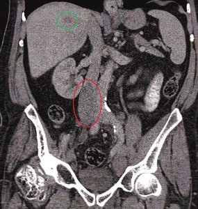
This case report identifies an IVC thrombosis in a patient with stage IV prostate cancer. The case demonstrates hypercoagulability as one of the many complications of malignancy. The patient presented clinically with bilateral pitting oedema to the groin and into the scrotum with dilated superficial abdominal veins. The prostate cancer was aggressive and unresponsive to anti-androgen therapy and brachytherapy. The latest staging CT and bone scans revealed diffuse disseminated disease and a caval thrombus. He is now receiving chemotherapy as an outpatient and unfortunately his prognosis is unfavourable.